. 2022 Mar 22;119(12):e2117334119.
doi: 10.1073/pnas.2117334119. Epub 2022 Mar 16.

Ribosomal RNA 2'- O-methylations regulate translation by impacting ribosome dynamics

Affiliations

Ribosomal RNA 2'- O-methylations regulate translation by impacting ribosome dynamics

Sohail Khoshnevis et al. Proc Natl Acad Sci U S A. .

Abstract

SignificanceThe presence of RNA chemical modifications has long been known, but their precise molecular consequences remain unknown. 2'-O-methylation is an abundant modification that exists in RNA in all domains of life. Ribosomal RNA (rRNA) represents a functionally important RNA that is heavily modified by 2'-O-methylations. Although abundant at functionally important regions of the rRNA, the contribution of 2'-O-methylations to ribosome activities is unknown. By establishing a method to disturb rRNA 2'-O-methylation patterns, we show that rRNA 2'-O-methylations affect the function and fidelity of the ribosome and change the balance between different ribosome conformational states. Our work links 2'-O-methylation to ribosome dynamics and defines a set of critical rRNA 2'-O-methylations required for ribosome biogenesis and others that are dispensable.

Keywords: 2′-O-methylation; rRNA modification; ribosome biogenesis; ribosome dynamics; translation regulation.


Conflict of interest statement

The authors declare no competing interest.

Figures

Fig. 1.
Fig. 1.
rRNA 2′-O-methylation sites change in a site-specific manner in bcd1-D72A cells. (A and D) The fraction of 2′-O-methylation (MethScore) at each modification site in 18S (A) and 25S (D) rRNA in bcd1-D72A cells relative to wild-type control cells was evaluated by RiboMethSeq. Data are shown as mean MethScore values for three independent biological replicates. (B and E) The position of each modification is marked on the 18S (B) and 25S (E) rRNA structure (Protein Data Bank [PDB] ID: 6GQV). The stable sites (MethScore > 0.8) are colored in blue, the variable sites (0.4 < MethScore < 0.8) are colored in green, and the hypo-2′-O-methylated sites (MethScore < 0.4) are colored in magenta. (C) Modifications around the decoding center (DC) within 18S rRNA. (F) Modifications around the PTC within 25S rRNA (PDB ID: 4V6I).
Fig. 2.
Fig. 2.
rRNA hypomethylation affects the function and fidelity of ribosomes. (A) Analysis of the incorporation rate of HPG into newly synthesized peptides in rapidly dividing yeast cells expressing wild-type or mutant Bcd1 over a time course (2 to 50 min). HPG-containing proteins were fluorescently labeled by addition of Alexa Fluor 488 and separated from unincorporated dye by SDS-PAGE and imaged (Top) before staining with Coomassie blue for total protein detection (Bottom). CHX stands for cycloheximide. (B) Quantification of the data shown in A. At each time point, the ratio of the newly synthesized protein to the total protein for bcd1-D72A cells is normalized to the wild-type cells. Three biological replicates were analyzed. (C) Schematic of the dual-luciferase plasmids used in this study. For all plasmids, Renilla luciferase is constitutively expressed, while the expression of firefly luciferase is dependent on a specific translational defect/element. (D) Expression of firefly and Renilla luciferase was measured in wild-type control or bcd1-D72A yeast harboring the indicated plasmids. The ratio of firefly luciferase to Renilla luciferase was normalized to the control plasmids and shown relative to wild type; three to four biological replicates were analyzed. (E) Doubling times of wild-type control and bcd1-D72A cells were measured in medium with or without translational inhibitors. The fold change, calculated by dividing the doubling time values of bcd1-D72A cells to wild-type control cells in each condition, is plotted. Three biological replicates were analyzed. In B, D, and E, column bars represent the mean values, and the error bars depict the SDs; Paromo, paromomycin; Apra, apramycin; HHT, homoharringtonine. Significance was determined using a t test; *P ≤ 0.05; **P ≤ 0.01; ***P ≤ 0.001; ****P ≤ 0.0001; n.s., nonsignificant.
Fig. 3.
Fig. 3.
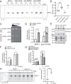
Hypo-2′-O-methylated ribosomes adopt a more rotated conformation in vivo. (A) In vivo RNA structure probing of cells expressing either wild-type or the D72A variant of Bcd1 with or without PGO treatment to probe the accessibility of SSU-G913. Cells expressing W255C or H256A variants of Rpl3 were used as controls for rotation status. Two biological replicates are shown in the figure. (B) Quantification of the SSU-G913 modification by PGO. Five biological replicates were analyzed. (C) bcd1-D72A cells are less sensitive to the overexpression of eEF2 than wild-type cells. Indicated cells were serially diluted on selective plates and grown for 48 h at 30 °C. (D) Quantification of the growth of BCD1 and bcd1-D72A cells expressing eEF2. The dot pattern indicates the expected doubling time of bcd1-D72A cells if there was no rescue of eEF2 overexpression by the bcd1-D72A mutation. (E) Western blot against free (S, supernatant) and ribosome-bound (P, pellet) eEF2 from formaldehyde-fixed whole-cell extracts from BCD1 or bcd1-D72A cells separated by centrifugation over a sucrose cushion. Rpl3 serves as an indicator of ribosome pelleting. (F) bcd1-D72A cells are less sensitive to sordarin (3 µg/mL) than wild-type cells. (G) Whereas rpl3-W255C cells have the same sensitivity to sordarin as the wild-type cells, rpl3-H256A shows less sensitivity to sordarin. In D, F, and G, four biological replicates were analyzed. (H) Probing the accessibility of SSU-G913 in NOP1 and nop1-ts using PGO. Three biological replicates are shown in the figure. (I) Quantification of the SSU-G913 modification by PGO. In B, D, F, G, and I, bars represent the mean values. The error bars in D, F, and G depict the SDs. Significance for all graphs was analyzed using a t test; *P ≤ 0.05; **P ≤ 0.01; ***P ≤ 0.001; ****P ≤ 0.0001.
Fig. 4.
Fig. 4.
Binding of eIF1 to hypomethylated 40S is weakened in vivo and in vitro. (A) Western blot against eIF1 for the fractions of sucrose gradients of formaldehyde-fixed whole-cell extracts from BCD1 (Top) or bcd1-D72A (Bottom) cells. The ratio of eIF1 in 43S–48S PIC relative to the total eIF1 is depicted under each blot. Two biological replicates were analyzed. N.D. stands for not determined. (B) Fraction of eIF1 bound to 40S plotted against the 40S concentration. Data were fitted with a nonlinear regression model in GraphPad Prism 8.0 to yield dissociation constants of 23 nM and 64 nM for wild-type and hypo-2′-O-methylated ribosomes, respectively; 95% confidence levels are shown in shades of gray. (C) Comparison of doubling times of BCD1 and bcd1-D72A cells harboring either an empty vector or vectors expressing SUI1 (eIF1) or TIF11 (eIF1A) in minimal medium containing glucose. Four biological replicates were analyzed. (D) Comparison of doubling times of BCD1 and bcd1-D72A cells in which the endogenous Rps3 is depleted and either wild-type or R116D or R117D variants of Rps3 are expressed from a plasmid. Four biological replicates were analyzed. (E) Probing the accessibility of SSU A579 in 40S ribosomal subunits purified from BCD1 and bcd1-D72A cells using DMS. Four biological replicates are shown in the figure. (F) Quantification of data shown in E for the SSU A579 modification by DMS. In C, D, and F, bars represent the mean values, and the error bars depict the SDs. Significance was analyzed using a t test. n.s., nonsignificant; ** P ≤ 0.01; ****P ≤ 0.0001.

References

    1. Helm M., Post-transcriptional nucleotide modification and alternative folding of RNA. Nucleic Acids Res. 34, 721–733 (2006). - PMC - PubMed
    1. Roundtree I. A., Evans M. E., Pan T., He C., Dynamic RNA modifications in gene expression regulation. Cell 169, 1187–1200 (2017). - PMC - PubMed
    1. Ayadi L., Galvanin A., Pichot F., Marchand V., Motorin Y., RNA ribose methylation (2′-O-methylation): Occurrence, biosynthesis and biological functions. Biochim. Biophys. Acta. Gene Regul. Mech. 1862, 253–269 (2019). - PubMed
    1. Sloan K. E., et al. , Tuning the ribosome: The influence of rRNA modification on eukaryotic ribosome biogenesis and function. RNA Biol. 14, 1138–1152 (2017). - PMC - PubMed
    1. Monaco P. L., Marcel V., Diaz J. J., Catez F., 2′-O-methylation of ribosomal RNA: Towards an epitranscriptomic control of translation? Biomolecules 8, 106 (2018). - PMC - PubMed

LinkOut - more resources