. 2022 Mar 16;18(3):e1010393.
doi: 10.1371/journal.ppat.1010393. eCollection 2022 Mar.

Differential viral RNA methylation contributes to pathogen blocking in Wolbachia-colonized arthropods

Affiliations

Differential viral RNA methylation contributes to pathogen blocking in Wolbachia-colonized arthropods

Tamanash Bhattacharya et al. PLoS Pathog. .

Abstract

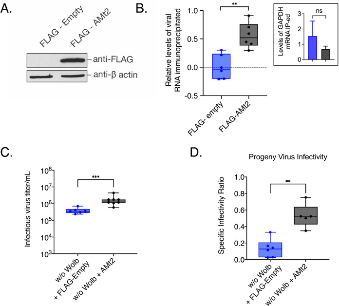
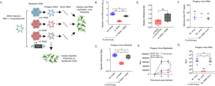
Arthropod endosymbiont Wolbachia pipientis is part of a global biocontrol strategy to reduce the replication of mosquito-borne RNA viruses such as alphaviruses. We previously demonstrated the importance of a host cytosine methyltransferase, DNMT2, in Drosophila and viral RNA as a cellular target during pathogen-blocking. Here we report a role for DNMT2 in Wolbachia-induced alphavirus inhibition in Aedes species. Expression of DNMT2 in mosquito tissues, including the salivary glands, is elevated upon virus infection. Notably, this is suppressed in Wolbachia-colonized animals, coincident with reduced virus replication and decreased infectivity of progeny virus. Ectopic expression of DNMT2 in cultured Aedes cells is proviral, increasing progeny virus infectivity, and this effect of DNMT2 on virus replication and infectivity is dependent on its methyltransferase activity. Finally, examining the effects of Wolbachia on modifications of viral RNA by LC-MS show a decrease in the amount of 5-methylcytosine modification consistent with the down-regulation of DNMT2 in Wolbachia colonized mosquito cells and animals. Collectively, our findings support the conclusion that disruption of 5-methylcytosine modification of viral RNA is a vital mechanism operative in pathogen blocking. These data also emphasize the essential role of epitranscriptomic modifications in regulating fundamental alphavirus replication and transmission processes.


Conflict of interest statement

The authors have declared no competing interests exist.

Figures

Fig 1
Fig 1. Virus and Wolbachia each differentially modulate expression of the RNA methyltransferase gene DNMT2 in mosquitoes.
(A) AMt2 expression was measured 5 days post bloodmeal in whole female mosquitoes using qRT-PCR with and without SINV. Error bars represent standard error of mean (SEM) of biological replicates. Two-way ANOVA with Tukey’s post-hoc test of multivariate comparisons. (B) AMt2 expression measured in dissected salivary gland tissues collected from female mosquitoes with and Wolbachia-free 5 days post bloodmeal with or without SINV. Error bars represent standard error of mean (SEM) of biological replicates. Two-way ANOVA with Tukey’s post-hoc test of multivariate comparisons. (C) Viral RNA levels were quantified in dissected salivary gland tissues with and Wolbachia-free using qRT-PCR at 5 days post infectious blood meal with SINV. Unpaired, student’s t-test, error bars represent standard error of mean (SEM) of biological replicates. For all panels: ****P < 0.0001; ***P < 0.001; **P < 0.01; ns = non-significant.
Fig 2
Fig 2. Overexpressing AMt2 in mosquito cells improve progeny virus infectivity.
(A) Western Blot of Aedes albopictus (C7/10) cells transfected with expression vector constructs with (FLAG-AMt2) or without (FLAG-empty) AMt2. Cytoplasmic lysates of cells were collected 48 hours post transfection and probed with anti-FLAG and anti-β actin antibodies. (B) Relative levels of SINV RNA recovered following AZA-IP of AMt2 in C7/10 cells was quantified using qRT-PCR. Wolbachia-free C7/10 mosquito cells were transfected with expression vectors FLAG-empty or FLAG-AMt2 for 48 hours prior to infection with SINV at MOI of 10. Cells were treated for approximately 18h with 5 μM 5-Azacytidine to covalently trap AMt2 with its target cellular RNA prior to RNA immunoprecipitation using anti-FLAG antibody. The horizontal dotted line represents the threshold set at 1 (log10). Inset bar graph show relative levels of GAPDH mRNA immunoprecipitated with anti-FLAG antibody. Unpaired two-tailed t-test with Welch’s correction, p = 0.0004, t = 4.216, df = 20 (C) Infectious progeny (PFU/mL) SINV produced from mosquito cells Wolbachia-free expressing either FLAG-empty (w/o Wolb) or FLAG-AMt2 (w/o Wolb + AMt2). Cells were transfected 48 hours prior to infection with SINV at MOI of 10. Infectious progeny viruses collected from supernatants 48 hours post-infection were quantified using plaque assays on BHK-21 cells. Unpaired two-tailed t-test with Welch’s correction, p = 0.0002, t = 5.404, df = 11.81. (D) Specific Infectivity Ratios of progeny SINV were calculated as described earlier [1]. Unpaired two-tailed t-test with Welch’s correction, p = 0.0084, t = 3.911, df = 5.820. For all panels error bars represent standard error of mean (SEM) of biological replicates and **P < 0.01; ****P < 0.0001.
Fig 3
Fig 3. Pharmacological inhibition of mosquito DNMT2 reduces virus replication and per-particle infectivity.
Inhibition of mosquito DNMT2 in Wolbachia-free Aedes albopictus derived C7/10 cells was carried out using MTase inhibitors 5-Azacytidine (5-AZAC). Dimethyl-sulfoxide (DMSO) was used as the negative control. In each case, cells were pretreated with 5 μM inhibitors overnight prior to infections with SINV at MOI of 10. Cell lysates and supernatants were harvested at 24 hours post infection to quantify cellular viral RNA levels and infectious titer, respectively. (A) Levels of SINV RNA in mosquito cells treated with MTase inhibitor 5-AZAC were determined using quantitative RT-PCR. Unpaired two-tailed t-test with Welch’s correction, SINV: p = 0.0012, t = 6.742, df = 4.892. (B) Infectious SINV titers produced from mosquito cells treated with MTase inhibitor 5-AZAC were determined using plaque assays on BHK-21 cells. Unpaired two-tailed t-test with Welch’s correction, SINV: p = 0.0339, t = 4.541, df = 2.322. (C) Specific infectivity ratios of progeny SINV was calculated as the ratio of infectious plaque forming units (B) over total viral genome copies present in collected cell supernatants as quantified by qRT-PCR. Unpaired two-tailed t-test with Welch’s correction, SINV: p = 0.0002, t = 12.59, df = 3.946. Error bars represent standard error of mean (SEM) of three independent experimental replicates. (D) Schematic representation of live cell experiments. CHIKV expressing mKate fluorescent reporter protein was grown in C7/10 Aedes albopictus cells in the presence (W+ virus) or absence (W- virus) of Wolbachia (strain wStri). These progeny viruses were then used to infect naïve C7/10 cells without (E) and with (F) Wolbachia (strain wStri) pretreated with MTase inhibitor or DMSO synchronously at a MOI of 1 particle/cell. Virus growth in cells was measured in real time by imaging and quantifying the number of red cells (Virus Positive Cells/Image) expressing the virus encoded mKate protein over a period of forty-eight hours, using live cell imaging. Color of the data points distinguish treatment conditions; blue represent C7/10 Wolbachia-free cells treated with DMSO, red represent C7/10 Wolbachia-colonized cells treated with DMSO, black represent both C7/10 cell types treated with 5 μM DAC5. Shape of data points represent the progeny virus type used to initiate infection; boxes represent viruses derived from W- cells, circles represent viruses derived from W+ cells. The Y-axis label red object count/Image represent virus-positive cells in a single field of view, four of which were collected and averaged/sample at each two-hour time point over the course of infection. Three-way ANOVA with Tukey’s post hoc test for multivariate comparisons. Error bars represent standard error of mean (SEM) of independent experimental replicates (n = 3). Three-way ANOVA with Tukey’s multivariate analyses, DAC5: p = 0.1793, Virus Source: p = 0.5060, Time: p < 0.0001, DAC5 X Time: p > 0.99, Virus Source X Time: p > 0.99, Virus Source X DAC5: p = 0.1039, Virus Source X DAC5 X Time: p = 0.9804 (Fig 3E). *P < 0.05; **P < 0.01, ****P < 0.0001. Three-way ANOVA with Tukey’s multivariate analyses, DAC5: p < 0.0001, Virus Source: p < 0.0001, Time: p < 0.0001, DAC5 X Time: p < 0.0001, Source X Time: p < 0.0001, Virus Source X DAC5: p = 0.1148, Virus Source X DAC5 X Time: p > 0.999 (Fig 3F). Graphical assets were made in BioRender (https://biorender.com).
Fig 4
Fig 4. AMt2 overexpression in Wolbachia-colonized cells rescues virus from endosymbiont-mediated inhibition.
C7/10 cells with Wolbachia were transfected with expression vectors FLAG-empty (w/ Wolb) or FLAG-AMt2 (w/ Wolb + AMt2) for 48 hours prior to infection with SINV-nLuc at MOI of 10. Wolbachia -free cells expressing FLAG-empty (w/o Wolb) were used as a positive control. (A) Schematic of experimental workflow. (B) Viral genome replication in C7/10 cells was quantified using qRT-PCR using extracted total RNA from infected cell lysates. One-way ANOVA with Tukey’s post-hoc test of multivariate comparison. (C) Specific Infectivity Ratios of progeny viruses produced from the aforementioned infection was calculated as described earlier [1]. Briefly, infectious progeny viruses collected from supernatants 48 hours post infection were quantified using plaque assays on BHK-21 cells, while total number of progeny virus particles was quantified via qRT-PCR of viral genome copies released into the supernatant. Error bars represent standard error of mean (SEM). One-way ANOVA with Tukey’s post-hoc test of multivariate comparison, w/ Wolb vs w/ Wolb + AMt2, p = 0.0003, w/o Wolb vs w/ Wolb, p < 0.0001. (D) C7/10 mosquito cells with Wolbachia were transfected with expression vectors FLAG-empty (w/ Wolb) or FLAG-AMt2 (w/ Wolb + AMt2) for 48 hours prior to quantification of endosymbiont titer via quantitative PCR using DNA from extracted cell lysates. Error bars represent standard error of mean (SEM). Unpaired, student’s t-test, p = 0.1316, t = 1.794, df = 5.097. Statistically non-significant values are indicated by ns. (E) Progeny viruses were used to synchronously infect naïve BHK-21 cells at equivalent MOIs of 5 particles/cell. Cell lysates were collected at indicated times post infection and luciferase activity (RLU), was used as a proxy for viral replication. Two-way ANOVA with Tukey’s post-hoc test of multivariate comparison, Time: p < 0.0001, Wolbachia/AMt2: p = 0.0003, Time x Wolbachia/AMt2: p < 0.0001. (F) Approximately 105 copies (determined using qRT-PCR) each of virion encapsidated RNA extracted from the aforementioned W+, W+ AMt2 and W- viruses were transfected into naïve BHK-21 cells and infectious titer was determined by the counting the number of plaques produced after 72 hours post transfection. Numbers above bars refer to the proportion of samples that formed quantifiable plaque-forming units on BHK-21 cells. One-way ANOVA with Tukey’s post-hoc test of multivariate comparison. (G) 105 copies each of virion encapsidated RNA extracted from the W+, W+ AMt2 and W- viruses were transfected into naïve BHK-21 cells and luciferase activity (RLU) was used as a proxy for viral replication at 9 hours post-transfection. Numbers above bars refer to the proportion of samples that produced luciferase signal above background levels, indicated by the dotted line. One-way ANOVA with Tukey’s post-hoc test of multivariate comparison, w/ Wolb vs w/ Wolb + AMt2: p < 0.00001, w/o Wolb vs w/ Wolb: p < 0.0001; w/o Wolb vs w/ Wolb + AMt2: p = 0.991. For all panels error bars represent standard error of mean (SEM). *P < 0.05; **P < 0.01; ****P < 0.0001. Graphical assets were made in BioRender (https://biorender.com).
Fig 5
Fig 5. Catalytically inactive DNMT2 is unable to rescue Wolbachia-mediated virus inhibition in mosquito cells.
(A) Multiple sequence alignment of the Motif IV region of DNMT2 derived from dipterans that are known to be colonized with native or non-native Wolbachia. The conserved catalytic cysteine (C) residue, depicted in red on the consensus sequence at the top, was mutated to a glycine (G) to abolish MTase activity of mosquito AMt2. Expression of the catalytic mutant (AMt2 C78G) was determined at 48 hours post transfection using Western Blot. C7/10 mosquito cells transfected with expression vector constructs with (FLAG-AMt2 C78G) or without (FLAG-empty) AMt2. Cytoplasmic lysates of cells were collected 48 hours post transfection and probed with anti-FLAG antibody. Bar graphs represent wild-type (example in Fig 2A) and C78G AMt2 mutant protein levels normalized to β-actin controls. (B) C7/10 mosquito cells with Wolbachia were transfected with expression vectors FLAG-empty (w/ Wolb) or FLAG-C78G AMt2 (w/ Wolb + AMt2 C78G) for 48 hours prior to infection with SINV at MOI of 10. Viral RNA was quantified using quantitative RT-PCR as before. Unpaired t-test with Welch’s correction, p = 0.1734, t = 1.920, df = 2.396. (C) C7/10 mosquito cells with and without Wolbachia were transfected with expression vectors FLAG-empty (w/o Wolb, w/ Wolb) and FLAG-C78G AMt2 (w/ Wolb + AMt2 C78G) for 48 hours prior to infection with SINV at MOI of 10. Specific Infectivity (SI) of progeny viruses produced after 72 hours post infection were quantified as before. One-way ANOVA with Dunnett’s test for multivariate analyses. w/o Wolb vs w/ Wolb, p = 0.035, w/o Wolb vs w/ Wolb + AMt2 C78G p = 0.032, w/ Wolb + AMt2 vs w/ Wolb + AMt2 C78G p = 0.9963. Error bars represent standard error of mean (SEM) of biological replicates. *P < 0.05, ns = non-significant.
Fig 6
Fig 6. Presence of Wolbachia is associated with altered virion RNA methylation.
(A) RNA isolated from progeny viruses derived from mosquito cells colonized with (W+ virus) or without (W- virus) Wolbachia were subjected to LC-MS/MS analyses to determine their nucleotide content. (B) Normalized 5-methyl cytosine (M5C) content of RNA isolated from W- and W+ viruses represented as a ratio of total unmodified cytosine content. Unpaired two-tailed t-test, p = 0.0013, t = 8.080, df = 4 (C) Normalized 6-methyl adenosine (M6A) content of RNA isolated from W- and W+ viruses represented as a ratio of total unmodified adenosine content. Unpaired two-tailed t-test, p = 0.666, t = 0.4643, df = 4. Error bars represent standard error of mean (SEM) of three independent virus preps from each cell type. **P < 0.01; ns = non-significant. (D) Model delineating the role of Aedes cytosine MTase AaDNMT2 in pathogen blocking. MTase (AMt2) expression is induced following alphavirus infection in mosquitoes constituting a proviral environment. Virion RNA produced under these conditions are m5C-methylated and efficiently disseminate across mosquito cells. Elevated MTase expression in salivary gland tissues result in the production of virions (W- virus) containing methylated viral RNA that are infectious in mammalian cells, which promotes efficient transmission. In contrast, MTase expression is reduced in mosquitoes colonized with Wolbachia and inhibition occurring at early stages of virus infection prevent virus-induced MTase upregulation. Consequently, virion RNA (W+ virus) is hypomethylated at cytosine residues, which compromise viral dissemination and transmission events. Graphical assets were made in BioRender (https://biorender.com).

References

    1. Bhattacharya T, Newton ILG, Hardy RW. Viral RNA is a target for Wolbachia-mediated pathogen blocking. PLoS Pathog. 2020;16(6):e1008513. doi: 10.1371/journal.ppat.1008513 - DOI - PMC - PubMed
    1. Díaz-Sánchez S, Hernández-Jarguín A, Torina A, Fernández de Mera I, Estrada-Peña A, Villar M, et al.. Biotic and abiotic factors shape the microbiota of wild-caught populations of the arbovirus vector Culicoides imicola. Insect molecular biology. 2018;27(6):847–61. doi: 10.1111/imb.12526 - DOI - PubMed
    1. Bellone R, Failloux A-B. Temperature in shaping mosquito-borne viruses transmission. Frontiers in Microbiology. 2020;11:2388. doi: 10.3389/fmicb.2020.584846 - DOI - PMC - PubMed
    1. Tabachnick WJ. Ecological effects on arbovirus-mosquito cycles of transmission. Current opinion in virology. 2016;21:124–31. doi: 10.1016/j.coviro.2016.09.008 - DOI - PubMed
    1. Hegde S, Rasgon JL, Hughes GL. The microbiome modulates arbovirus transmission in mosquitoes. Current opinion in virology. 2015;15:97–102. doi: 10.1016/j.coviro.2015.08.011 - DOI - PMC - PubMed

Publication types