. 2022 May 18;110(10):1671-1688.e6.
doi: 10.1016/j.neuron.2022.02.011. Epub 2022 Mar 15.

Loss of mouse Stmn2 function causes motor neuropathy

Affiliations

Loss of mouse Stmn2 function causes motor neuropathy

Irune Guerra San Juan et al. Neuron. .

Erratum in

  • Loss of mouse Stmn2 function causes motor neuropathy.
    San Juan IG, Nash LA, Smith KS, Leyton-Jaimes MF, Qian M, Klim JR, Limone F, Dorr AB, Couto A, Pintacuda G, Joseph BJ, Whisenant DE, Noble C, Melnik V, Potter D, Holmes A, Burberry A, Verhage M, Eggan K. San Juan IG, et al. Neuron. 2022 Dec 7;110(23):4031. doi: 10.1016/j.neuron.2022.11.003. Neuron. 2022. PMID: 36480942 No abstract available.

Abstract

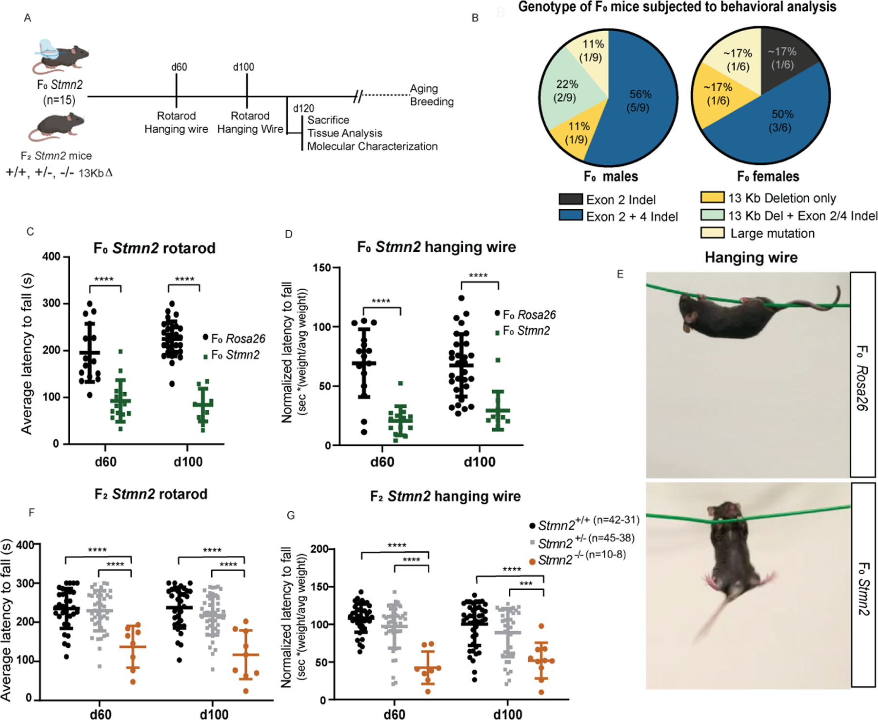
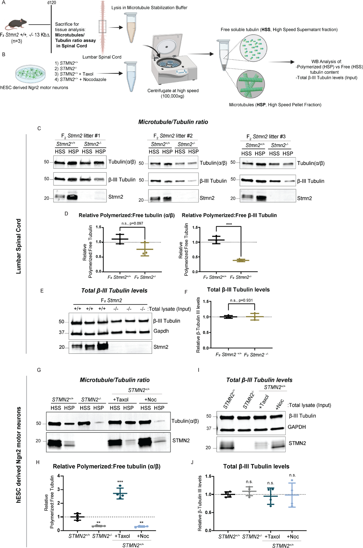
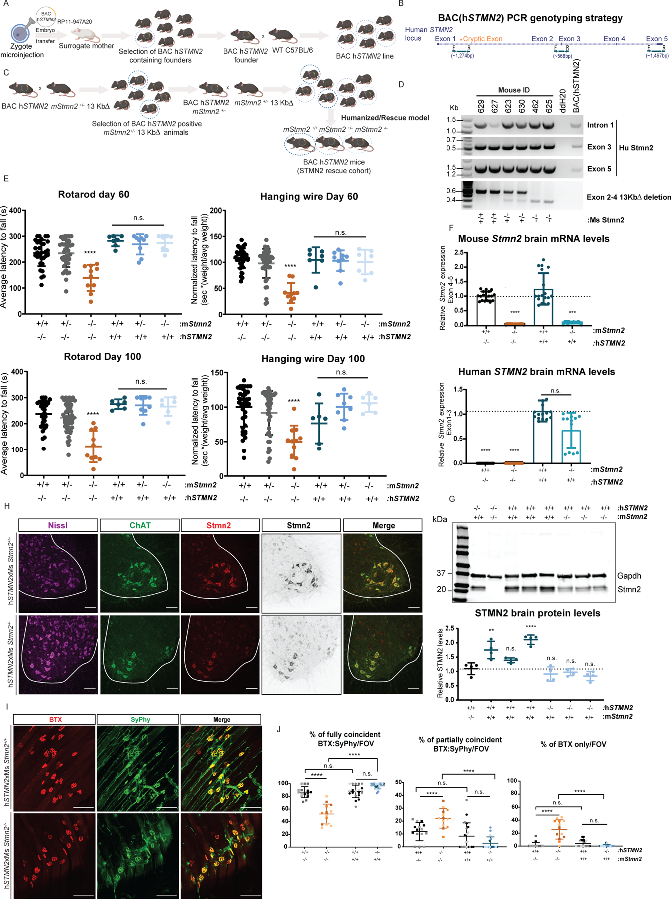
Amyotrophic lateral sclerosis (ALS) is characterized by motor neuron degeneration accompanied by aberrant accumulation and loss of function of the RNA-binding protein TDP43. Thus far, it remains unresolved to what extent TDP43 loss of function directly contributes to motor system dysfunction. Here, we employed gene editing to find whether the mouse ortholog of the TDP43-regulated gene STMN2 has an important function in maintaining the motor system. Both mosaic founders and homozygous loss-of-function Stmn2 mice exhibited neuromuscular junction denervation and fragmentation, resulting in muscle atrophy and impaired motor behavior, accompanied by an imbalance in neuronal microtubule dynamics in the spinal cord. The introduction of human STMN2 through BAC transgenesis was sufficient to rescue the motor phenotypes observed in Stmn2 mutant mice. Collectively, our results demonstrate that disrupting the ortholog of a single TDP43-regulated RNA is sufficient to cause substantial motor dysfunction, indicating that disruption of TDP43 function is likely a contributor to ALS.

Keywords: ALS; CRISPR; SCG10; TARDBP; TDP43; microtubules; motor neuron; motor neuropathy; neuromuscular junction; stathmin 2.


Conflict of interest statement

Declaration of interests K.E. is a cofounder of Q-State Biosciences, Quralis, and Enclear Therapies and currently head of research and early development at BioMarin Pharmaceutical. J.R.K. is an employee of Faze Medicines and a shareholder in Faze Medicines and QurAlis. K.E., I.G.S.J., J.R.K., and F.L. are authors on a pending patent that describes methods and compositions for restoring STMN2 levels (WO/2020/150290). K.E. is an author on a pending patent that describes compounds and methods for treating neurodegenerative diseases (WO2020107037).

Figures

Figure 1.
Figure 1.. CRISPR/Cas9 editing of Stmn2
(A) Diagram of breeding strategy to generate Stmn2 F0, Rosa26 gRNA, and Stmn2 F2 mice. (B) Schematic representation of the STMN2 locus in which gRNAs were targeted to exons 2 and 4 to create a predicted 13 Kb deletion. Primers were designed to flank the region of the deletion region to confirm the presence or absence of mutations. (C) PCR genotyping of F2 Stmn2 mice exhibiting WT (+/+), heterozygous (+/−), and homozygous (−/−) mutations. (D) F2 Stmn2 brain mRNA levels flanking Exons 1 to 3 (left) and Exons 4 to 5 (right). (E) Spinal cord and brain Stmn2 protein levels from Western blots including the housekeeping protein GAPDH. **** p < 0.0001. In all figures: results are shown as a mean with error bars calculated as standard deviation. Detailed information (average, SD, n and detailed statistics) is shown in STAR Methods.
Figure 2.
Figure 2.. Stmn2 mutant mice display motor deficits
(A) Diagram of the experimental strategy to examine Stmn2 LOF on motor behavior in Stmn2 F0 mice. (B) Stmn2 F0 genotype subjected to behavioral analysis. (C) Rotarod and (D) hanging wire performance at p60 and p100 in F0 Stmn2 mutant mice and Rosa26 F0 controls, (P60: n=15 Rosa26 F0, n=13 F0 Stmn2; p100: n=29 Rosa26 F0, n=10 F0 Stmn2) (E) Graphical representation of Rosa26 F0 (top) and Stmn2 F0 (bottom) mice performing the hanging wire test. F2 Stmn2 performance on (F) rotarod and (G) hanging wire at p60 and p100. **** p < 0.0001.
Figure 3.
Figure 3.. Loss of Stmn2 leads to denervation in the hindlimb gastrocnemius
(A-B) Diagram of the experimental strategy to examine innervation in the GA of mice. (C) Whole mount preparations from Stmn2 F2 mutant mice and littermate controls were stained with fluorescently conjugated BTX and anti-SyPhy antibodies at p21 (top panel) and p120 (bottom panel) (D) and quantified the extent of co-localization per NMJ per field of view (FOV). ns, p>0.05, **p < 0.01, **** p < 0.0001. (p21–23: n=5 Stmn2+/+and n=4 Stmn2−/− mice; p120 n=3 Stmn2+/+ and −/−). Data points for each animal are represented by different colors.
Figure 4.
Figure 4.. Stmn2 loss-driven denervation results in NMJ fragmentation and muscle injury
(A) Diagram of the experimental strategy to examine level of fragmentation in the NMJs within the GA of mice. (B) Diagram exhibiting NMJ denervation over time. (C) GA isolated from Stmn2 F2 and littermate controls, stained with fluorescent conjugated BTX. (D) Quantification of fragmented NMJs per FOV in Stmn2 F2 and littermate controls. (E) Diagram of the experimental strategy to examine the level of centralized myonuclei in the GA. (F) Representative cross-sectional images of F2 Stmn2 mutants and control littermates. (G) Quantification of centralized myonuclei F2 Stmn2 and control littermates per FOV. ns, p>0.05, *** p < 0.001, **** p < 0.0001. (p21–23: n=5 Stmn2+/+ and n=4 Stmn2−/− mice; p120: n=3 Stmn2+/+ and −/− mice). Data points for each animal are represented by different colors.
Figure 5.
Figure 5.. Stmn2 is selectively expressed in motor neurons within the adult ventral spinal cord
(A) Diagram of the experimental strategy to examine Stmn2 expression in the lumbar spinal cord. (B) Stmn2 F0 genotypes in mice used for spinal cord analysis. (C) Stmn2 F0 mutant mice and Rosa26 controls cords stained with antibodies specific to Chat and Stmn2 proteins Quantification of (D) Chat positive and Stmn2 positive neurons in each hemi-section of Stmn2 F0 mutant mice and Rosa26 F0 controls. (E) Percentage of Chat positive motor neurons expressing Stmn2 protein quantified per hemi-section in Stmn2 F0 mice and Rosa26 F0 controls. (F) Ventral horn spinal cord sections from F2 Stmn2 mice and control littermates were stained with anti-Stmn2 antibodies and anti-Chat antibodies to detect motor neurons. Quantification of (G) Chat positive and Stmn2 positive neurons quantified per hemi-section in F2 Stmn2 mice and littermate controls. (H) Percent of Chat positive motor neurons containing Stmn2 protein quantified per hemi-section in F2 Stmn2 mice and littermate controls. **** p < 0.0001. Scale bars, 50 μm.
Figure 6.
Figure 6.. Absence of neural inflammation and Tdp-43 pathology in Stmn2 mutant cord.
(A) Diagram of the experimental strategy to examine astrocyte and microglia activation within the spinal cord along with Tdp-43 localization. (B) Diagram of cell types analyzed in the spinal cords. (C) Stmn2 F0 genotype corresponding to the spinal cord analysis. Hemi-sections of the lumbar spinal cord from Stmn2 F0 mice and Rosa26 F0 controls were stained for (D) GFAP, (E) Iba1, and (F) Tdp-43 and quantified in motor neurons (F). Graphs of cell counts (G), ns, p>0.05 (n=3 animals/genotype, n=5 sections/animal, for Tdp43 localization analysis: n=100 Chat+ neurons counted/animal)
Figure 7.
Figure 7.. Loss of Stmn2 in the murine spinal cord and in cultured human motor neurons impairs microtubule dynamics.
(A-B) Diagram of the experimental strategy to examine free and polymerized tubulin in Stmn2 F2 and control littermates, as well as hPSC derived Ngn2 motor neurons. (C-D) Quantified western blot analysis of the ratio of polymerized (HSP, High Speed Pellet) and free (HSS, High Speed Supernatant) tubulin (α/β and β-III) fractions isolated from the lumbar spinal cord of Stmn2−/− mice and normalized to that of Stmn2+/+ controls littermates. (E-F) Levels of overall β III tubulin in the tissue lysates (Input fraction), n=3 animals/genotype. Respective littermate controls displayed. Average of n=3 technical replicates represented. (G-H) Quantified western blot analysis of the ratio of polymerized (HSP) and free (HSS) tubulin (α/β) fractions isolated from STMN2−/− hPSC-derived Ngn2 motor neurons, normalized to corresponding controls STMN2+/+ hMNs. STMN2+/+ hMNs were also treated with microtubule regulator drugs, Taxol (1μM for 72 hours) and Nocodazole (10 μM for 45 minutes) as experimental controls. (I-J) Levels of overall β III tubulin in the cultured motor neuron lysates (Input fraction). n=2 biologically independent experiments (2 technical replicates represented for each experiment).
Figure 8.
Figure 8.. Introduction of hSTMN2 gene rescues LOF-associated motor deficits.
(A) Diagram of breeding strategy to generate BAChSTMN2 transgenic line and subsequent hSTMN2xmStmn2+/+, +/−, −/− lines in (B). (C) Schematic representation of the STMN2 locus in which primers were designed to flank beginning (intron 1), middle (Exon 3) and end (Exon 5). (D) PCR genotyping of hSTMN2 exhibiting presence of the hSTMN2 gene, and of WT (+/+), heterozygous (+/−), and homozygous (−/−) mutations of the mouse Stmn2 gene (E) Rotarod (left panel) and hanging wire (right panel) at p60 and p100 in hSTMN2xmStmn2+/+ and −/− compared to Stmn2 mutant mice. (p60: hSTMN2xmStmn2+/+ n=7, +/− n=9, −/− n=7; p100 hSTMN2xmStmn2+/+ n=6, +/− n=8, −/− n=6). (F) mouse Stmn2 Exons 4–5 (left) and human STMN2 flanking Exons 1–3 (right) in transgenic mice compared to Stmn2 mutant and wild-type littermate mice. (G) Brain Stmn2 protein levels from Western blots including the housekeeping protein GAPDH. (H) Representative stain of hSTMN2xmStmn2+/+ and −/− cords labeled with anti-Chat and anti-Stmn2 and counterstained for Nissl. (I) Whole mount preparations from hSTMN2xmStmn2+/+ and −/− mice were stained with fluorescently conjugated BTX and anti-SyPhy at p120 (J) and quantified the extent of co-localization per NMJ per FOV compared to Stmn2 mutant mice. * p < 0.05, **p < 0.01, *** p < 0.001, **** p < 0.0001 (n=3 animals/genotype, hSTMN2xmStmn2+/+ and −/−). Scale bars, 100 μm. Results from Stmn2 mutant mice in (E,J) are those generated in Figures 2 and 3.

References

    1. Al-Chalabi A, Calvo A, Chio A, Colville S, Ellis CM, Hardiman O, Heverin M, Howard RS, Huisman MHB, Keren N, et al. (2014). Analysis of amyotrophic lateral sclerosis as a multistep process: a population-based modelling study. Lancet Neurol 13, 1108–1113. 10.1016/S14744422(14)70219-4. - DOI - PMC - PubMed
    1. Alami NH, Smith RB, Carrasco MA, Williams LA, Winborn CS, Han SSW, Kiskinis E, Winborn B, Freibaum BD, Kanagaraj A, et al. (2014). Axonal transport of TDP-43 mRNA granules is impaired by ALS-causing mutations. Neuron 81, 536–543. 10.1016/j.neuron.2013.12.018. - DOI - PMC - PubMed
    1. Arnold ES, Ling SC, Huelga SC, Lagier-Tourenne C, Polymenidou M, Ditsworth D, Kordasiewicz HB, McAlonis-Downes M, Platoshyn O, Parone PA, et al. (2013). ALS-linked TDP-43 mutations produce aberrant RNA splicing and adult-onset motor neuron disease without aggregation or loss of nuclear TDP-43. Proceedings of the National Academy of Sciences of the United States of America 110, E736–745. 10.1073/pnas.1222809110 - DOI - PMC - PubMed
    1. Ash PEA, Zhang Y-J, Roberts CM, Saldi T, Hutter H, Buratti E, Petrucelli L, and Link CD (2010). Neurotoxic effects of TDP-43 overexpression in C. elegans. Human molecular genetics 19, 3206–3218. 10.1093/hmg/ddq230. - DOI - PMC - PubMed
    1. Balice-Gordon RJ, and Lichtman JW (1990). In vivo visualization of the growth of pre- and postsynaptic elements of neuromuscular junctions in the mouse. J Neurosci 10(3), 894–908. 10.1523/JNEUROSCI.10-03-00894.1990 - DOI - PMC - PubMed

Publication types