

Effects of Prenatal Exposure to Titanium Dioxide Nanoparticles on DNA Methylation and Gene Expression Profile in the Mouse Brain
- PMID: 35295148
- PMCID: PMC8915839
- DOI: 10.3389/ftox.2021.705910
Effects of Prenatal Exposure to Titanium Dioxide Nanoparticles on DNA Methylation and Gene Expression Profile in the Mouse Brain
Abstract
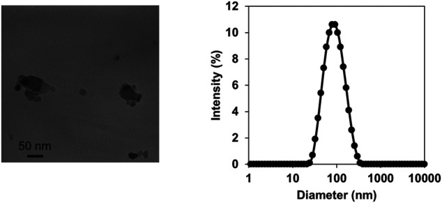
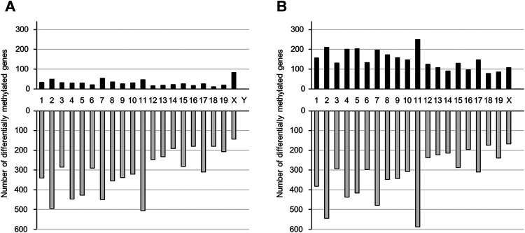
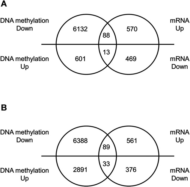
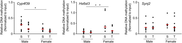
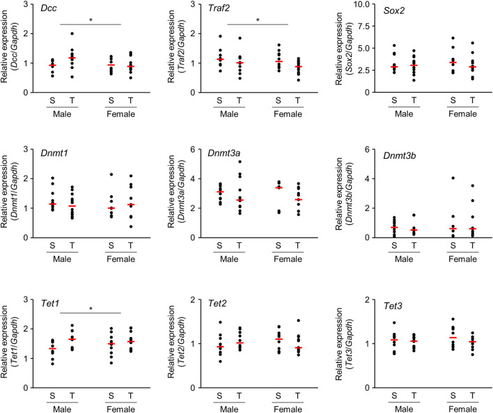
Background and Objectives: Titanium dioxide nanoparticles (TiO2-NP) are important materials used in commercial practice. Reportedly, TiO2-NP exposure during pregnancy can affect the development of the central nervous system in mouse offspring; however, the underlying mechanism remains unknown. In the present study, we investigated the impact of prenatal TiO2-NP exposure on global DNA methylation and mRNA expression patterns in the brains of neonatal mice. Materials and Methods: Pregnant C57BL/6J mice were intratracheally administered a TiO2-NP suspension (100 μg/mouse) on gestational day 10.5, and brains were collected from male and female offspring at day 1 postpartum. After extraction of methylated DNA by immunoprecipitation, the DNA methylation profile was analyzed using a mouse CpG island microarray. Total RNA was obtained, and mRNA expression profiles were comprehensively assessed using microarray analysis. Results: Among genes in the CpG island microarray, DNA methylation was increased in 614 and 2,924 genes and decreased in 6,220 and 6,477 genes in male and female offspring, respectively. Combined with mRNA microarray analysis, 88 and 89 genes were upregulated (≥1.5-fold) accompanied by demethylation of CpG islands, whereas 13 and 33 genes were downregulated (≤0.67-fold) accompanied by methylation of CpG islands in male and female offspring mice, respectively. Gene Set Enrichment Analysis (GSEA) revealed that these genes were enriched in gene ontology terms related to the regulation of transcription factors, cell proliferation, and organism development. Additionally, MeSH terms related to stem cells and morphogenesis were enriched. Conclusion: Prenatal TiO2-NP exposure induced genome-wide alterations in DNA methylation and mRNA expression in the brains of male and female offspring. Based on GSEA findings, it can be speculated that prenatal TiO2-NP exposure causes adverse effects on brain functions by altering the DNA methylation state of the fetal brain, especially neural stem cells, resulting in the subsequent abnormal regulation of transcription factors that modulate development and differentiation.
Keywords: DNA methylation; brain; gene expression; prenatal exposure; titanium dioxide nanoparticle.
Copyright © 2021 Tachibana, Kawazoe, Onoda, Umezawa and Takeda.
Conflict of interest statement
The authors declare that the research was conducted in the absence of any commercial or financial relationships that could be construed as a potential conflict of interest.
Figures
Similar articles
-
Prenatal diesel exhaust exposure disrupts the DNA methylation profile in the brain of mouse offspring.J Toxicol Sci. 2015 Feb;40(1):1-11. doi: 10.2131/jts.40.1. J Toxicol Sci. 2015. PMID: 25560391
-
Dose-dependent induction of astrocyte activation and reactive astrogliosis in mouse brain following maternal exposure to carbon black nanoparticle.Part Fibre Toxicol. 2017 Feb 2;14(1):4. doi: 10.1186/s12989-017-0184-6. Part Fibre Toxicol. 2017. PMID: 28148272 Free PMC article.
-
Different effects of titanium dioxide nanoparticles instillation in young and adult mice on DNA methylation related with lung inflammation and fibrosis.Ecotoxicol Environ Saf. 2019 Jul 30;176:1-10. doi: 10.1016/j.ecoenv.2019.03.055. Epub 2019 Mar 21. Ecotoxicol Environ Saf. 2019. PMID: 30903973
-
Maternal exposure to titanium dioxide nanoparticles during pregnancy and lactation alters offspring hippocampal mRNA BAX and Bcl-2 levels, induces apoptosis and decreases neurogenesis.Exp Toxicol Pathol. 2017 Jul 5;69(6):329-337. doi: 10.1016/j.etp.2017.02.006. Epub 2017 Feb 27. Exp Toxicol Pathol. 2017. PMID: 28254502
-
Effect of titanium dioxide nanoparticles on DNA methylation in multiple human cell lines.Nanotoxicology. 2020 May;14(4):534-553. doi: 10.1080/17435390.2020.1723730. Epub 2020 Feb 7. Nanotoxicology. 2020. PMID: 32031460
Cited by
-
Multigenerational inheritance of breathing deficits following perinatal exposure to titanium dioxide nanoparticles in the offspring of mice.Discov Nano. 2024 Jan 23;19(1):16. doi: 10.1186/s11671-023-03927-0. Discov Nano. 2024. PMID: 38261116 Free PMC article.
-
Neurotoxicity of Titanium Dioxide Nanoparticles: A Comprehensive Review.Int J Nanomedicine. 2023 Dec 5;18:7183-7204. doi: 10.2147/IJN.S442801. eCollection 2023. Int J Nanomedicine. 2023. PMID: 38076727 Free PMC article. Review.
-
Metals Exposures and DNA Methylation: Current Evidence and Future Directions.Curr Environ Health Rep. 2022 Dec;9(4):673-696. doi: 10.1007/s40572-022-00382-4. Epub 2022 Oct 25. Curr Environ Health Rep. 2022. PMID: 36282474 Free PMC article. Review.
-
Protective role of the ginsenoside Rg1 against methimazole-induced gestational hypothyroidism on reflexive behaviors, conditioned fear and cortical antioxidant levels in mice offspring.IBRO Neurosci Rep. 2024 Mar 30;16:485-496. doi: 10.1016/j.ibneur.2024.03.010. eCollection 2024 Jun. IBRO Neurosci Rep. 2024. PMID: 38634016 Free PMC article.
-
A Novel Staining Method for Detection of Brain Perivascular Injuries Induced by Nanoparticle: Periodic Acid-Schiff and Immunohistochemical Double-Staining.Front Toxicol. 2022 Mar 21;4:825984. doi: 10.3389/ftox.2022.825984. eCollection 2022. Front Toxicol. 2022. PMID: 35391824 Free PMC article.
References
LinkOut - more resources
Full Text Sources
Miscellaneous