Review
. 2022 Feb 8:4:814071.
doi: 10.3389/ftox.2022.814071. eCollection 2022.

Using the Isolated Rat Placenta to Assess Fetoplacental Hemodynamics

Affiliations
Review

Using the Isolated Rat Placenta to Assess Fetoplacental Hemodynamics

K L Garner et al. Front Toxicol. .

Abstract

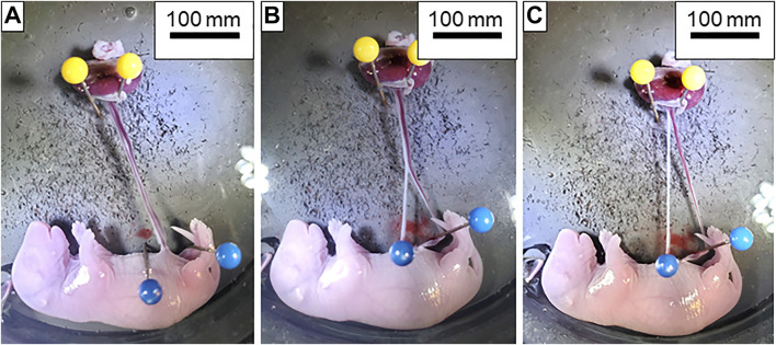
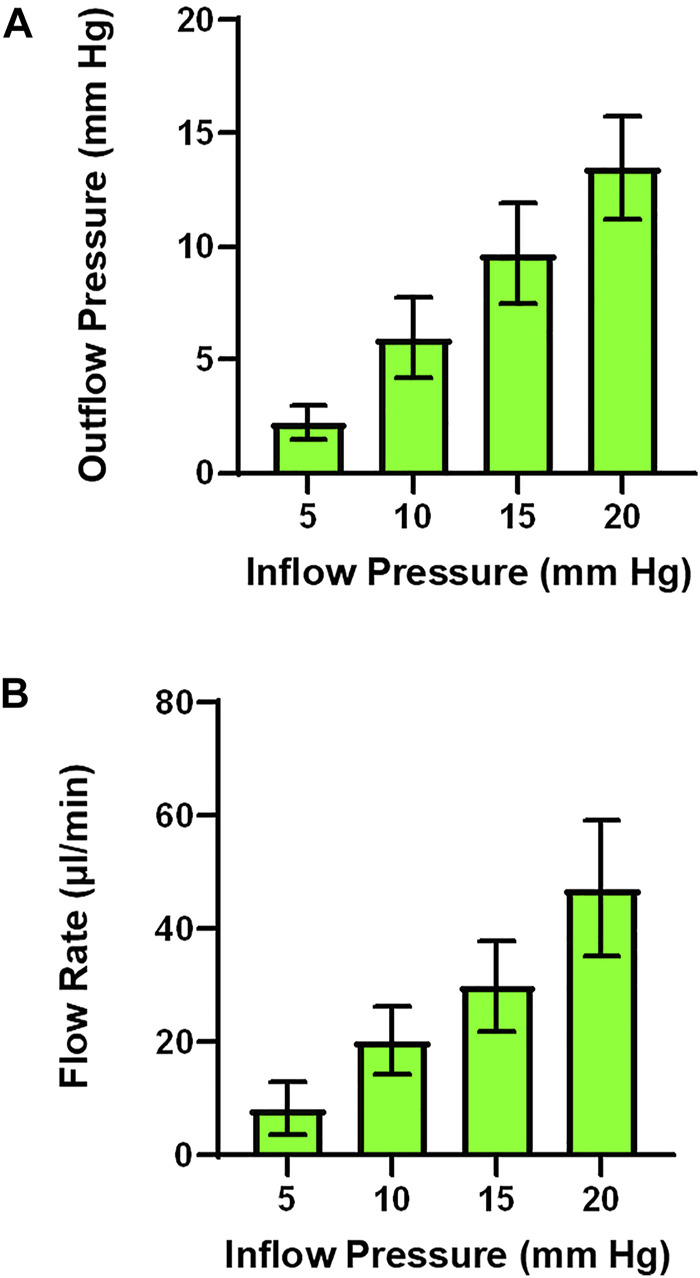
Placental health is critical to fetal growth and maternal health during gestation. However, investigating placental flow in an ex-vivo isolated system where inflow is independently controlled has yet to be developed in the rat. Here, we describe a novel technique, isolated perfused placenta technique that allows for analysis of placental pressure outflow pressure, placental flow in rats at gestational day 20. Using this method, we successfully perfused placentas from dams and were able to observe increases in outflow pressure and flow as the inflow pressure to the placenta was increased in a step wise fashion. This method will help to advance the functional analysis of placental flow and therefore placental resistance and efficiency.

Keywords: fetus; hemodynamics; placenta; protocol; reproduction; technique; validation.


Conflict of interest statement

The authors declare that the research was conducted in the absence of any commercial or financial relationships that could be construed as a potential conflict of interest.

Figures

FIGURE 1
FIGURE 1
Dissection of the umbilical artery and vein. (A) Isolated fetoplacental unit with the umbilical artery and vein completely intact. (B) Dissection of the umbilical artery (white vessel located on the left) and vein (red vessel on the right) beginning at the insertion into the fetus. (C) Umbilical artery and vein completely separated from fetus to the placenta and ready for transfer to the chamber.
FIGURE 2
FIGURE 2
Dissection of placental and chorionic vessels. (A) From left to right: umbilical vein; umbilical artery; vitelline artery and other chorionic vessels. Amnionic membranes can also be seen surrounding the vessels prior to removal. Black arrow indicates the umbilical artery and vein pair while the white arrow shows the vitelline artery and chorionic vessels. (B) Placental unit with membranes removed and umbilical artery and vein isolated. Note the chorionic vessels with the white arrow at the top of the figure indicating how closely these two are prior to dissection. Vessel insertion and branching into the placenta can also be seen.
FIGURE 3
FIGURE 3
Chamber set up for isolated placental perfusion. The placental unit with separated umbilical artery and vein is placed into the Living Systems chamber with PSS. The umbilical artery is canulated onto the inflow glass pipette while the umbilical vein is canulated onto the outflow pipette and tied with braided silk suture. Red arrows indicate the inflow (left, 1) and outflow (right, 2) pipettes.
FIGURE 4
FIGURE 4
Preparation of the placental unit prior to starting an experiment. (A) The umbilical artery is canulated onto the inflow pressure canula while the umbilical vein is left united in order to clear the blood from the placenta. Note the pooling blood in the bottom of the chamber as PSS is flowing through the placenta. (B) Both umbilical artery and vein are placed onto and tied to the inflow and outflow cannulas, respectively. No pressure is being applied to the system. (C) The placenta is fully pressurized. Note the increase in size and volume of the vessels within the chamber. Again, red arrows indicate inflow (left, 1) and outflow (right, 2) pipettes.
FIGURE 5
FIGURE 5
Representative trace of placental flow. (A) Input (umbilical artery) perfusion pressure is increased in a stepwise fashion to measure placental resistance. (B) Output pressure is measured and a drop in pressure across the placenta is expected and indicative of placental health. (C) Alterations in placenta flow (μL/min) can be seen concomitantly with the change in inflow and outflow pressure, indicating placental resistance.
FIGURE 6
FIGURE 6
Outflow pressure and flow rate across inflow pressures. (A) As inflow pressure is increased in a stepwise fashion outflow pressure increases with both normal superfusate. (B) Alternatively, flow rate can be measured as the result of increasing inflow pressure. (n = 3 placentas).
FIGURE 7
FIGURE 7
Graphical representation of the isolated rat placenta model and perfusion pathways. Numbering in the figure represents the following: 1) input pressure pipette; 2) umbilical artery; 3) arteriolar waste delivery network; 4) venular nutrient network return; 5) umbilical vein; 6) output pressure pipette. Arrows show direction of the perfusate flow.

Similar articles

Cited by

References

    1. Abukabda A. B., Bowdridge E. C., McBride C. R., Batchelor T. P., Goldsmith W. T., Garner K. L., et al. (2019). Maternal Titanium Dioxide Nanomaterial Inhalation Exposure Compromises Placental Hemodynamics. Toxicol. Appl. Pharmacol. 367, 51–61. 10.1016/j.taap.2019.01.024 - DOI - PMC - PubMed
    1. Brosens J. J., Pijnenborg R., Brosens I. A. (2002). The Myometrial Junctional Zone Spiral Arteries in Normal and Abnormal Pregnancies: A Review of the Literature. Am. J. Obstet. Gynecol. 187 (5), 1416–1423. 10.1067/mob.2002.127305 - DOI - PubMed
    1. Conings S., Amant F., Annaert P., Van Calsteren K. (2017). Integration and Validation of the Ex Vivo Human Placenta Perfusion Model. J. Pharmacol. Toxicol. Methods 88 (Pt 1), 25–31. 10.1016/j.vascn.2017.05.002 - DOI - PubMed
    1. Fournier S. B., D’Errico J. N., Adler D. S., Kollontzi S., Goedken M. J., Fabris L., et al. (2020). Nanopolystyrene Translocation and Fetal Deposition after Acute Lung Exposure during Late-Stage Pregnancy. Part. Fibre Toxicol. 17 (1), 55. 10.1186/s12989-020-00385-9 - DOI - PMC - PubMed
    1. Fowden A. L., Sferruzzi-Perri A. N., Coan P. M., Constancia M., Burton G. J. (2009). Placental Efficiency and Adaptation: Endocrine Regulation. J. Physiol. 587 (Pt 14), 3459–3472. 10.1113/jphysiol.2009.173013 - DOI - PMC - PubMed

LinkOut - more resources