. 2022 Feb 28:13:841124.
doi: 10.3389/fmicb.2022.841124. eCollection 2022.

Light Triggered Enhancement of Antibiotic Efficacy in Biofilm Elimination Mediated by Gold-Silver Alloy Nanoparticles

Affiliations

Light Triggered Enhancement of Antibiotic Efficacy in Biofilm Elimination Mediated by Gold-Silver Alloy Nanoparticles

Cinthia Alves-Barroco et al. Front Microbiol. .

Abstract

Bacterial biofilm is a tri-dimensional complex community of cells at different metabolic stages involved in a matrix of self-produced extracellular polymeric substances. Biofilm formation is part of a defense mechanism that allows the bacteria to survive in hostile environments, such as increasing resistance or tolerance to antimicrobial agents, causing persistent infections hard to treat and impair disease eradication. One such example is bovine mastitis associated with Streptococcus dysgalactiae subsp. dysgalactiae (SDSD), whose worldwide health and economic impact is on the surge. As such, non-conventional nanobased approaches have been proposed as an alternative to tackle biofilm formation and to which pathogenic bacteria fail to adapt. Among these, metallic nanoparticles have gained significant attention, particularly gold and silver nanoparticles, due to their ease of synthesis and impact against microorganism growth. This study provides a proof-of-concept investigation into the use of gold-silver alloy nanoparticles (AuAgNPs) toward eradication of bacterial biofilms. Upon visible light irradiation of AuAgNPs there was considerable disturbance of the biofilms' matrix. The hindering of structural integrity of the biofilm matrix resulted in an increased permeability for entry of antibiotics, which then cause the eradication of biofilm and inhibit subsequent biofilm formation. Additionally, our results that AuAgNPs inhibited the formation of SDSD biofilms via distinct stress pathways that lead to the downregulation of two genes critical for biofilm production, namely, brpA-like encoding biofilm regulatory protein and fbpA fibronectin-binding protein A. This study provides useful information to assist the development of nanoparticle-based strategies for the active treatment of biofilm-related infections triggered by photoirradiation in the visible.

Keywords: Streptococcus; biofilm inhibition; biofilms; eradication mature biofilms; gold-silver alloy nanoparticles.


Conflict of interest statement

The authors declare that the research was conducted in the absence of any commercial or financial relationships that could be construed as a potential conflict of interest.

Figures

FIGURE 1
FIGURE 1
Schematic description of experimental setup. From the initial planktonic bacterial growth, biofilms were allowed to form. These were then washed to remove non-adherent bacteria and exposed to AuAgNP When indicated, these were irradiated and further cultured in presence of AuAgNPs and Ciprofloxacin (10 μg/mL).
FIGURE 2
FIGURE 2
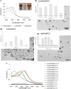
(A) Ultraviolet-spectrum of produced nanoparticles. AuNPs solution show an intense red color corresponding to a LSPR peak at 520 nm; AgNPs solution showed the typical faint yellow color corresponding to the LSPR peak at 430 nm; the alloy AuAgNPs resulted orange with an LSPR peak at 484 nm. (B1–3) Respective TEM images NPs with indication of the size distribution. (C) Ultraviolet spectra of NPs in different pH solutions correlating to the colloid stability of the different nanoconjugates.
FIGURE 3
FIGURE 3
Biofilm destruction (%). (i) AuAgNPs; (ii) ciprofloxacin (CIP); (iii) AuAgNPs + CIP; (iv) visible light irradiation (VLI); (v) AuAgNPs + VLI; (vi) VLI + CIP; (vii) AuAgNPs + VLI + CIP. Nanoparticles were at 10 nM and VLI at 2.02 W cm–2. Data represented as the mean ± standard deviation (SD) of three independent measurements. The following formula calculated the percentage of biofilm reduction: Reduced cell viability (%) = 100–{[(log10 CFU/mL control – log10 CFU/mL Treat)/log10 CFU/mL control] × 100}, where CFU/mL control corresponds to the number of colonies forming units per ml per milliliter of untreated biofilms, and CFU/mL Treat corresponds to the number of colonies forming units per ml per milliliter of treated biofilms. *p < 0.05, Significant differences regarding each condition as a single strategy or when compared to treatment with two conditions simultaneously. **p < 0.05 Significant differences AuAgNPs + CIP or AuAgNPs + VLI + CIP when compared to treatment with other conditions.
FIGURE 4
FIGURE 4
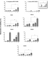
Percent inhibition of biofilm formation of (i) AuNPs; (ii) AuNPs + visible light irradiation (VLI); (iii) AgNPs; (iv) AgNPs + VLI; (v) alloy AuAgNPs; (vi) alloy AuAgNPs + VLI. To assess biofilm formation, the absorbance was measured at 570 nm. All nanoparticles were at 10 nM and VLI at 2.02 W cm–2. *p < 0.05.
FIGURE 5
FIGURE 5
Transcriptional profiling of (A) brpA-like (encoding biofilm regulatory protein) and (B) fbpA (fibronectin-binding protein A) in the SDSD strains AuAgNPs combined with visible light irradiation (VLI) relative to the untreated. 16S rRNA gene was used as the internal control for normalization. Relative expression was calculated by the 2–ΔΔCt method. (i) AuAgNP and (ii) AuAgNP and VLI. Nanoparticles were at 10 nM and VLI at 2.02 W cm–2.
FIGURE 6
FIGURE 6
Transmission electron microscopy microphotographs of SDSD VSD22 cells (A) untreated; (B,C) treated with AuAgNPs (10 nM) combined with visible light irradiation at 2.02 W cm–2 for 60 s; (D–F) treated with AuAgNPs alone. Blue arrow shows glycocalyx from untreated cells; Yellow arrow shows cell envelope not defined with wrinkling of the cell wall; Red arrows show nanoparticles adhered to the cell envelope; White arrow shows aggregation of the cytoplasmic components.

References

    1. Adeyemi O. S., Sulaiman F. A. (2015). Evaluation of metal nanoparticles for drug delivery systems. J. Biomed. Res. 29 145–149. 10.7555/JBR.28.20130096 - DOI - PMC - PubMed
    1. Alhmoud H., Cifuentes-Rius A., Delalat B., Lancaster D. G., Voelcker N. H. (2017). Gold-decorated porous silicon nanopillars for targeted hyperthermal treatment of bacterial infections. ACS Appl. Mater. Interfaces 9 33707–33716. 10.1021/acsami.7b13278 - DOI - PubMed
    1. Alves-barroco C., Caço J., Roma-Rodrigues C., Fernandes A. R., Bexiga R., Oliveira M., et al. (2021). New Insights on Streptococcus dysgalactiae subsp. dysgalactiae Isolates. Front. Microbiol. 12:1–16. 10.3389/fmicb.2021.686413 - DOI - PMC - PubMed
    1. Alves-barroco C., Rivas-García L., Fernandes A. R., Baptista P. V. (2020a). Tackling Multidrug Resistance in Streptococci – From Novel Biotherapeutic Strategies to Nanomedicines. Front. Microbiol. 11:1–21. 10.3389/fmicb.2020.579916 - DOI - PMC - PubMed
    1. Alves-barroco C., Paquete-Ferreira J., Santos-Silva T., Fernandes A. R. (2020b). Singularities of Pyogenic Streptococcal Biofilms – From Formation to Health Implication. Front. Microbiol. 11:1–22. 10.3389/fmicb.2020.584947 - DOI - PMC - PubMed

LinkOut - more resources