

Fabrication of In Situ Grown Hydroxyapatite Nanoparticles Modified Porous Polyetheretherketone Matrix Composites to Promote Osteointegration and Enhance Bone Repair
- PMID: 35295654
- PMCID: PMC8919038
- DOI: 10.3389/fbioe.2022.831288
Fabrication of In Situ Grown Hydroxyapatite Nanoparticles Modified Porous Polyetheretherketone Matrix Composites to Promote Osteointegration and Enhance Bone Repair
Abstract
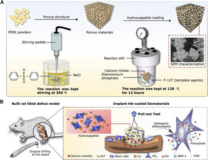
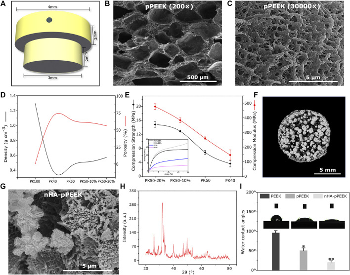
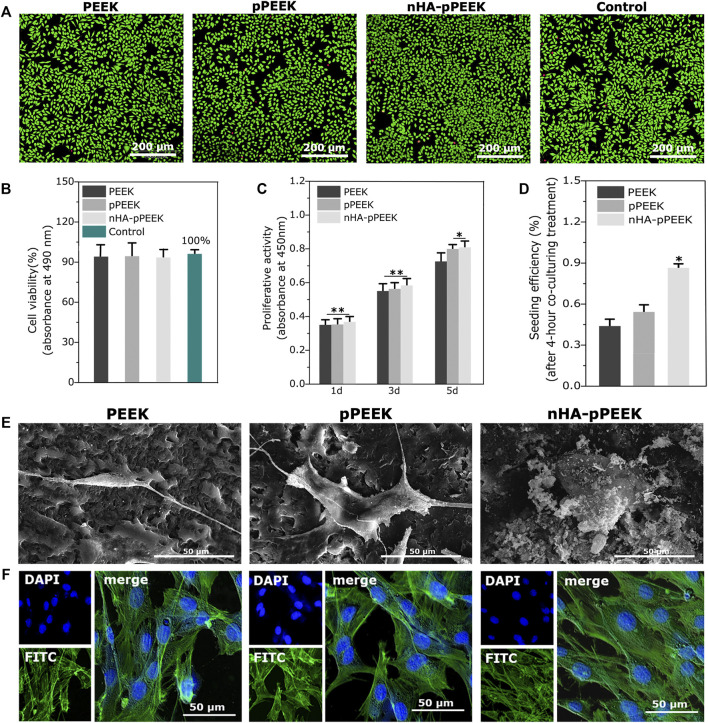
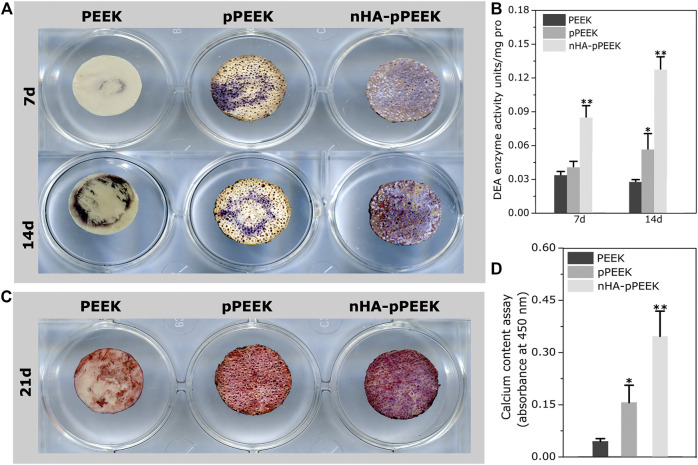
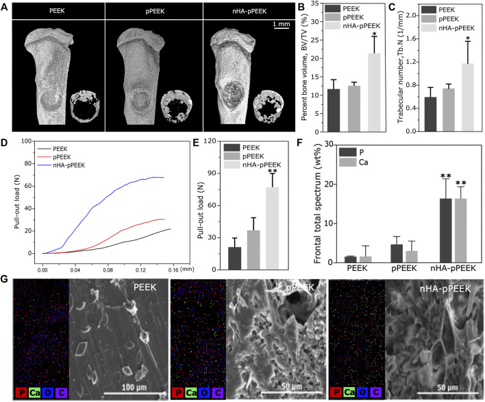
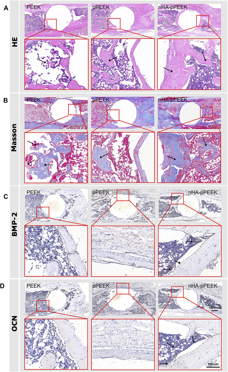
The repairment of critical-sized bone defects is a serious problem that stimulates the development of new biomaterials. In this study, nanohydroxyapatite (nHA)-doped porous polyetheretherketone (pPEEK) were successfully fabricated by the thermally induced phase separation method and hydrothermal treatment. Structural analysis was performed by X-ray diffraction. The water contact angles and scanning electron microscopy were measured to assess physical properties of surfaces. The mechanical strength of the composites is also determined. Microcomputed tomography is used to characterize the nHA content of the composites. The in vitro bioactivity of the composites with or without nHA was investigated by using murine pre-osteoblasts MC3T3-E1, and the results of cytotoxicity and cell proliferation assays revealed that the cytocompatibility of all specimens was good. Adherence assays were employed to examine the adhesion and morphology of cells on different materials. However, nHA-doped composites induced cell attachment and cell spreading more significantly. Osteogenic differentiation was investigated using alkaline phosphatase activity and alizarin red staining, and these in vitro results demonstrated that composites containing nHA particles enhanced osteoblast differentiation. Its effectiveness for promoting osteogenesis was also confirmed in an in vivo animal experiment using a tibial defective rat model. After 8 weeks of implantation, compared to the pure PEEK and pPEEK without nHA groups, the nHA-pPEEK group showed better osteogenic activity. The results indicate that the nHA-pPEEK composites are possibly a well-designed bone substitute for critical-sized bone defects by promoting bone regeneration and osteointegration successfully.
Keywords: animal model; differentiation; nanohydroxyapatite; osteointegration; polyetheretherketone.
Copyright © 2022 Wang, Qi, Liu, Zhu, Liu and Zhu.
Conflict of interest statement
The authors declare that the research was conducted in the absence of any commercial or financial relationships that could be construed as a potential conflict of interest.
Figures
Similar articles
-
Polyetheretherketone Hybrid Composites with Bioactive Nanohydroxyapatite and Multiwalled Carbon Nanotube Fillers.Polymers (Basel). 2016 Dec 8;8(12):425. doi: 10.3390/polym8120425. Polymers (Basel). 2016. PMID: 30974701 Free PMC article.
-
Performance of 3D printed porous polyetheretherketone composite scaffolds combined with nano-hydroxyapatite/carbon fiber in bone tissue engineering: a biological evaluation.Front Bioeng Biotechnol. 2024 Jan 25;12:1343294. doi: 10.3389/fbioe.2024.1343294. eCollection 2024. Front Bioeng Biotechnol. 2024. PMID: 38333080 Free PMC article.
-
Preparation, characterization, and in vitro osteoblast functions of a nano-hydroxyapatite/polyetheretherketone biocomposite as orthopedic implant material.Int J Nanomedicine. 2014 Aug 18;9:3949-61. doi: 10.2147/IJN.S67358. eCollection 2014. Int J Nanomedicine. 2014. PMID: 25170265 Free PMC article.
-
Advancements in nanohydroxyapatite: synthesis, biomedical applications and composite developments.Regen Biomater. 2024 Nov 5;12:rbae129. doi: 10.1093/rb/rbae129. eCollection 2025. Regen Biomater. 2024. PMID: 39776858 Free PMC article. Review.
-
Nano-Hydroxyapatite as a Delivery System for Promoting Bone Regeneration In Vivo: A Systematic Review.Nanomaterials (Basel). 2021 Sep 29;11(10):2569. doi: 10.3390/nano11102569. Nanomaterials (Basel). 2021. PMID: 34685010 Free PMC article. Review.
Cited by
-
Gallic acid-grafted chitosan antibacterial hydrogel incorporated with polydopamine-modified hydroxyapatite for enhancing bone healing.Front Bioeng Biotechnol. 2023 Jun 2;11:1162202. doi: 10.3389/fbioe.2023.1162202. eCollection 2023. Front Bioeng Biotechnol. 2023. PMID: 37334266 Free PMC article.
-
Bone Tissue Engineering and Nanotechnology: A Promising Combination for Bone Regeneration.Biology (Basel). 2024 Apr 2;13(4):237. doi: 10.3390/biology13040237. Biology (Basel). 2024. PMID: 38666849 Free PMC article. Review.
-
Biomimetic, biodegradable and osteoinductive treated dentin matrix/α-calcium sulphate hemihydrate composite material for bone tissue engineering.Regen Biomater. 2023 Jun 19;10:rbad061. doi: 10.1093/rb/rbad061. eCollection 2023. Regen Biomater. 2023. PMID: 37501676 Free PMC article.
-
Nanostructures in Orthopedics: Advancing Diagnostics, Targeted Therapies, and Tissue Regeneration.Materials (Basel). 2024 Dec 17;17(24):6162. doi: 10.3390/ma17246162. Materials (Basel). 2024. PMID: 39769763 Free PMC article. Review.
References
-
- Chen E. E. M., Zhang W., Ye C. C. Y., Gao X., Jiang L. L. J., Zhao T. T. F., et al. (2017a). Knockdown of SIRT7 Enhances the Osteogenic Differentiation of Human Bone Marrow Mesenchymal Stem Cells Partly via Activation of the Wnt/β-Catenin Signaling Pathway. Cell Death Dis 8, e3042. 10.1038/cddis.2017.429 - DOI - PMC - PubMed
-
- Chen Y., Liu X., Liu R., Gong Y., Wang M., Huang Q., et al. (2017b). Zero-order Controlled Release of BMP2-Derived Peptide P24 from the Chitosan Scaffold by Chemical Grafting Modification Technique for Promotion of Osteogenesis In Vitro and Enhancement of Bone Repair In Vivo . Theranostics 7, 1072–1087. 10.7150/thno.18193 - DOI - PMC - PubMed
-
- Chubrik A., Senatov F., Kolesnikov E., Orlova P., Poponova M., Grunina T., et al. (2020). Highly Porous PEEK and PEEK/HA Scaffolds with Escherichia Coli-Derived Recombinant BMP-2 and Erythropoietin for Enhanced Osteogenesis and Angiogenesis. Polym. Test. 87, 106518. 10.1016/j.polymertesting.2020.106518 - DOI
LinkOut - more resources
Full Text Sources