. 2022 Mar 7:2022:5938688.
doi: 10.1155/2022/5938688. eCollection 2022.

Anti-PD-L1 Antibody Enhances T Cell Immune Responses and Reduces Resistance of Breast Cancer Cells to Radiotherapy

Affiliations

Anti-PD-L1 Antibody Enhances T Cell Immune Responses and Reduces Resistance of Breast Cancer Cells to Radiotherapy

Lei-Ming Guo et al. Oxid Med Cell Longev. .

Abstract

Immune escape is a frequent occurrence, which limits the duration of antitumor immune responses to radiotherapy. Here, we aimed to ascertain the roles and underlying mechanisms of programmed death ligand 1 (PD-L1) in tolerance of breast cancer (BC) to radiotherapy. We first quantified microRNA-21 (miR-21) and PD-L1 expression in BC tissues and cells, followed by identification of the interactions between miR-21, PD-L1, and programmed cell death protein 4 (PDCD4). miR-21 knock-in mice were used to construct tumor-bearing models, which were then treated with anti-PD-L1 antibody and irradiation, followed by measurement of tumor growth and tumor immune escape. Finally, we evaluated the synergistic effects of radiotherapy and anti-PD-L1 antibody in vivo. The results showed increased miR-21 expression in BC tissues and cells, which was positively correlated with PD-L1 expression. The treatment with radiotherapy or anti-PD-L1 antibody in the miR-21 knock-in mice diminished tumor weight and volume, along with decreased CD3+CD8+ positive cells, serum IL-2 and IFN-γ levels, and lower PD-L1 expression, but augmented apoptosis of T and BC cells. Moreover, miR-21 significantly augmented PD-L1 expression via PI3K/Akt pathway activation by targeting PDCD4 in BC cells. Thus, radiotherapy and anti-PD-L1 antibody synergistically accelerated the therapeutic effect against BC in mice, thereby implicating a close interplay between radiotherapy, T cells, and the miR-21/PDCD4/PI3K/Akt/PD-L1 axis.


Conflict of interest statement

The authors have declared that no competing interests exist.

Figures
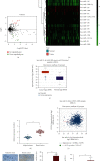
Figure 1
Figure 1
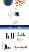
miR-21 expression is increased and positively correlates to PD-L1 expression in BC tissues of patients. (a) Volcano map of differentially expressed miRNAs in 50 BC tissue samples and in 3 adjacent normal tissue samples in the GSE44124 dataset (The x-axis represents the difference log10 p value and the ordinate represents logFoldChange. Each point in the graph represents a gene, the red point represents the upregulated gene with logFoldChange greater than 1 and the green point represents the downregulated gene with logFoldChange less than -1). (b) Heat map of differentially expressed miRNAs in 50 BC tissue samples and in 3 adjacent normal tissue samples in the GSE44124 dataset. (c) miR-21 differential expression in BC tissue samples and normal breast tissue samples using the TCGA database. (d) Expression of miR-21 in BC and adjacent normal tissues determined by RT-qPCR (n = 50). (e) Correlation between PD-L1 and miR-21 expression in BC tissue samples. (f) PD-L1 protein level in BC tissues measured by immunohistochemistry (n = 50, scale bar = 50 μm; the nucleus was stained blue, and the positive cells were stained yellow-brown). (g) PD-L1 protein level in BC tissues measured by Western blot analysis (n = 50). p < 0.05 compared with adjacent normal tissues. Data (mean ± standard deviation) from two groups were compared using paired t-test or independent sample t-test. Each sample was evaluated three times independently.
Figure 2
Figure 2
miR-21 knock-in increases tolerance of BC cells to radiotherapy and promotes tumor immune escape in mice. WT or miR-21+/+ mice were either treated or not treated with radiotherapy and anti-PD-L1 antibody. (a) Tumor volume curve of the mice. (b) Tumor weight of the mice. (c) The number of CD3+CD8+ cells in peripheral bloods and tumor tissues of mice detected by flow cytometry. (d) T lymphocyte apoptosis in peripheral blood and tumor tissues of mice measured using Annexin V-FITC/PI. (e) Level of IL-2 in peripheral blood and tumor tissues of mice measured using ELISA. (f) Level of IFN-γ in peripheral blood and tumor tissues of mice measured using ELISA. p < 0.05 compared with WT mice; #p < 0.05 compared with WT mice treated with radiotherapy. Each sample was evaluated three times independently. Data (mean ± standard deviation) from two groups were compared using independent sample t-test while data for tumor volume were compared using repeated measures ANOVA. There were 10 mice in each group.
Figure 3
Figure 3
miR-21 potentiates PD-L1 expression via activation of the PI3K/Akt pathway by targeting PDCD4 in BC cells. (a) Venn diagram depicting the intersection of target genes of miR-21 predicted with PicTar, miRDB, TargetScan, and starBase databases. (b) Protein-protein interaction network of the miR-21 downstream target genes. (c) Correlation between PDCD4 and miR-21 expression in BC tissue samples from the TCGA database. (d) Binding site between miR-21 and PDCD4 predicted using the TargetScan database. (e) The binding of miR-21 to PDCD4 confirmed by dual luciferase reporter gene assay. (f) Expression of miR-21, PDCD4, and PD-L1 after alteration of miR-21 and overexpression of PDCD4 determined by RT-qPCR in BC cells. p < 0.05 compared with cells treated with mimic NC. #p < 0.05 compared with cells treated with inhibitor NC and p < 0.05 compared with cells treated with miR-21 mimic. (g) The silencing efficiency of si-PDCD4-1 and si-PDCD4-2 in BC cells determined by RT-qPCR. p < 0.05 compared with cells treated with si-NC. (h) Western blot analysis of PDCD4, PI3K, Akt, and PD-L1 proteins, and the extent of PI3K and Akt phosphorylation in BC cells. p < 0.05 compared with cells treated with oe-NC. #p < 0.05 compared with cells treated with si-NC and p < 0.05 compared with cells treated with si-PDCD4. Data (mean ± standard deviation) from two groups were compared using independent sample t-test while those from multiple groups were compared using one-way ANOVA. All cell experiments were repeated 3 times independently.
Figure 4
Figure 4
Anti-PD-L1 antibody combined with radiotherapy represses BC cell growth and promotes apoptosis in vivo. miR-21+/+ mice were either treated or not treated with radiotherapy or radiotherapy + anti-PD-L1 antibody. (a) Tumor volume curve of miR-21+/+ mice following different treatment protocols. (b) Tumor weight of miR-21+/+ mice following different treatment protocols. (c) The number of PD-L1 positive cells in tumor tissues of miR-21+/+ mice following different treatment protocols, detected by immunofluorescence staining. (d) The positive rate of Ki67 protein expression in tumor tissues of miR-21+/+ mice following different treatment protocols, detected by immunohistochemistry (10 mice in each group, 3 sections for each mouse). (e) Cell apoptosis in tumor tissues of miR-21+/+ mice following different treatment protocols, measured using TUNEL staining. (f) Protein levels of apoptosis-related factors in tumor tissues of miR-21+/+ mice following different treatment protocols, determined by Western blot analysis. p < 0.05, compared with miR-21+/+ mice without treatment; #p < 0.05, compared with miR-21+/+ mice treated with radiotherapy or anti-PD-L1 antibody. Each sample was evaluated three times independently. Data (mean ± standard deviation) from two groups were compared using independent sample t-test. There were 10 mice in each group.
Figure 5
Figure 5
Anti-PD-L1 antibody combined with radiotherapy facilitates T cell immune response in miR-21+/+ mice in vivo. miR-21+/+ mice were either treated or not treated with radiotherapy or radiotherapy+anti-PD-L1 antibody. (a) The number of CD3+CD8+ cells in the peripheral blood of miR-21+/+ mice following different treatment protocols detected by flow cytometry. (b) T lymphocyte apoptosis rate in peripheral blood of miR-21+/+ mice following different treatment protocols measured using Annexin V-FITC/PI. (c) Levels of IL-2 in the peripheral blood of miR-21+/+ mice following different treatment protocols measured using ELISA. (d) Levels of IFN-γ in peripheral blood of miR-21+/+ mice following different treatment protocols measured using ELISA. p < 0.05 compared with miR-21+/+ mice without treatment; #p < 0.05 compared with miR-21+/+ mice treated with radiotherapy or anti-PD-L1 antibody. Each sample was evaluated three times independently. Data (mean ± standard deviation) from two groups were compared using independent sample t-test. The experiments were repeated 3 times independently.
Figure 6
Figure 6
Mechanistic investigations indicated that anti-PD-L1 antibody augmented T cell immune response and alleviated the tolerance of BC to radiotherapy by regulating the miR-21/PDCD4/PI3K/Akt/PD-L1 axis.

References

    1. Fiordelisi M. F., Auletta L., Meomartino L., et al. Preclinical molecular imaging for precision medicine in breast cancer mouse models. Contrast Media & Molecular Imaging . 2019;2019, article 8946729 doi: 10.1155/2019/8946729. - DOI - PMC - PubMed
    1. Nakhjavani M., Hardingham J. E., Palethorpe H. M., Price T. J., Townsend A. R. Druggable molecular targets for the treatment of triple negative breast cancer. Journal of Breast Cancer . 2019;22(3):341–361. doi: 10.4048/jbc.2019.22.e39. - DOI - PMC - PubMed
    1. DeSantis C. E., Ma J., Gaudet M. M., et al. Breast cancer statistics, 2019. CA: a Cancer Journal for Clinicians . 2019;69(6):438–451. doi: 10.3322/caac.21583. - DOI - PubMed
    1. Roge M., Thureau S., Dampierre J., Dubray B., Rivera S. Prophylactic nodal radiotherapy for breast cancer. Cancer Radiothérapie . 2019;23(8):904–912. doi: 10.1016/j.canrad.2019.08.006. - DOI - PubMed
    1. Toma C. L., Serbescu A., Alexe M., Cervis L., Ionita D., Bogdan M. A. The bronchoalveolar lavage pattern in radiation pneumonitis secondary to radiotherapy for breast cancer. Maedica . 2010;5(4):250–257. - PMC - PubMed

Substances