. 2022 Feb 28:12:816477.
doi: 10.3389/fphar.2021.816477. eCollection 2021.

β-Sitosterol Inhibits Rheumatoid Synovial Angiogenesis Through Suppressing VEGF Signaling Pathway

Affiliations

β-Sitosterol Inhibits Rheumatoid Synovial Angiogenesis Through Suppressing VEGF Signaling Pathway

Kai Qian et al. Front Pharmacol. .

Abstract

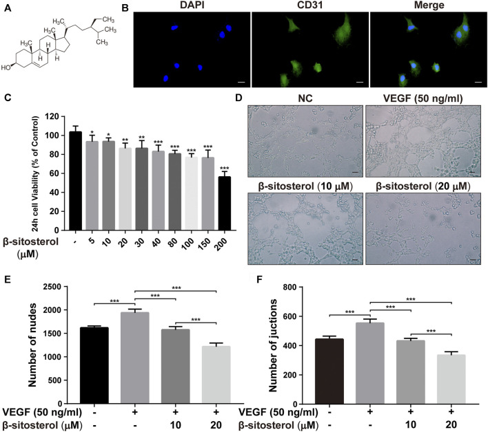
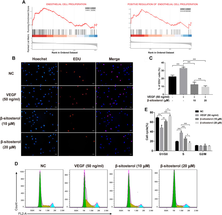
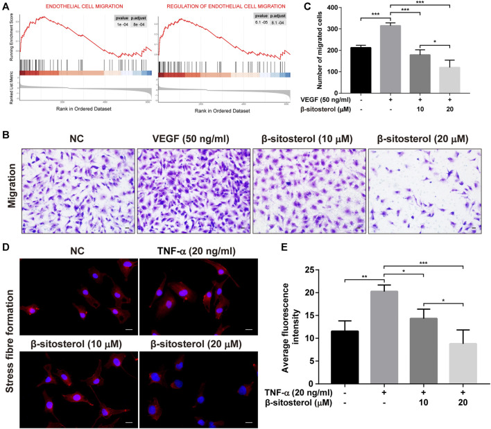
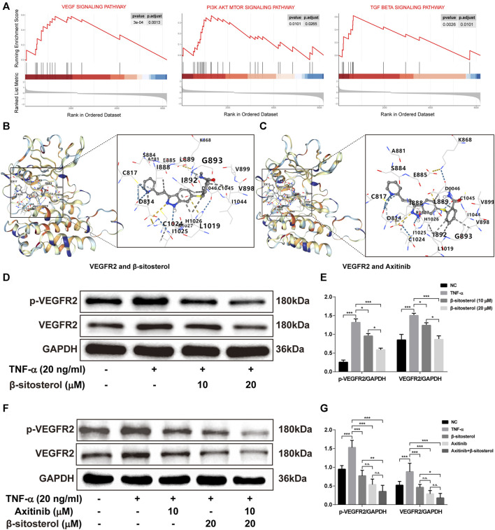
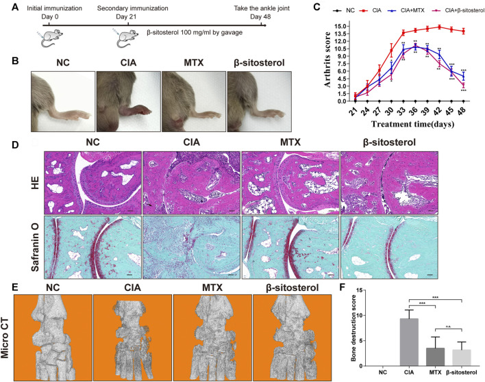
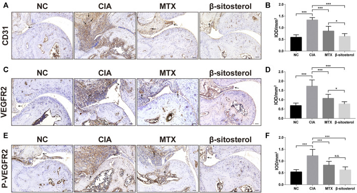
Background: Rheumatoid arthritis (RA) is a chronic disabling inflammatory disease that causes synovial angiogenesis in an invasive manner and leads to joint destruction. Currently available pharmacotherapy for RA has unwanted side effects and limitations. Although anti-angiogenic therapy is regarded as a new potential treatment for RA, only a few anti-angiogenic drugs are available. An increasing number of studies have shown that β-sitosterol (BSS) may exert inhibitory effects against angiogenesis. However, the mechanisms involved are still unclear. Methods: Based on the results of the gene set enrichment analysis (GSEA) of the transcriptome data of endothelial cells from RA patients, we evaluated the pharmacological effects of BSS on the tube formation, cell proliferation, and migration of human umbilical vein endothelial cells (HUVECs). Furthermore, the effects of BSS treatment on vascular endothelial growth factor receptor 2 (VEGFR2) were determined using molecular docking and Western blotting. Additionally, in the presence or absence of BSS, synovial angiogenesis and joint destruction of the ankle were investigated in collagen-induced arthritis (CIA) mice. The effect of BSS treatment on VEGFR2/p-VEGFR2 expression was verified through immunohistochemical staining. Results: The immunohistochemistry results revealed that BSS treatment inhibited angiogenesis both in vitro and in vivo. In addition, the results of 5-ethynyl-2'-deoxyuridine and cell cycle analysis showed that BSS treatment suppressed the proliferation of HUVECs, while the Transwell migration and stress fiber assays demonstrated that BSS treatment inhibited the migration of HUVECs. Notably, the inhibitory effect of BSS treatment on VEGFR2/p-VEGFR2 was similar to that of axitinib. In CIA mice, BSS also exerted therapeutic effects on the ankles by reducing the degree of swelling, ameliorating bone and cartilage damage, preventing synovial angiogenesis, and inhibiting VEGFR2 and p-VEGFR2 expression. Conclusion: Therefore, our findings demonstrate that BSS exerts an inhibitory effect on synovial angiogenesis by suppressing the proliferation and migration of endothelial cells, thereby alleviating joint swelling and bone destruction in CIA mice. Furthermore, the underlying therapeutic mechanisms may involve the inhibition of VEGF signaling pathway activation.

Keywords: VEGFR2; angiogenesis; collagen-induced arthritis; endothelial cells; rheumatoid arthritis; β-sitosterol.


Conflict of interest statement

The authors declare that the research was conducted in the absence of any commercial or financial relationships that could be construed as a potential conflict of interest.

Figures

FIGURE 1
FIGURE 1
BSS inhibits angiogenesis in HUVECs. (A) BSS chemical formula (quoted from sigma official website). (B) CD31 fluorescent staining identification (×630, scale bar = 20 μm). (C) Effects of different concentrations of BSS on CCK-8 in HUVECs after 24 h (n = 3). (D) Representative images of tube formation at 6 h in HUVECs (×200, scale bar = 50 μm), Y-axis shows the nodes (E) and junction numbers (F) (n = 3). All data are shown as the mean ± SEM. *p < 0.05, **p < 0.01, ***p < 0.001.
FIGURE 2
FIGURE 2
Inhibitory effect of BSS on HUVECs proliferation induced by VEGF. (A) GSEA analysis of ECs proliferation in the GSE121894 dataset. (B) Effect of BSS on proliferation of HUVECs (VEGF 20 ng/ml for 24 h) and analysis of proliferation rate (C) by EdU (×200, scale bar = 50 μm) (n = 3). (D) Effect of BSS on cell cycle of HUVECs and analysis of cell cycle data (E) by flow cytometry (n = 3). All data are shown as the mean ± SEM. *p < 0.05, **p < 0.01, ***p < 0.001, n.s. = not significant.
FIGURE 3
FIGURE 3
Inhibitory effect of BSS on HUVECs migration induced by VEGF or TNF-α. (A) GSEA analysis of ECs migration in GSE121894 dataset. (B) Representative images of cell migration in a modified Boyden chamber following HUVECs induced by VEGF stimulation (×200, scale bar = 50 μm). (C) Y-axis shows the number of migrated cells (n = 3). (D) Representative images of stress fiber formation on TNF-α stimulation (20 ng/ml for 24 h) (×630, scale bar = 20 μm), Nuclei are stained with DAPI (blue). (E) Y-axis shows fluorescence intensity quantified by ImageJ (n = 3). All data are shown as the mean ± SEM. *p < 0.05, **p < 0.01, ***p < 0.001.
FIGURE 4
FIGURE 4
The effect of VEGF signaling pathway in TNF-α-induced HUVECs regulated by BSS. (A) GSEA analysis of KEGG signaling pathway in the GSE121894 dataset. The structural interactions between VEGFR2 and BSS (B) and between VEGFR2 and axitinib (C) were described by molecular simulation, and binding affinity of VEGFR2 with BSS and axitinib is shown in Table 1. (D) p-VEGFR2 and VEGFR2 protein levels in HUVECs (on TNF-α 20 ng/ml for 1 h) intervened with BSS dosage groups were detected by immunoblotting and the relative expression levels (E) of proteins were corrected by GAPDH (n = 3). (F) p-VEGFR2 and VEGFR2 protein levels in HUVECs intervened with BSS and axitinib were detected by immunoblotting, and the relative expression levels (G) of proteins were corrected by GAPDH (n = 3). All data are shown as the mean ± SEM. *p < 0.05, **p < 0.01, ***p < 0.001, n.s. = not significant.
FIGURE 5
FIGURE 5
Effects of BSS on experimental arthritis. (A) Animal experiment process, (B) the mice foot swelling photos, (C) Arthritis scores were shown. (D) Ankle HE and safranin O-fast green staining in the mice from each group (n = 6). (E) Micro-CT detection of bone destruction in the ankle joint of each group of mice, and analysis of (F) bone destruction scores in each group (n = 6). *p < 0.05, **p < 0.01, ***p < 0.001, n.s. = not significant.
FIGURE 6
FIGURE 6
Effect of BSS on immunohistochemistry of CD31, VEGFR2, and P-VEGFR2 on experimental arthritis. (A) CD31 immunohistochemical staining of the ankle joints in each group, and the optical density value (B) analysis using ImageJ (n = 6) (×200, scale bar = 50 μm). (C) VEGFR2 immunohistochemical staining of the ankle joints in each group, and the optical density value (D) analysis using ImageJ (n = 6) (×200, scale bar = 50 μm). (E) p-VEGFR2 immunohistochemical staining of the ankle joints in each group, and (F) the optical density value analysis using ImageJ (n = 6) (×200, scale bar = 50 μm). All data are shown as the mean ± SEM. *p < 0.05, **p < 0.01, ***p < 0.001, n.s. = not significant.

References

    1. Apte R. S., Chen D. S., Ferrara N. (2019). VEGF in Signaling and Disease: Beyond Discovery and Development. Cell 176, 1248–1264. 10.1016/j.cell.2019.01.021 - DOI - PMC - PubMed
    1. Babu S., Jayaraman S. (2020). An Update on β-sitosterol: A Potential Herbal Nutraceutical for Diabetic Management. Biomed. Pharmacother. 131, 110702. 10.1016/j.biopha.2020.110702 - DOI - PubMed
    1. Bottini N., Firestein G. S. (2013). Duality of Fibroblast-like Synoviocytes in RA: Passive Responders and Imprinted Aggressors. Nat. Rev. Rheumatol. 9, 24–33. 10.1038/nrrheum.2012.190 - DOI - PMC - PubMed
    1. Brand D. D., Latham K. A., Rosloniec E. F. (2007). Collagen-induced Arthritis. Nat. Protoc. 2, 1269–1275. 10.1038/nprot.2007.173 - DOI - PubMed
    1. Carpentier G., Berndt S., Ferratge S., Rasband W., Cuendet M., Uzan G., et al. (2020). Angiogenesis Analyzer for ImageJ - A Comparative Morphometric Analysis of "Endothelial Tube Formation Assay" and "Fibrin Bead Assay". Sci. Rep. 10, 11568. 10.1038/s41598-020-67289-8 - DOI - PMC - PubMed