. 2022 Feb 24;25(3):103978.
doi: 10.1016/j.isci.2022.103978. eCollection 2022 Mar 18.

The role of long noncoding RNA Nron in atherosclerosis development and plaque stability

Affiliations

The role of long noncoding RNA Nron in atherosclerosis development and plaque stability

Meng Du et al. iScience. .

Abstract

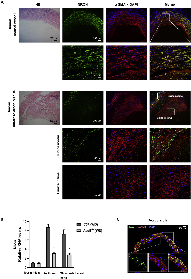
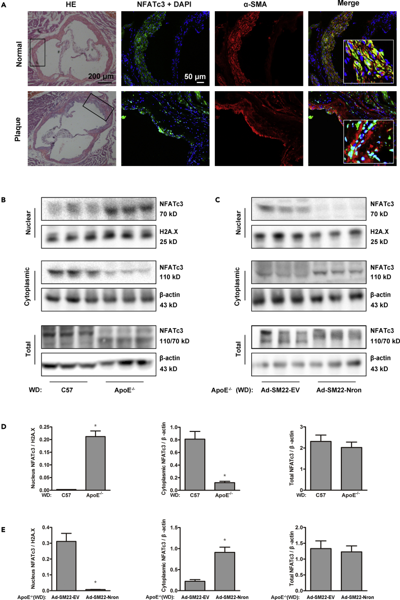
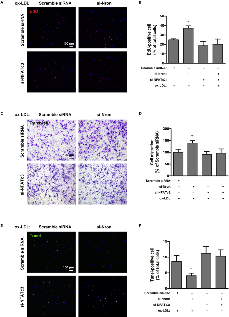
The major clinical consequences of atherosclerosis such as myocardial infarction or stroke are because of thrombotic events associated with acute rupture or erosion of an unstable plaque. Here, we identify an lncRNA Noncoding Repressor of NFAT (Nron) as a critical regulator of atherosclerotic plaque stability. Nron overexpression (OE) in vascular smooth muscle cells (VSMC) induces a highly characteristic architecture of more-vulnerable plaques, while Nron knockdown (KD) suppresses the development of atherosclerosis and favors plaque stability. Mechanistically, Nron specifically binds to and negatively regulates NFATc3, thus inhibiting the proliferation and promoting the apoptosis of VSMCs. Moreover, we also provide evidence that Nron increases the production and secretion of VEGFA from VSMCs, which functions as a paracrine factor to enhance intra-plaque angiogenesis. All of these effects contribute to plaque instability. Genetic or pharmacological inhibition of Nron may have potential for future therapy of atherosclerosis.

Keywords: Functional aspects of cell biology; Molecular biology; Pathophysiology.


Conflict of interest statement

The authors declare no potential conflicts of interest.

Figures

None
Graphical abstract
Figure 1
Figure 1
The expression of Nron is downregulated in atherosclerotic lesions from human and mice (A) RNA FISH analysis of NRON in human normal vessels (upper panel) and carotid atherosclerotic lesions (lower panel). For colocalization analysis, sections were co-stained for NRON (green) and α-SMA (red; smooth muscle cell marker). 4′,6-diamidino-2-phenylindole (DAPI) was used for nucleus staining (blue). (B) 6-week male ApoE−/- or wild type mice were fed a western diet (WD) for 12 weeks. Relative expression of Nron in the aortic arch, thoracoabdominal aorta, and myocardium were determined. (C) RNA FISH analysis of Nron in the aortic arch from wild type mice. For colocalization analysis, sections were co-stained for Nron (green) and α-SMA (red; smooth muscle cell marker). DAPI was used for nucleus staining (blue).
Figure 2
Figure 2
Nron overexpression induces a highly characteristic architecture of more-vulnerable plaques 6-Week male ApoE−/- mice were fed a western diet for 16 weeks. Mice were intravenously administered with adenovirus (Ad-SM22-EV or Ad-SM22-Nron) every two weeks in the last 8 weeks before being sacrificed. (A) Oil Red O staining of thoracoabdominal aorta, and lipid accumulation was quantified as percentage of total surface area of aorta. (B) Images and quantification of Oil Red O staining in lesions of aortic sinus. (C) Representative images of aortic sinus for H&E (HE), Masson’s trichrome (Masson), Elastica van Gieson (EVG), smooth muscle cells (α-SMA), and macrophages (F4/80). Arrows in EVG staining panels indicate ruptures of elastic fibers of tunica media. (D) Quantification of collagen content, fibrous cap area, necrotic core area, ruptures of elastic fibers, macrophage content, and smooth muscle cell content. Data are expressed as mean ± SEM (n = 12 per group). Statistical analysis was performed using Student’s t test. ∗p <0.05 vs. Ad-SM22-EV group.
Figure 3
Figure 3
Knockdown of Nron suppresses the development of atherosclerosis and increases the stability of atherosclerotic plaques 6-Week male ApoE−/- mice were fed a western diet for 16 weeks. Mice received a single intravenous injection of adeno-associated virus 2/8 (rAAV-shNC or rAAV-shNron) after feeding for 4 weeks. (A) Oil Red O staining of thoracoabdominal aorta, and lipid accumulation was quantified as percentage of total surface area of aorta. (B) Images and quantification of Oil Red O staining in lesions of aortic sinus. (C) Representative images of aortic sinus for H&E (HE), Masson’s trichrome (Masson), Elastica van Gieson (EVG), smooth muscle cells (α-SMA), and macrophages (F4/80). (D) Quantification of collagen content, fibrous cap area, necrotic core area, ruptures of elastic fibers, macrophage content, and smooth muscle cell content. Data are expressed as mean ± SEM (n = 12 per group). Statistical analysis was performed using Student’s t test. ∗p <0.05 vs. rAAV-shNC group.
Figure 4
Figure 4
Nron binds to NFATc3 in VSMCs (A–D) 6-week male ApoE−/- mice were fed a western diet for 16 weeks. Mice were intravenously administered with adenovirus (Ad-SM22-EV or Ad-SM22-Nron) every two weeks in the last 8 weeks before being sacrificed. Images (A) show Ki67 staining (green) of nuclei in sections of aortic sinus, and the number of proliferating cells was quantified (B) in VSMC-rich areas (red; α-SMA staining). Images (C) show TUNEL staining (green) of nuclei in sections of aortic sinus, and the number of apoptotic cells was quantified (D) in VSMC-rich areas (red; α-SMA staining). Data are expressed as mean ± SEM (n = 8 per group). ∗p <0.05 vs. Ad-SM22-EV group. (E) Silver-stained SDS-PAGE gel analysis of proteins in VSMCs that are bound to biotinylated lncRNA-Nron. The highlighted regions were analyzed by mass spectrometry, identifying Interleukin enhancer-binding factor 2 (ILF2), a subunit of NFAT, as a protein unique to Nron. (F) Immunoblotting analysis of proteins in VSMCs bound to biotinylated Nron using anti-NFATc3 antibody. (G) RNA immunoprecipitation (RIP) analysis to determine the recovery of Nron in VSMCs using anti-NFATc3 antibody. IgG served as control. Data represent the mean ± SEM of three independent experiments. ∗p <0.05 vs. IgG group. Statistical analysis was performed using Student’s t test.
Figure 5
Figure 5
NFATc3 is activated and translocates into the nucleus of VSMCs in atherosclerotic lesions (A) Immunofluorescence assay of NFATc3 in aortic sinus of C57BL/6 mice (normal) and ApoE−/- mice (plaque). For colocalization analysis, sections were co-stained for NFATc3 (green) and α-SMA (red; smooth muscle cell marker). DAPI was used for nucleus staining (blue). (B) Representative immunoblot for NFATc3 expression in nucleus and cytoplasm in aorta of C57BL/6 mice and ApoE−/- mice fed with WD. (C) Representative immunoblot for NFATc3 expression in nucleus and cytoplasm in aorta of WD-fed ApoE−/- mice infected with Ad-SM22-EV or Ad-SM22-Nron. (D) Quantification of band density in panel (B). (E) Quantification of band density in panel (C) Data are expressed as mean ± SEM (n = 6 per group). ∗p <0.05 vs. C57 group or Ad-SM22-EV group.
Figure 6
Figure 6
Nron knockdown combined with ox-LDL treatment induces nuclear translocation of NAFTc3 in VSMCs Nron was knocked down in cultured mouse VSMCs with or without ox-LDL treatment. (A) Immunofluorescence assay of NFATc3 (green) in VSMCs. DAPI was used for nucleus staining (blue). (B) Representative immunoblot for NFATc3 expression in nucleus and cytoplasm of VSMCs. (C) Quantification of band density in panel (B). (D) The activity of NFAT-driven luciferase reporter was analyzed. Data were shown as mean ± SEM from three independent experiments. One-way ANOVA with Bonferroni test was used for data analysis. ∗p <0.05 vs. Scramble siRNA group.
Figure 7
Figure 7
The effect of Nron KD on the proliferation, migration, and apoptosis of VSMC is dependent on NFATc3 activation Nron or NFATc3 was knocked down in cultured mouse VSMCs treated with ox-LDL. (A and B) VSMC proliferation was evaluated by the EdU assay. (C and D) VSMC migration was examined using a transwell assay. (E and F) VSMC apoptosis was evaluated by TUNEL staining. Data represent the mean ± SEM of three independent experiments. One-way ANOVA with Bonferroni test was used for data analysis. ∗p <0.05 vs. Scramble siRNA group.
Figure 8
Figure 8
Nron contributes to neovascularization in the atherosclerotic plaques (A) 6-week male ApoE−/- mice were fed a western diet for 16 weeks. Mice were intravenously administered with adenovirus (Ad-SM22-EV or Ad-SM22-Nron) every two weeks in the last 8 weeks before being sacrificed. Left panel: Sections of the aortic sinus were subjected to immunofluorescence staining with antibodies against CD31. Positive staining of the endothelium on the lumen and in adventitial capillaries served as an internal control. The arrowhead indicates the neovascularization in the plaques. Right panel: Quantitative data of the total number of vasa vasorum numbers in the plaques. (B–D) Mouse VSMCs cultured in vitro was infected with Ad-SM22-EV or Ad-SM22-Nron. The mRNA levels of pro-angiogenic factors (B), the protein level of VEGFA (C), and the activities of NFAT-driven or VEGFA promoter luciferase reporter (D) were determined. (E–G) Mouse VSMCs cultured in vitro were transfected with si-Nron or scramble siRNA. The mRNA levels of pro-angiogenic factors (E), the protein level of VEGFA (F), and the activities of NFAT-driven and VEGFA promoter luciferase reporter (G) were determined. (H and I) Tube formation was determined in mouse aortic endothelial cells (MAECs). MAECs were transfected with VEGFR2 siRNA or scramble siRNA, and then incubated with medium from Nron OE VSMCs or control VSMCs. Representative images (H) and quantitative data (I) of branch length and branch points were presented. Data were shown as mean ± SEM from three independent experiments. Student’s t test was used for panel (B, D, E, and G). One-way ANOVA with Bonferroni test was used for panel (I) ∗p <0.05.

Similar articles

Cited by

References

    1. Allahverdian S., Chaabane C., Boukais K., Francis G.A., Bochaton-Piallat M.L. Smooth muscle cell fate and plasticity in atherosclerosis. Cardiovasc. Res. 2018;114:540–550. doi: 10.1093/cvr/cvy022. - DOI - PMC - PubMed
    1. Clarke M.C., Figg N., Maguire J.J., Davenport A.P., Goddard M., Littlewood T.D., Bennett M.R. Apoptosis of vascular smooth muscle cells induces features of plaque vulnerability in atherosclerosis. Nat. Med. 2006;12:1075–1080. doi: 10.1038/nm1459. - DOI - PubMed
    1. Crabtree G.R., Clipstone N.A. Signal transmission between the plasma membrane and nucleus of T lymphocytes. Annu. Rev. Biochem. 1994;63:1045–1083. doi: 10.1146/annurev.bi.63.070194.005145. - DOI - PubMed
    1. de Nooijer R., Bot I., von der Thusen J.H., Leeuwenburgh M.A., Overkleeft H.S., Kraaijeveld A.O., Dorland R., van Santbrink P.J., van Heiningen S.H., Westra M.M., et al. Leukocyte cathepsin S is a potent regulator of both cell and matrix turnover in advanced atherosclerosis. Arterioscler. Thromb. Vasc. Biol. 2009;29:188–194. doi: 10.1161/ATVBAHA.108.181578. - DOI - PubMed
    1. Du M., Yang L., Liu B., Mao X., Liang M., Huang K. Inhibition of NFAT suppresses foam cell formation and the development of diet-induced atherosclerosis. FASEB J. 2021;35:e21951. doi: 10.1096/fj.202100947R. - DOI - PubMed

LinkOut - more resources