

In Vitro Quantitative Evaluation of Postprocessing Filter for Metal Artifact Reduction in Cone Beam Computed Tomography Images of Titanium and Zirconium Dioxide Implants
- PMID: 35295958
- PMCID: PMC8920685
- DOI: 10.1155/2022/1362473
In Vitro Quantitative Evaluation of Postprocessing Filter for Metal Artifact Reduction in Cone Beam Computed Tomography Images of Titanium and Zirconium Dioxide Implants
Abstract
Objective: To evaluate a postprocessing filter of a new imaging-processing software for analysis of metal artifact reduction.
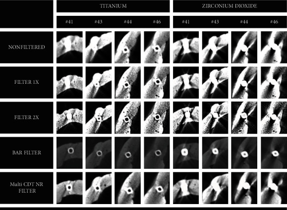
Methods: Eight artificial edentulous mandibles (phantoms), where titanium and zirconium dioxide implants had been installed in four different regions (i.e., incisors, canine, premolars, and molars). CBCT volume was acquired, and then, four types of filters were applied to the images: BAR filter and Multi-CDT NR filter (e-Vol DX) and Sharpening Filters 1x and 2x (OnDemand). Artifact was assessed by measuring the standard deviation (SD) of the gray values of filtered and unfiltered images. The comparison between implant material, teeth, and filters was performed by using ANOVA, whereas multiple comparisons were performed by using Bonferroni's test. The level of significance adopted was 5%.
Results: The results showing higher SD values, which suggests a worse image, were obtained with titanium implants compared to zirconium dioxide ones. With regard to the four filters used, it can be seen that the lowest SD values were obtained with BAR and Multi-CDT NR filters and the highest with Sharpening Filters 1x and 2x, with no statistical difference between them, except regarding the molar region in titanium implants.
Conclusion: The highest SD values were seen in zirconium dioxide implants, mainly in the region of anterior teeth. The BAR filter was found to be the most effective as its SD value decreased significantly, indicating that the image quality was improved.
Copyright © 2022 Andre Luiz Ferreira Costa et al.
Conflict of interest statement
The authors declare that there is no conflict of interest about this study.
Figures


References
-
- Harris D., Horner K., Grondahl K., et al. E.A.O. guidelines for the use of diagnostic imaging in implant dentistry 2011. A consensus workshop organized by the European Association for Osseointegration at the Medical University of Warsaw. Clinical Oral Implants Research . 2012;23(11):1243–1253. doi: 10.1111/j.1600-0501.2012.02441.x. - DOI - PubMed
-
- de-Azevedo-Vaz S. L., Peyneau P. D., Ramirez-Sotelo L. R., de Faria Vasconcelos K., Campos P. S., Haiter-Neto F. Efficacy of a cone beam computed tomography metal artifact reduction algorithm for the detection of peri-implant fenestrations and dehiscences. Oral Surgery, Oral Medicine, Oral Pathology, Oral Radiology . 2016;121(5):550–556. doi: 10.1016/j.oooo.2016.01.013. - DOI - PubMed
MeSH terms
Substances
LinkOut - more resources
Full Text Sources
