. 2022 Apr 12;145(15):1140-1153.
doi: 10.1161/CIRCULATIONAHA.121.056929. Epub 2022 Mar 17.

MicroRNA-210 Controls Mitochondrial Metabolism and Protects Heart Function in Myocardial Infarction

Affiliations

MicroRNA-210 Controls Mitochondrial Metabolism and Protects Heart Function in Myocardial Infarction

Rui Song et al. Circulation. .

Abstract

Background: Ischemic heart disease remains a leading cause of death worldwide. In this study, we test the hypothesis that microRNA-210 protects the heart from myocardial ischemia-reperfusion (IR) injury by controlling mitochondrial bioenergetics and reactive oxygen species (ROS) flux.

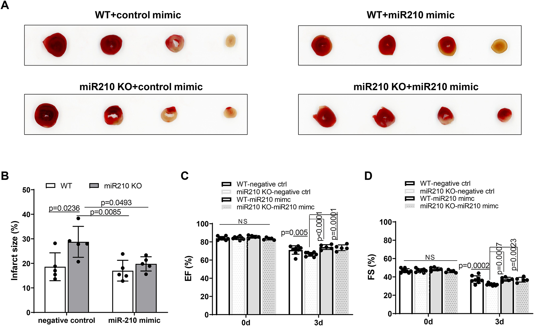
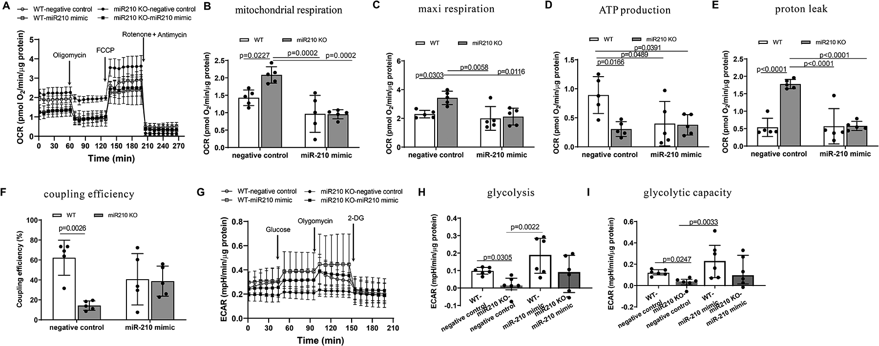
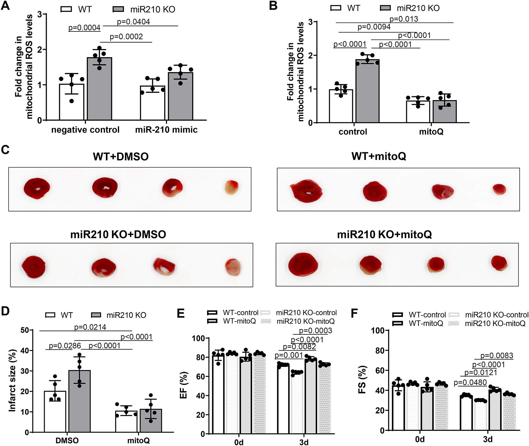
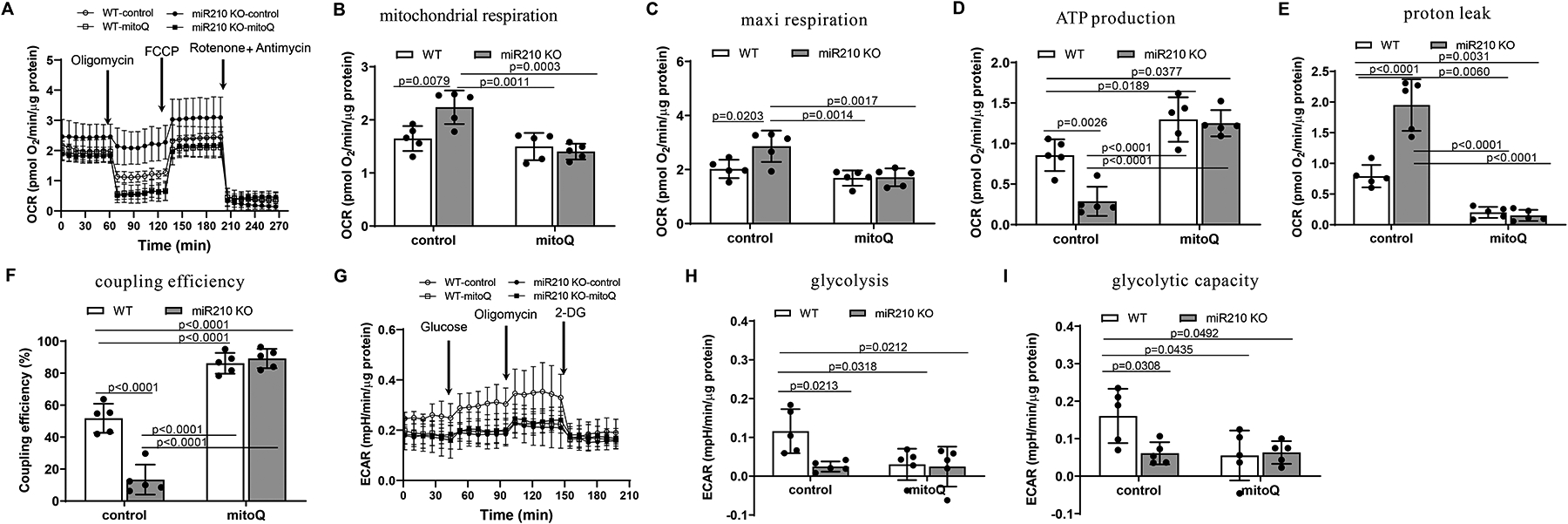
Methods: Myocardial infarction in an acute setting of IR was examined through comparing loss- versus gain-of-function experiments in microRNA-210-deficient and wild-type mice. Cardiac function was evaluated by echocardiography. Myocardial mitochondria bioenergetics was examined using a Seahorse XF24 Analyzer.

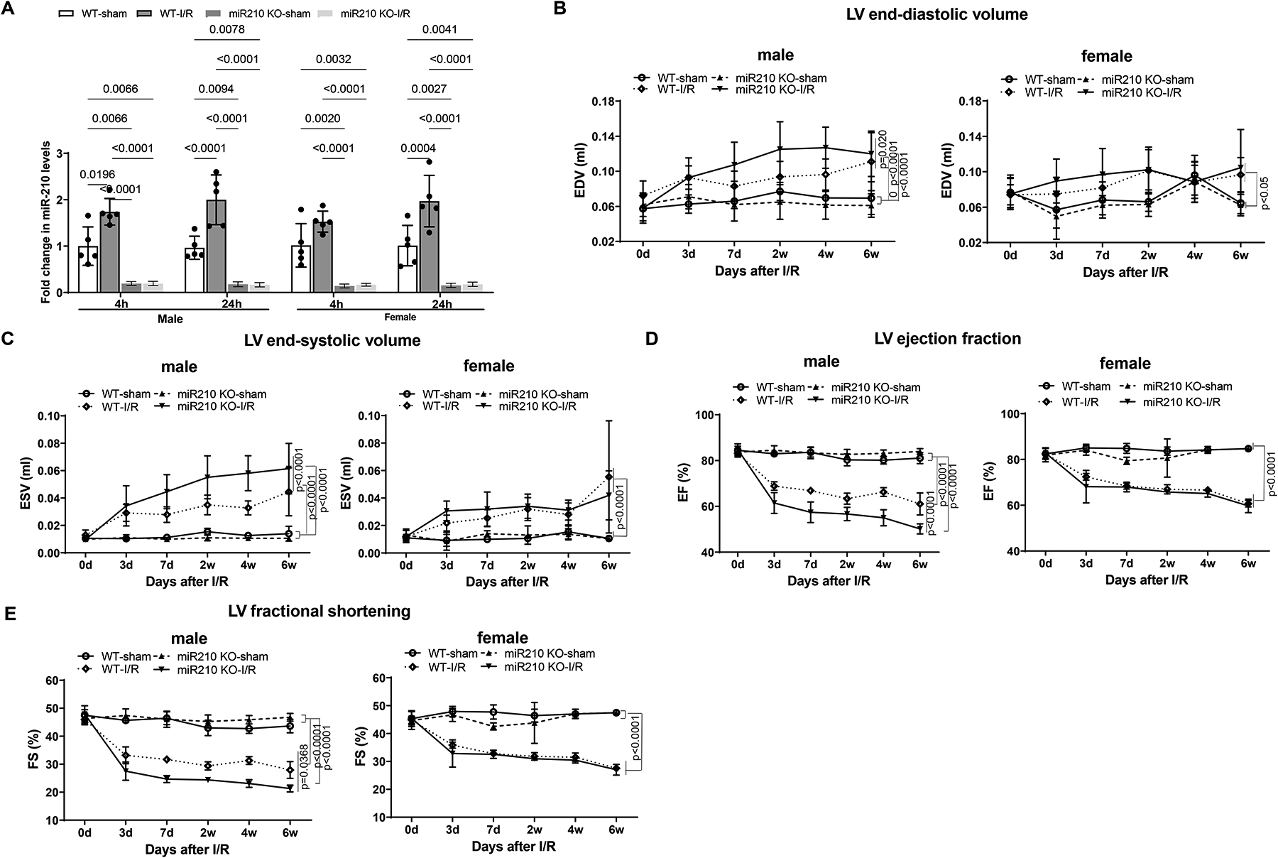
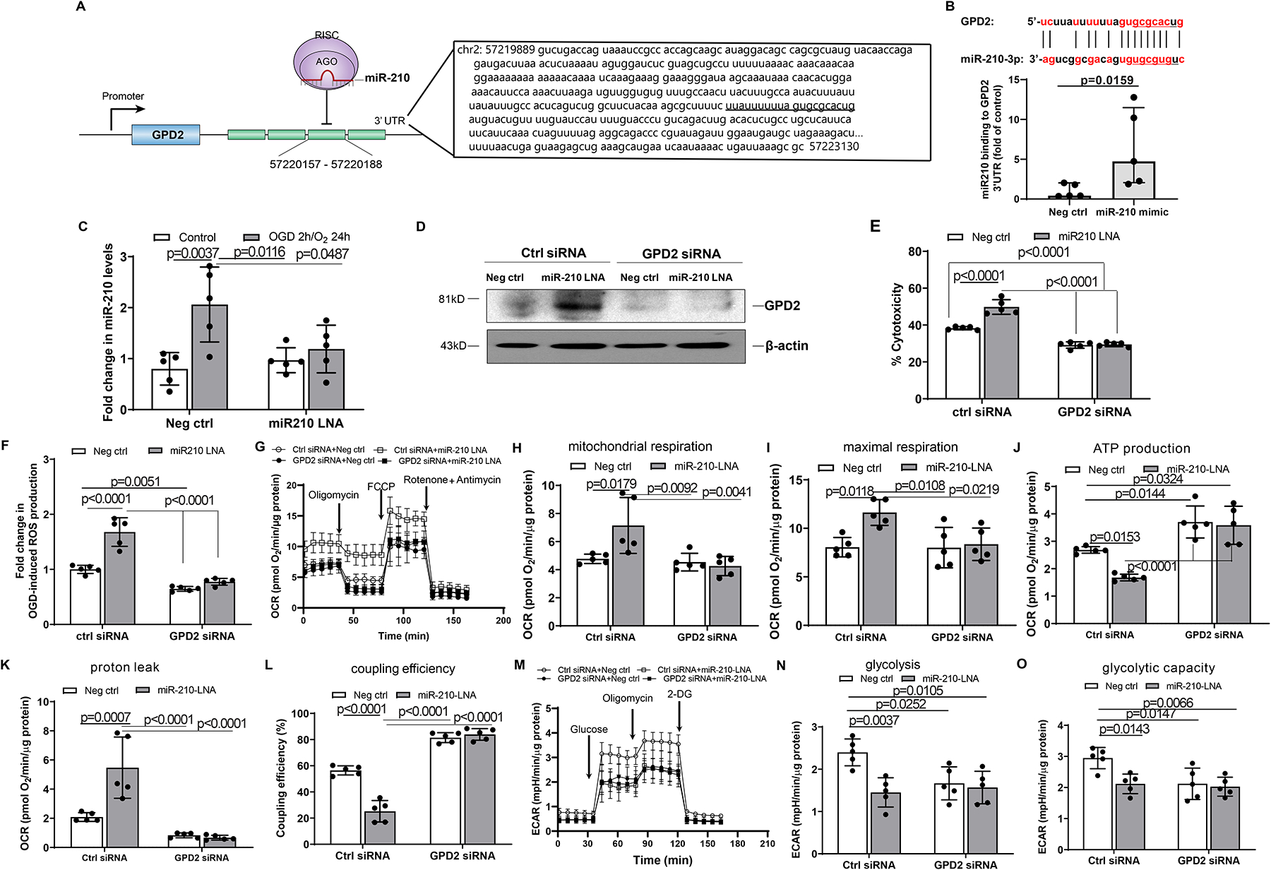
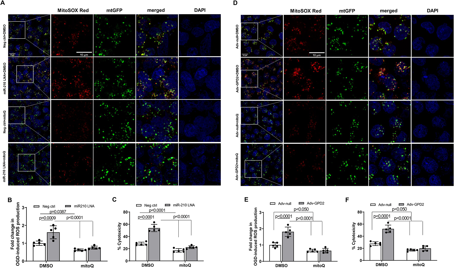
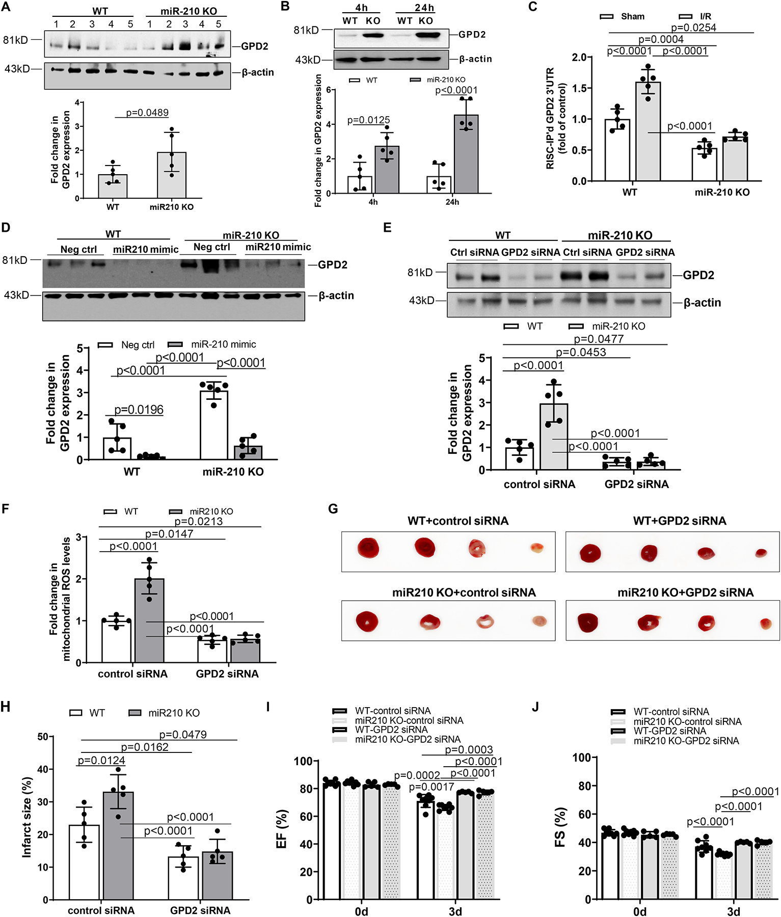
Results: MicroRNA-210 deficiency significantly exaggerated cardiac dysfunction up to 6 weeks after myocardial IR in male, but not female, mice. Intravenous injection of microRNA-210 mimic blocked the effect and recovered the increased myocardial IR injury and cardiac dysfunction. Analysis of mitochondrial metabolism revealed that microRNA-210 inhibited mitochondrial oxygen consumption, increased glycolytic activity, and reduced mitochondrial ROS flux in the heart during IR injury. Inhibition of mitochondrial ROS with MitoQ consistently reversed the effect of microRNA-210 deficiency. Mechanistically, we showed that mitochondrial glycerol-3-phosphate dehydrogenase is a novel target of microRNA-210 in the heart, and loss-of-function and gain-of-function experiments revealed that glycerol-3-phosphate dehydrogenase played a key role in the microRNA-210-mediated effect on mitochondrial metabolism and ROS flux in the setting of heart IR injury. Knockdown of glycerol-3-phosphate dehydrogenase negated microRNA-210 deficiency-induced increases in mitochondrial ROS production and myocardial infarction and improved left ventricular fractional shortening and ejection fraction after the IR treatment.

Conclusions: MicroRNA-210 targeting glycerol-3-phosphate dehydrogenase controls mitochondrial bioenergetics and ROS flux and improves cardiac function in a murine model of myocardial infarction in the setting of IR injury. The findings suggest new insights into the mechanisms and therapeutic targets for treatment of ischemic heart disease.

Keywords: energy metabolism; glycerolphosphate dehydrogenase; microRNA-210; myocardial infarction.


Figures

Figure 1.
Figure 1.. MiR-210 deficiency exaggerates cardiac dysfunction after myocardial infarction in male mice.
MiR-210 knockout (KO) and their wild-type (WT) littermate control mice with C57Bl/6 background at 2 months old underwent myocardial infarction (MI) induced by in vivo heart ischemia and reperfusion (IR) treatment via ligation of the mid-left anterior descending coronary artery for 30 min followed by reperfusion. Sham-operated animals without ligation of LAD coronary artery served as naïve controls. Hearts were isolated after 4 h or 24 h reperfusion and miR-210 in the heart was measured (A). Cardiac function was evaluated by echocardiography prior to or up to 6 weeks after in vivo IR treatment (B-E). Data are means ± SD, with n=5–6 animals per group, and analyzed by three-way ANOVA (A) or two-way repeated-measures ANOVA (B-E) followed by Tukey’s test. P values are shown in the Figure.
Figure 2.
Figure 2.. MiR-210 mimic exerts a protective effect on IR-induced MI and cardiac dysfunction.
Male miR-210 knockout (KO) and their wild-type (WT) littermate control mice with C57Bl/6 background at 2 months old underwent myocardial infarction (MI) induced by in vivo heart ischemia and reperfusion (IR) treatment via ligation of the mid-left anterior descending coronary artery for 30 min followed by reperfusion. MiR-210 mimic (10 nmol/kg) or negative control was intravenously administered 1 h prior to the IR treatment. MI (A, B) and functional echocardiography (C, D) were evaluated 72 h after IR. Data are means ± SD, with n=5–8 animals per group, and analyzed by two-way ANOVA followed by Tukey’s test. P values are shown in the Figure.
Figure 3.
Figure 3.. MiR-210 controls mitochondrial bioenergetics in the acute heart IR.
Male miR-210 knockout (KO) and their wild-type (WT) littermate control mice with C57Bl/6 background at 2 months old underwent myocardial infarction (MI) induced by in vivo heart ischemia and reperfusion (IR) treatment via ligation of the mid-left anterior descending coronary artery for 30 min followed by reperfusion. MiR-210 mimic (10 nmol/kg) or negative control was intravenously administered 1 h prior to the IR treatment. The hearts were harvested 24 h after IR and mitochondrial oxygen consumption rate (OCR) (A-F) and extracellular acidification rate (ECAR) (G-I) were analyzed by Seahorse XFe24 Analyzer in isolated cardiac muscle fiber bundles. Seahorse real-time traces and averaged data for OCR (A) and ECAR (G) including the chemical agents that dissect the metabolic responses are shown. Data are means ± SD, and analyzed by two-way ANOVA followed by Tukey’s test (A-E) or median and IQRs with non-parametric Kruskal–Wallis test followed by Dunn’s test (F, G), with n=5–6 animals per group. P values are shown in the Figure.
Figure 4.
Figure 4.. MiR-210 suppresses mitochondrial ROS in acute heart IR injury and cardiac dysfunction.
Male miR-210 knockout (KO) and their wild-type (WT) littermate control mice with C57Bl/6 background at 2 months old underwent myocardial infarction (MI) induced by in vivo heart ischemia and reperfusion (IR) treatment via ligation of the mid-left anterior descending coronary artery for 30 min followed by reperfusion. MiR-210 mimic (10 nmol/kg) or negative control was intravenously administered 1 h prior to the IR treatment. MitoQ (4 mg/kg) or vehicle control (DMSO, 1%) was intravenously applied 15 min prior to the IR. Mitochondria were isolated from the hearts 24 h after IR and mitochondria-derived ROS was analyzed with MitoSOX Red (A, B). MI (C, D) and functional echocardiography (E, F) were evaluated 72 h after IR. Data are means ± SD, with n=5 animals per group, and analyzed by two-way ANOVA followed by Tukey’s test. P values are shown in the Figure.
Figure 5.
Figure 5.. MitoQ rescues the effect of miR-210 deficiency on mitochondrial bioenergetics.
Male miR-210 knockout (KO) and their wild-type (WT) littermate control mice with C57Bl/6 background at 2 months old underwent myocardial infarction (MI) induced by in vivo heart ischemia and reperfusion (IR) treatment via ligation of the mid-left anterior descending coronary artery for 30 min followed by reperfusion. MitoQ (4 mg/kg) or vehicle control (DMSO, 1%) was intravenously applied 15 min prior to IR. The hearts were harvested 24 h after IR and mitochondrial oxygen consumption rate (OCR) (A-F) and extracellular acidification rate (ECAR) (G-I) were analyzed by Seahorse XFe24 Analyzer in isolated cardiac muscle fiber bundles. Seahorse real-time traces and averaged data for OCR (A) and ECAR (G) including the chemical agents that dissect the metabolic responses are shown. Data are means ± SD, with n=5 animals per group, and analyzed by two-way ANOVA followed by Tukey’s test. P values are shown in the Figure.
Figure 6.
Figure 6.. MiR-210 targeting GPD2 inhibits mitochondrial bioenergetics in modulating cardiomyocyte hypoxic injury.
Sequence of GPD2 3’UTR with miR-210 targeting sites and a schematic depiction of miR-210 silencing the GPD2 mRNA translation are shown in panel A. The binding of miR-210 to GPD2 3’UTR was assessed by RISC-IP assay in mouse neonatal cardiomyocytes (B). Mouse neonatal cardiomyocytes were treated with 50 nM miR-210-LNA or scramble LNA (Neg. Ctrl) for 24 h, followed by subjecting to 2 h OGD and 24 h reoxygenation. Control cells were cultured in the normoxic condition for the same durations. The levels of miR-210 were measured 24 h after reoxygenation (C). Mouse neonatal cardiomyocytes were treated with 100 nM GPD2 siRNA and 50 nM miR-210-LNA or scramble LNA (Neg. Ctrl) for 24 h, and then were subjected to OGD followed by reoxygenation (D-O). GPD2 protein abundance was determined with Western blots (D). Cell injury was measured with LDH release assay (E). Mitochondria-derived ROS was analyzed with MitoSOX Red (F). Mitochondrial oxygen consumption rate (OCR) (G-L) and extracellular acidification rate (ECAR) (M-O) were analyzed by Seahorse XFe24 Analyzer. Data are median and IQRs with Mann-Whitney U test (B), or means ± SD and analyzed by two-way ANOVA followed by Tukey’s test (C, E-O), with n=5 independent cultures per group. P values are shown in the Figure.
Figure 7.
Figure 7.. MitoQ inhibits miR-210 LNA and GPD2 overexpression-induced mtROS generation and cardiomyocyte hypoxic injury.
Mouse neonatal cardiomyocytes were treated with miR-210 LNA (50 nM) or scramble LNA (Neg. Ctrl), Adv-GPD2 (1.6 × 10⁷ pfu/ml) or Adv-null in the presence of MitoQ (250 nM) or vehicle control DMSO overnight, followed by subjecting to 2 h OGD and 24 h reoxygenation. Mitochondria-derived ROS was visualized with MitoSOX Red and fluorescence was colocalized with mitochondria, as visualized with Mitochondria-GFP (mtGFP) using fluorescent confocal microscopy (A and D). Panels B and E show the quantitative data of mtROS. Cell injury was measured with LDH release assay (C and F). Data are means ± SD with n=5 independent cultures per group, and analyzed by two-way ANOVA followed by Tukey’s test. P values are shown in the Figure.
Figure 8.
Figure 8.. Knockdown of GPD2 protects the heart and negates the effect of miR-210 deficiency on IR injury.
Male miR-210 knockout (KO) and their wild-type (WT) littermate control mice with C57Bl/6 background at 2 months old underwent myocardial infarction (MI) induced by in vivo heart ischemia and reperfusion (IR) treatment via ligation of the mid-left anterior descending coronary artery for 30 min followed by reperfusion. GPD2 protein abundance was measured in the heart before IR (A) and after IR (B) in WT and KO mice. Panel C shows the binding of endogenous miR-210 to GPD2 3’UTR in vivo in the heart after ischemia for 30 min and reperfusion for 24 h in WT and miR-210 KO mice. Panel D shows that intravenous administration of miR-210 mimic 1 h prior to the IR treatment decreases GPD2 protein abundance in the heart of WT mice and blocks the effect of miR-210 deficiency on GPD2 protein in the setting of acute heart IR. GPD2 siRNAs (2 mg/kg) or control siRNAs were intravenously applied 24 h prior to the IR treatment, and hearts were isolated 24 h after IR. GPD2 protein abundance (E) was determined with Western blots and mitochondria-derived ROS (F) was analyzed with MitoSOX Red. MI (G, H) and functional echocardiography (I, J) were evaluated 72 h after IR. Data are means ± SD, with n=5–8 animals per group, and analyzed by t-test (A) and two-way ANOVA followed by Tukey’s test (B-J). P values are shown in the Figure.

Comment in

References

    1. Mortality GBD, Causes of Death C. Global, regional, and national age-sex specific all-cause and cause-specific mortality for 240 causes of death, 1990–2013: a systematic analysis for the Global Burden of Disease Study 2013. Lancet. 2015;385:117–171. doi: 10.1016/S0140-6736(14)61682-2 - DOI - PMC - PubMed
    1. Hu S, Huang M, Li Z, Jia F, Ghosh Z, Lijkwan MA, Fasanaro P, Sun N, Wang X, Martelli F, et al. MicroRNA-210 as a novel therapy for treatment of ischemic heart disease. Circulation. 2010;122:S124–131. doi: 10.1161/CIRCULATIONAHA.109.928424 - DOI - PMC - PubMed
    1. Ong SG, Lee WH, Huang M, Dey D, Kodo K, Sanchez-Freire V, Gold JD, Wu JC. Cross talk of combined gene and cell therapy in ischemic heart disease: role of exosomal microRNA transfer. Circulation. 2014;130:S60–69. doi: 10.1161/CIRCULATIONAHA.113.007917 - DOI - PMC - PubMed
    1. Patterson AJ, Song MA, Choe D, Xiao D, Foster G, Zhang L. Early Detection of Coronary Artery Disease by Micro-RNA Analysis in Asymptomatic Patients Stratified by Coronary CT Angiography. Diagnostics (Basel). 2020;10:875. doi: 10.3390/diagnostics10110875 - DOI - PMC - PubMed
    1. Karakas M, Schulte C, Appelbaum S, Ojeda F, Lackner KJ, Munzel T, Schnabel RB, Blankenberg S, Zeller T. Circulating microRNAs strongly predict cardiovascular death in patients with coronary artery disease-results from the large AtheroGene study. Eur Heart J. 2017;38:516–523. doi: 10.1093/eurheartj/ehw250 - DOI - PubMed

Publication types

MeSH terms