. 2022 Mar 16;19(1):19.
doi: 10.1186/s12989-022-00460-3.

Surface functionalization and size modulate the formation of reactive oxygen species and genotoxic effects of cellulose nanofibrils

Affiliations

Surface functionalization and size modulate the formation of reactive oxygen species and genotoxic effects of cellulose nanofibrils

Kukka Aimonen et al. Part Fibre Toxicol. .

Abstract

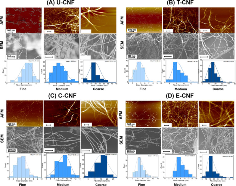
Background: Cellulose nanofibrils (CNFs) have emerged as a sustainable and environmentally friendly option for a broad range of applications. The fibrous nature and high biopersistence of CNFs call for a thorough toxicity assessment, but it is presently unclear which physico-chemical properties could play a role in determining the potential toxic response to CNF. Here, we assessed whether surface composition and size could modulate the genotoxicity of CNFs in human bronchial epithelial BEAS-2B cells. We examined three size fractions (fine, medium and coarse) of four CNFs with different surface chemistry: unmodified (U-CNF) and functionalized with 2,2,6,6-tetramethyl-piperidin-1-oxyl (TEMPO) (T-CNF), carboxymethyl (C-CNF) and epoxypropyltrimethylammonium chloride (EPTMAC) (E-CNF). In addition, the source fibre was also evaluated as a non-nanosized material.

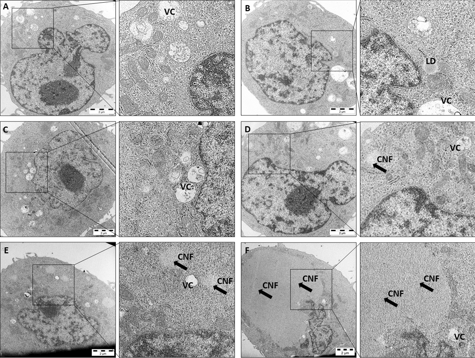
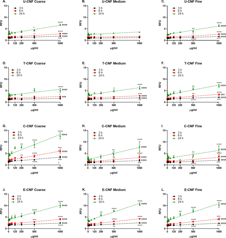
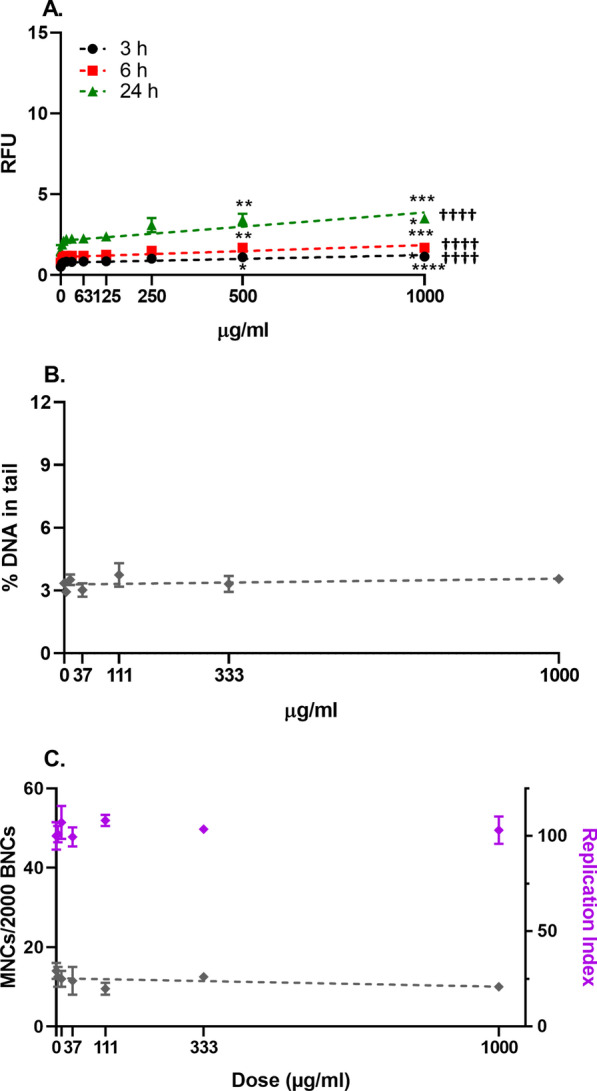
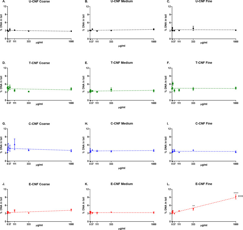
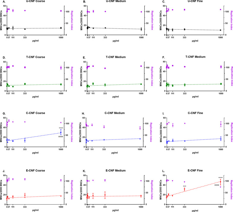
Results: The presence of the surface charged groups in the functionalized CNF samples resulted in higher amounts of individual nanofibrils and less aggregation compared with the U-CNF. T-CNF was the most homogenous, in agreement with its high surface group density. However, the colloidal stability of all the CNF samples dropped when dispersed in cell culture medium, especially in the case of T-CNF. CNF was internalized by a minority of BEAS-2B cells. No remarkable cytotoxic effects were induced by any of the cellulosic materials. All cellulosic materials, except the medium fraction of U-CNF, induced a dose-dependent intracellular formation of reactive oxygen species (ROS). The fine fraction of E-CNF, which induced DNA damage (measured by the comet assay) and chromosome damage (measured by the micronucleus assay), and the coarse fraction of C-CNF, which produced chromosome damage, also showed the most effective induction of ROS in their respective size fractions.

Conclusions: Surface chemistry and size modulate the in vitro intracellular ROS formation and the induction of genotoxic effects by fibrillated celluloses. One cationic (fine E-CNF) and one anionic (coarse C-CNF) CNF showed primary genotoxic effects, possibly partly through ROS generation. However, the conclusions cannot be generalized to all types of CNFs, as the synthesis process and the dispersion method used for testing affect their physico-chemical properties and, hence, their toxic effects.

Keywords: Cellulose nanofibrils; Functionalization; Genotoxicity; High aspect ratio; Nanofibrillated celluloses; Reactive oxygen species; Surface chemistry.


Conflict of interest statement

The authors declare that they have no competing interests.

Figures

Fig. 1
Fig. 1
Atomic force microscopy (AFM) and scanning electron microscopy (SEM) images, and fibril diameter distributions (fitted log-normal models) of the three size fractions of U-CNF (A), T-CNF (B), C-CNF (C) and E-CNF (D) (N = 100 independent observations)
Fig. 2
Fig. 2
Transmission electron microscopic (TEM) images of BEAS-2B cells without treatment (A), and after a 6-h exposure to 111 µg/ml of coarse (B) and fine (C) U-CNF, fine C-CNF (D), and fine E-CNF (E, F). One cell shows a large mass of fine E-CNF inside the cytoplasm (F). CNFs are indicated by black arrows. VC: vacuoles; LD: lipid droplets (N = 15–20 independent observations)
Fig. 3
Fig. 3
Cellular reactive oxygen species (ROS) production assessed by the fluorescent probe DCFDA in BEAS-2B cells after 3, 6 and 24-h exposure to coarse (AJ), medium (BK) and fine (CL) size fractions of U-CNF (AC), T-CNF (DF), C-CNF (GI) and E-CNF (JL). Data are expressed as relative fluorescence units (RFU) and presented as the mean ± se (N = 2 independent experiments). Asterisks designate statistically significant differences compared with the untreated cultures at *p < 0.05, **p < 0.01, ***p < 0.001 and ****p < 0.0001. Daggers designate statistically significant linear regression at †††p < 0.001 and ††††p < 0.0001. The positive control, H2O2 (2 mM), induced a statistically significant increase in ROS production over the negative control values in all the experiments performed (7.9 ± 0.4-fold increase; p < 0.003) confirming the validity of the experiments (data not shown)
Fig. 4
Fig. 4
Cellular reactive oxygen species (ROS) production (A), DNA strand breaks (B) and micronucleus induction (C) in BEAS-2B cells after 3-h (A), 6-h (A), 24-h (A, B) and 48-h (C) exposure to pulp fibres. Data are presented as the mean ± se. Asterisks designate statistically significant differences compared with the untreated cultures at *p < 0.05, **p < 0.01, ***p < 0.001 and ****p < 0.0001. Daggers designate statistically significant linear regression at ††††p < 0.0001
Fig. 5
Fig. 5
DNA strand breaks assessed by the comet assay in BEAS-2B cells after a 24-h exposure to coarse (AJ), medium (BK) and fine (CL) size fractions of U-CNF (AC), T-CNF (DF), C-CNF (GI) and E-CNF (JL). Data are expressed as percentage of DNA in tail and presented as the mean ± se (N = 2 independent experiments). Asterisks designate statistically significant differences compared with the untreated cultures at **p < 0.01 and ****p < 0.0001. Daggers designate statistically significant linear regression at ††††p < 0.0001. The positive control, H2O2 (20 mM), induced a statistically significant increase in the percentage of DNA in tail over the negative control values in all the experiments performed (15.2 ± 1.1-fold increase; p < 0.02) confirming the validity of the experiments (data not shown)
Fig. 6
Fig. 6
Frequency of micronucleated cells in 2000 binucleated cells (MNCs/2000 BNCs) and replication index (RI) after a 48-h exposure of BEAS-2B cells to coarse (AJ), medium (BK) and fine (CL) size fractions of U-CNF (AC), T-CNF (DF), C-CNF (GI) and E-CNF (JL). Data are presented as the mean ± se (N = 2 independent replicates). Asterisks designate statistically significant differences compared with the untreated cultures at ***p < 0.001 and ****p < 0.0001. Daggers designate statistically significant linear regression at ††††p < 0.0001. The positive control, MMC (150 ng/ml), induced a statistically significant increase in the frequency of micronucleated cells over the negative control values in all the experiments performed (21.59 ± 1.80-fold increase; p < 0.01; 27.6 ± 3.5% RI) confirming the validity of the experiments (data not shown)

References

    1. Moon RJ, Martini A, Nairn J, Simonsen J, Youngblood J. Cellulose nanomaterials review: structure, properties and nanocomposites. Chem Soc Rev. 2011;40(7):3941–3994. doi: 10.1039/C0CS00108B. - DOI - PubMed
    1. Bai L, Greca LG, Xiang W, Lehtonen J, Huan S, Nugroho RWN, et al. Adsorption and assembly of cellulosic and lignin colloids at oil/water interfaces. Langmuir. 2019;35(3):571–588. doi: 10.1021/acs.langmuir.8b01288. - DOI - PMC - PubMed
    1. Tan K, Heo S, Foo M, Chew IM, Yoo C. An insight into nanocellulose as soft condensed matter: challenge and future prospective toward environmental sustainability. Sci Total Environ. 2019;650:1309–1326. doi: 10.1016/j.scitotenv.2018.08.402. - DOI - PubMed
    1. Ventura C, Pinto F, Lourenço AF, Ferreira PJT, Louro H, Silva MJ. On the toxicity of cellulose nanocrystals and nanofibrils in animal and cellular models. Cellulose. 2020;27(10):5509–5544. doi: 10.1007/s10570-020-03176-9. - DOI
    1. Li T, Chen C, Brozena AH, Zhu JY, Xu L, Driemeier C, et al. Developing fibrillated cellulose as a sustainable technological material. Nature. 2021;590(7844):47–56. doi: 10.1038/s41586-020-03167-7. - DOI - PubMed

Publication types

LinkOut - more resources