. 2022 Mar 16;13(3):246.
doi: 10.1038/s41419-022-04684-1.

Dual BTK/SYK inhibition with CG-806 (luxeptinib) disrupts B-cell receptor and Bcl-2 signaling networks in mantle cell lymphoma

Affiliations

Dual BTK/SYK inhibition with CG-806 (luxeptinib) disrupts B-cell receptor and Bcl-2 signaling networks in mantle cell lymphoma

Elana Thieme et al. Cell Death Dis. .

Abstract

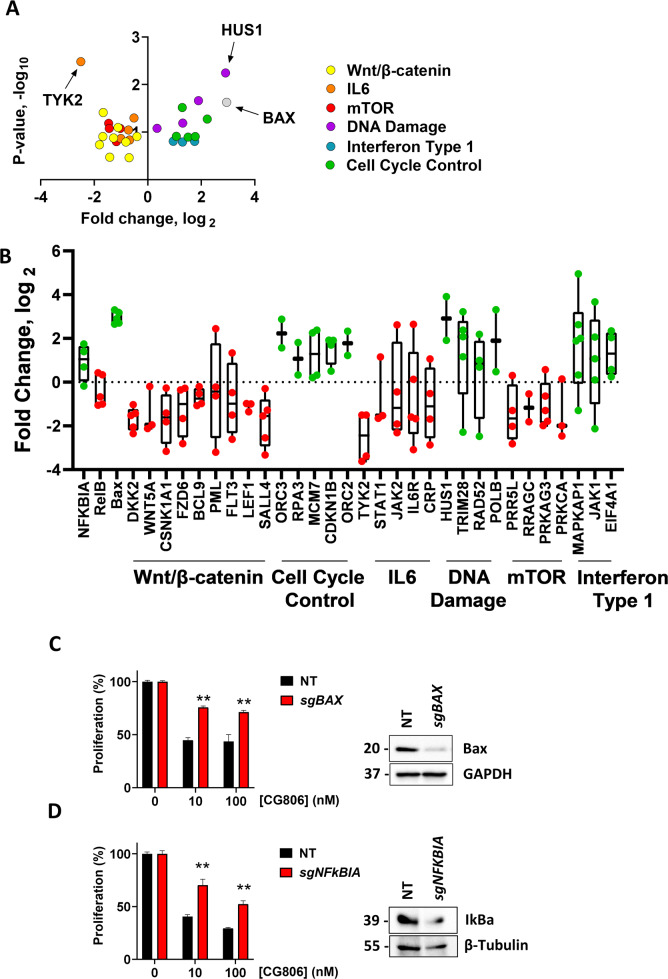
Aberrant B-cell receptor (BCR) signaling is a key driver in lymphoid malignancies. Bruton tyrosine kinase (BTK) inhibitors that disrupt BCR signaling have received regulatory approvals in therapy of mantle cell lymphoma (MCL). However, responses are incomplete and patients who experience BTK inhibitor therapy failure have dire outcomes. CG-806 (luxeptinib) is a dual BTK/SYK inhibitor in clinical development in hematologic malignancies. Here we investigated the pre-clinical activity of CG-806 in MCL. In vitro treatment with CG-806 thwarted survival of MCL cell lines and patient-derived MCL cells in a dose-dependent manner. CG-806 blocked BTK and SYK activation and abrogated BCR signaling. Contrary to ibrutinib, CG-806 downmodulated the anti-apoptotic proteins Mcl-1 and Bcl-xL, abrogated survival of ibrutinib-resistant MCL cell lines, and partially reversed the pro-survival effects of stromal microenvironment-mimicking conditions in primary MCL cells. Dual BTK/SYK inhibition led to mitochondrial membrane depolarization accompanied by mitophagy and metabolic reprogramming toward glycolysis. In vivo studies of CG-806 demonstrated improved survival in one of the two tested aggressive MCL PDX models. While suppression of the anti-apoptotic Bcl-2 family proteins and NFκB signaling correlated with in vivo drug sensitivity, OxPhos and MYC transcriptional programs were upregulated in the resistant model following treatment with CG-806. BAX and NFKBIA were implicated in susceptibility to CG-806 in a whole-genome CRISPR-Cas9 library screen (in a diffuse large B-cell lymphoma cell line). A high-throughput in vitro functional drug screen demonstrated synergy between CG-806 and Bcl-2 inhibitors. In sum, dual BTK/SYK inhibitor CG-806 disrupts BCR signaling and induces metabolic reprogramming and apoptosis in MCL. The Bcl-2 network is a key mediator of sensitivity to CG-806 and combined targeting of Bcl-2 demonstrates synergy with CG-806 warranting continued exploration in lymphoid malignancies.


Conflict of interest statement

AVD has received consulting fees from AstraZeneca, Abbvie, BeiGene, Genentech, TG Therapeutics, Bayer Oncology, and Pharmacyclics and has ongoing research funding from AstraZeneca, Takeda Oncology, Bayer Oncology, Genentech, SecuraBio, MEI, TG Therapeutics, and Bristol Myers Squibb. JWT received research support from Agios, Aptose, Array, AstraZeneca, Constellation, Genentech, Gilead, Incyte, Janssen, Petra, Seattle Genetics, Syros, Takeda, and Tolero.

Figures

Fig. 1
Fig. 1. CG-806 induces apoptosis and attenuates B-cell receptor signaling in MCL.
A Cells were treated with indicated doses of CG-806 for 24 h. Apoptosis was determined by Annexin-V-FITC staining. Data are shown as mean ± SEM. B Parental and ibrutinib-resistant MCL cell lines were treated with the indicated doses of CG-806 for 48 h. Cell proliferation was assessed using a colorimetric tetrazolium-based assay. Mean ± SEM is shown. C Cells were treated with drugs as indicated. Cell lysates were subjected to immunoblotting. D Mino cells were treated with the indicated concentrations of drugs for 24 h. E Primary MCL cells (from three individual patients) were treated with vehicle control, ibrutinib or CG-806 for 1 h, then were stimulated with 5 µg/mL IgM for 30 min. Whole-cell lysates were analyzed using immunoblotting. Representative blots are shown. F Primary MCL cells (n = 5 individual samples tested in technical duplicates) were co-cultured with stroma for 24 h. Cells were then treated with 1 µM CG-806 or ibrutinib for 48 h. Apoptosis of the CD19+ B-cell population was quantified using Annexin-V staining. *p < 0.05 and **p < 0.01 vs. untreated control.
Fig. 2
Fig. 2. CG-806 induces mitochondrial damage and metabolic reprogramming.
A Cells were treated with CG-806 (1 µM), venetoclax (Ven, 0.1 µM), or combination for 24 h and were analyzed for mitochondrial depolarization with JC-1 dye using flow cytometry. Data are presented as mean ± SEM. A representative dot plot image is also shown (depolarized mitochondria are boxed). B, C MCL cells were treated with 1 µM CG-806 or DMSO control for 24 h and subjected to Seahorse analysis measuring oxygen consumption rate (OCR) and extracellular acidification rate (ECAR). D Cell lines were treated with drug for 24 h as indicated and mitochondrial mass was assessed with Mitobright Green using flow cytometry. Data are presented as mean ± SEM. E Mino cells were treated with 1 µM CG-806 vs. control for 24 h and stained with Mtphagy dye (red) and Lysodye (green). Live cells were imaged with confocal microscopy. A representative image is shown. White arrows point to the mitophagy puncta. MFI was quantified, and colocalization was measured and calculated using Zen software. Data are presented as mean ± SEM and pixel count. *p < 0.05 and **p < 0.01 vs. control.
Fig. 3
Fig. 3. CG-806 restricts MCL development and BCR signaling in vivo.
AC Mice were inoculated with MCL cells as described in the methods. Two distinct MCL PDX models were used. Once circulating CD5/CD19+ MCL cells were detected in the peripheral blood, mice began daily treatment with CG-806, ibrutinib, or vehicle control via oral gavage in the indicated doses. Kaplan–Meier survival curves are shown, significance determined by log-rank test. At the end of the experiment, mice were euthanized 2 h after the final drug dose, and spleen-resident tumor cells were analyzed for proliferation and were subjected to immunoblotting. D NF-κB activation in splenocytes from MCL-A (N = 8) and MCL-B (N = 5) mice was quantified using the TransAm NF-κB Activation Assay. E mRNA expression of the indicated gene transcripts was quantified using RT-PCR and normalized to GAPDH. Data are shown as a box and whisker plot with dots representing expression values from four mice. *p < 0.05 and **p < 0.01 vs. vehicle control.
Fig. 4
Fig. 4. Transcriptional effects of dual SYK/BTK inhibition in vivo.
Spleen-resident tumor cells of the MCL-B PDX model were harvested 2 h after the final treatment with CG-806 at 308 mg/kg or control as described in the methods. Gene set enrichment of RNA was calculated using gsea2–2.24. N = 3. A Representation of the top 8 pathways that were differentially regulated in the treatment vs. control samples. B Enrichment plots of select gene sets. Differentially regulated genes were defined by log2fc ≥ 0.58/≤−0.58 and Padj <0.05.
Fig. 5
Fig. 5. Genome-wide CRISPR-Cas9 library screen implicates BAX and NFKBIA loss in resistance to CG-806.
Genome-wide loss of function CRISPR library screening was carried out in OCI-LY3 cells as described in the methods. Data were analyzed using a MaGeCK pipeline analysis. A Volcano plot of mid fold change vs. mid p value of all sgRNA per gene in select genes grouped by the association in pathways identified using webgestalt software. Highly differential and significant genes are identified with black arrows. B Box and whisker plot spanning min/max fold change of CG-806 vs control with individual sgRNA per gene depicted as points. Genes for which a majority of sgRNA demonstrate positive fold change are shown in green, negative in red. C, D BAX and NFKBIA knockout was established in Jeko-1 cells using RNP electroporation as described in the methods. Whole-cell lysates were subjected to immunoblotting. Cells were treated with CG-806 or vehicle control at the indicated concentrations for 48 h. Cell proliferation was assessed using a colorimetric tetrazolium-based assay. Mean±SE is shown. *p < 0.05 and **p < 0.01 vs. NT control.
Fig. 6
Fig. 6. Concurrent Bcl-2 inhibition sensitizes NHL cells to dual SYK/BTK inhibitor G-806.
A U-2932 cells were subjected to functional screening assay as described in the methods. IC50 values were calculated (represented in the color gradient). Drugs that exhibit synergy with CG-806 are shown. B Visualization of calculated synergy map based on the dose-response matrix. JeKo-1 and JeKo-1R cells were treated with CG-806, venetoclax, or a combination of the two in an 7 × 7 matrix as indicated for 48 h. Cell proliferation was measured using a colorimetric tetrazolium-based assay; % viable cells were normalized to DMSO-treated control. C Primary MCL cells (N = 4 individual samples analyzed in technical duplicates) were co-cultured with HS-5 stroma for 24 h. Cells were then treated with drugs for 24 h. Apoptosis of the CD19+ B-cell population was quantified using Annexin-V staining. *p < 0.05 and **p < 0.01 vs. vehicle control.

References

    1. Nabrinsky E, Danilov AV, Koller PB. High-risk mantle cell lymphoma in the era of novel agents. Curr Hematol Malig Rep. 2021;16:8–18. - PubMed
    1. Hill HA, Qi X, Jain P, Nomie K, Wang Y, Zhou S, et al. Genetic mutations and features of mantle cell lymphoma: a systematic review and meta-analysis. Blood Adv. 2020;4:2927–38. - PMC - PubMed
    1. Saba NS, Liu D, Herman SE, Underbayev C, Tian X, Behrend D, et al. Pathogenic role of B-cell receptor signaling and canonical NF-kappaB activation in mantle cell lymphoma. Blood. 2016;128:82–92. - PMC - PubMed
    1. Gordon M, Danilov AV. The evolving role of Bruton’s tyrosine kinase (BTK) inhibitors in chronic lymphocytic leukemia. Ther Adv Hematol. 2021;12:2040620721989588. - PMC - PubMed
    1. Martin P, Maddocks K, Leonard JP, Ruan J, Goy A, Wagner-Johnston N, et al. Postibrutinib outcomes in patients with mantle cell lymphoma. Blood. 2016;127:1559–63. - PubMed

Publication types

MeSH terms