. 2022 May;27(5):2492-2501.
doi: 10.1038/s41380-022-01501-1. Epub 2022 Mar 16.

Corticosteroid sensitization drives opioid addiction

Affiliations

Corticosteroid sensitization drives opioid addiction

Stephanie A Carmack et al. Mol Psychiatry. 2022 May.

Abstract

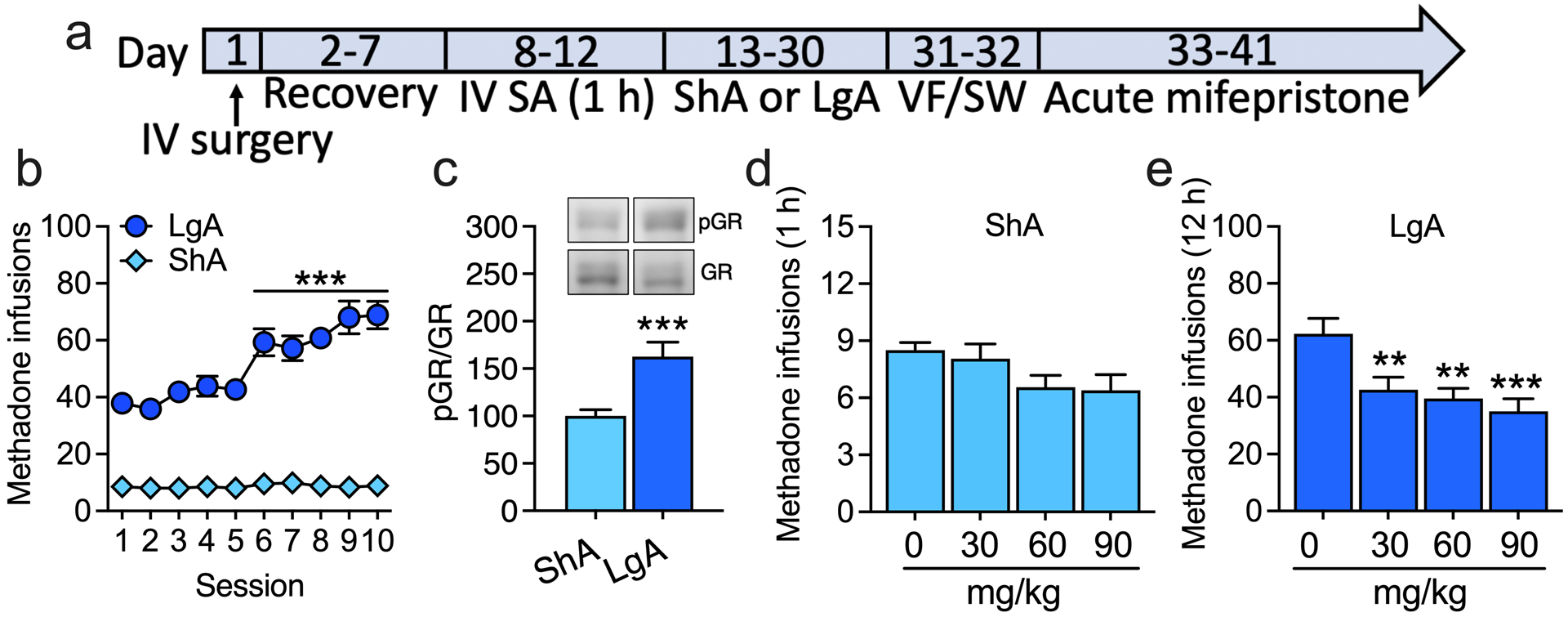
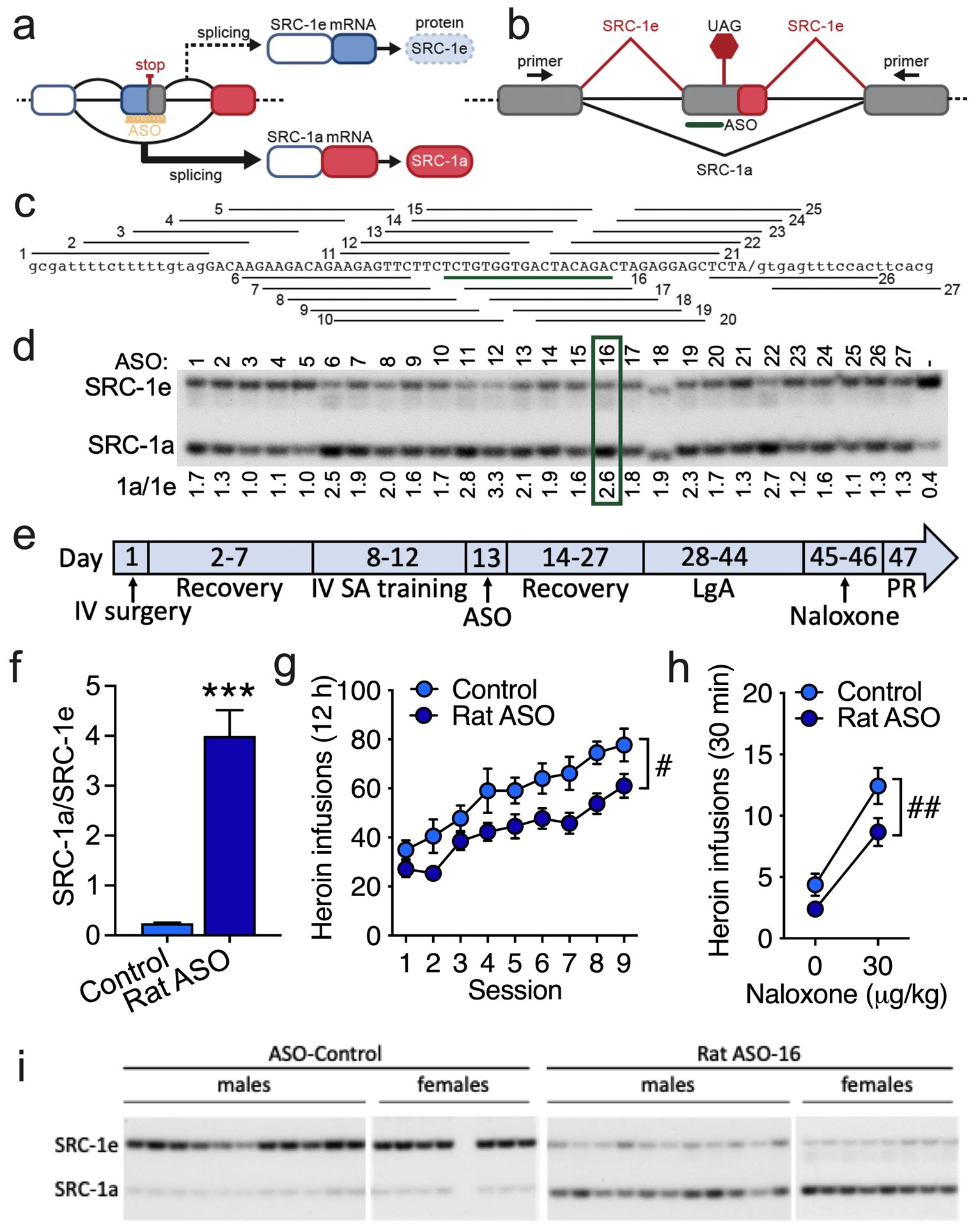
The global crisis of opioid overdose fatalities has led to an urgent search to discover the neurobiological mechanisms of opioid use disorder (OUD). A driving force for OUD is the dysphoric and emotionally painful state (hyperkatifeia) that is produced during acute and protracted opioid withdrawal. Here, we explored a mechanistic role for extrahypothalamic stress systems in driving opioid addiction. We found that glucocorticoid receptor (GR) antagonism with mifepristone reduced opioid addiction-like behaviors in rats and zebrafish of both sexes and decreased the firing of corticotropin-releasing factor neurons in the rat amygdala (i.e., a marker of brain stress system activation). In support of the hypothesized role of glucocorticoid transcriptional regulation of extrahypothalamic GRs in addiction-like behavior, an intra-amygdala infusion of an antisense oligonucleotide that blocked GR transcriptional activity reduced addiction-like behaviors. Finally, we identified transcriptional adaptations of GR signaling in the amygdala of humans with OUD. Thus, GRs, their coregulators, and downstream systems may represent viable therapeutic targets to treat the "stress side" of OUD.


Conflict of interest statement

Competing interests: The authors declare no competing interests.

Figures

Figure 1.
Figure 1.. Chronic glucocorticoid receptor antagonism blunts the development of heroin addiction-like behaviors.
(a) Male and female rats were surgically implanted with intravenous (IV) catheters. After recovery from surgery, the rats were trained to self-administer heroin (unit dose: 60 μg/kg/0.1 ml for males; 180 μg/kg/0.1 ml for females) in 1 h short-access (ShA) self-administration (SA) sessions under a fixed-ratio 1 (FR1) schedule of reinforcement (each active lever press was reinforced with heroin). The rats were subcutaneously implanted with pellets that released the GR antagonist mifepristone (Mifep; 200 mg for continuous 21-day delivery) or vehicle pellets. Twenty-four hours later, the rats were allowed ShA or long-access (LgA; 12 h) to heroin self-administration (FR1) for 9 sessions. This was followed by a two-session naloxone (vs. saline) challenge test (FR1) that lasted 30 min because of the short-acting effect of naloxone [14] and by a progressive-ratio (PR) test, in which the number of lever presses that was required to receive a heroin infusion progressively increased. (b) Heroin infusions (± SEM) in rats under LgA FR1 heroin self-administration sessions. (c) Heroin infusions (± SEM) in rats in the LgA group in a PR test. (d) Heroin infusions (± SEM) in rats in the LgA group in 30 min sessions (FR1) following vehicle (saline) administration (0 μg/kg naloxone, subcutaneous) or naloxone administration (30 μg/kg naloxone, subcutaneous). * p < 0.05, **p < 0.01, ***p < 0.001, difference from Mifep. Vehicle: n = 17 (10 males, 7 females). Mifep: n = 22 (14 males, 8 females).
Figure 2.
Figure 2.. Chronic glucocorticoid receptor antagonism decreases the excitability of CeA CRF cells.
(a) Male Wistar CRH-Cre rats were prepared with intravenous (IV) catheters and trained (1 h ShA sessions; FR1) for intravenous (IV) heroin self-administration. Via intracerebral surgery (IC), the rats received bilateral infusions of AAV1-CAG-FLEX-eGFP (0.5 μl/side; AAV) in the CeA (anterior/posterior: −2.06; medial/lateral: ±4.55; dorsal/ventral: −8.45). The rats were implanted with Mifep pellets and allowed LgA (12 h) to heroin self-administration. Eight hours after their last self-administration session (i.e., acute opioid withdrawal), the rats were euthanized, and coronal slices were prepared for electrophysiology (Ephys). Neurons that expressed green fluorescent protein (CRF+) were patched. (b) Input-output curves of the number of action potentials of CRF neurons in rats in the LgA group that received chronic treatment with Mifep (n = 9 cells) or no treatment (n = 10 cells). * p < 0.05, difference from Mifep. (c) Representative current-clamp traces of action potential firing in CeA CRF neurons induced by 0, 200, and 300 pA stimulus currents.
Figure 3.
Figure 3.. Acute glucocorticoid receptor antagonism reverses escalated IV methadone self-administration.
(a) Male and female rats were prepared with intravenous (IV) catheters and trained to self-administer (SA) methadone (unit dose: 300 μg/kg/0.1 ml) in 1 h ShA sessions (FR1) and split into ShA and LgA (12 h) groups. We used the unit dose of 300 μg/kg/0.1 ml based on studies that reported that rats dose-dependently self-administered methadone and that methadone substitutes for morphine [–23]. We used the 300 μg/kg/0.1 ml dose for both sexes based on their similar response to methadone-induced antinociception and hyperalgesia during withdrawal [24]. Once rats in the LgA group significantly escalated their methadone self-administration, rats in both the ShA and LgA groups were acutely administered with mifepristone (0, 30, 60, and 90 mg/kg; intraperitoneal, 90 min before testing) in a within-subjects Latin-square design. (b) Methadone infusions (± SEM) in rats in the ShA and LgA groups. ***p < 0.0001, difference from session 1. ShA: n = 18 (10 males, 8 females). LgA: n = 18 (9 males, 9 females). To confirm that the rats in the LgA group exhibited greater signs of opioid dependence than rats in the ShA group, we tested the rats in the von Frey (VF) test to measure hyperalgesia during spontaneous withdrawal and naloxone-precipitated signs of somatic withdrawal (STW). (c) Phosphorylation of GRs at Ser232 in the CeA in rats in the LgA group relative to rats in the ShA group. The optical densities of total GR bands were normalized to the housekeeping protein GAPDH. Because there was no significant difference in total GR levels between the ShA and LgA groups, we normalized the optical densities of phosphorylated GR bands to the optical densities of total GR bands. ***p < 0.001, difference from ShA. ShA: n = 14 (7 males, 7 females). LgA: n = 12 (6 males, 6 females). (d, e) Methadone infusions (± SEM) in (d) rats in the ShA group and (e) rats in the LgA group. **p < 0.01, ***p = 0.0002, difference from 0 mg/kg (Dunnett’s post hoc comparisons). ShA: n = 18. LgA: n = 18.
Figure 4.
Figure 4.. Increasing the SRC-1a isoform in the CeA reduces heroin self-administration in rats under LgA conditions.
(a) Schematic representation of the divergent activities of SRC-1 isoforms in gene transcription. (b) Schematic diagram of the alternative splicing pattern of SRC-1. Exons are boxes, and introns are lines. SRC-1 splicing is shown as red diagonal lines, and SRC-1a is shown in black. The SRC-1e termination codon is shown (UAG). A representative antisense oligonucleotide (ASO) that is aligned to the targeted region is shown in green. The location of the primers that were used to detect SRC-1 spliced isoforms in the polymerase chain reaction (PCR) are shown in exons that flank the alternatively spliced exon. (c) General alignment of the 27 ASOs that were individually tested for activity in blocking SRC-1e splicing. ASO 16, which was selected for further testing, is highlighted in green. (d) Analysis of individual ASO activity in rat PC-12 cells. SRC-1e-specific exon skipping assessed by the separation of radioactive RT-PCR products by polyacrylamide gel electrophoresis. The quantification of products by phosphorimaging analysis (SRC-1a/SRC-1e) is shown below the gel image. The green box indicates the ASO selected for further testing. The sequence of the most active ASOs and their alignment to the SRC-1 sequences with which they base pair, the SRC-1 amplicons from the CeA in rats that were treated with the SRC-1-targetted or control ASO, and the quantification of RT-PCR products are shown in Figure S12. (e) Male rats were prepared with intravenous (IV) catheters and trained to self-administer (SA) heroin (60 μg/kg/0.1 ml) in 1 h ShA sessions (FR1). The rats received bilateral infusions (16.66 μg in 0.5 μl/side) of the SRC-1-targeted antisense oligonucleotide (ASO) or control ASO in the CeA (anterior/posterior: −2.06; medial/lateral: ±4.55; dorsal/ventral: −8.45) and were allowed LgA heroin self-administration (FR1). This regimen was followed by a PR test (Figure S14) and a naloxone challenge test. (f) Ratio of SRC-1a and SRC-1e splice variants in the CeA in rats that were treated with the SRC-1-specific ASO or non-targeted control ASO. ***p < 0.001, difference from controls. Control ASO: n = 18 (11 males, 7 females). Rat SRC-1 ASO: n = 18 (11 males, 7 females). (g) Heroin infusions (± SEM; FR1) in rats that received intra-CeA infusions of SRC-1-specific ASO (Rat SRC-1 ASO) or non-targeted ASO (Control) and were tested in LgA self-administration sessions. #p < 0.05, overall treatment effect. Control ASO: n = 19 (11 males, 8 females). Rat SRC-1 ASO: n = 19 (11 males, 8 females). (h) Heroin self-administration (± SEM) following saline administration (0 μg/kg naloxone, subcutaneous) or naloxone-precipitated withdrawal (30 μg/kg, subcutaneous) in rats. ##p < 0.01, overall treatment effect. Control ASO: n = 18 (11 males, 7 females). Rat SRC-1 ASO: n = 18 (11 males, 7 females). (i) Polyacrylamide gel of Src-1a/1e RT-PCR products quantified in (f).
Figure 5.
Figure 5.. Transcriptional evidence of the activation of GR signaling in individuals with histories of opioid dependence.
Gene expression was profiled from the CeA in opioid-dependent individuals and unaffected controls by RNA sequencing. Demographic data are shown in Table S5. A representative coronal brain section of the human CeA is shown in Figure S15. We used the Gene Set Enrichment Analysis (GSEA) algorithm [33], a computational method that determines whether a gene set shows significant concordant differential expression between two conditions. The gene expression signature in humans with a history of opioid dependence vs. unaffected control individuals showed significant enrichement of the WP_GLUCOCORTICOID_RECEPTOR_PATHWAY gene set from the Broad Institute’s MSigDB database [58], indicating transcriptional adaptations of GR signaling in the amygdala in humans with OUD.
Figure 6.
Figure 6.. Glucocorticoid receptor-dependent plasticity in the CeA mediates opioid addiction-like behaviors.
The collective data suggest that excessive HPA axis activation during repeated episodes of opioid withdrawal leads to long-lasting neuroadaptations in extrahypothalamic brain regions. In CeA neurons, corticosteroids bind to GRs. Bound GRs dimerize and translocate to the nucleus where the coregulator SRC-1e is preferentially recruited. The GR/SRC-1e complex causes gene transactivation, including the expression of stress-related neuropeptides, such as CRF. Increases in the expression and release of CRF during opioid withdrawal may drive excessive drug seeking and taking. Blocking GRs and increasing the SRC-1a isoform constitute viable approaches to reverse allostatic changes in stress-related brain regions in opioid addiction.

Similar articles

Cited by

References

    1. Strang J, Volkow ND, Degenhardt L, Hickman M, Johnson K, Koob GF, et al. Opioid use disorder. Nat Rev Dis Primers. 2020;6:3. - PubMed
    1. Understanding the Epidemic | Drug Overdose | CDC Injury Center. 2019. https://www.cdc.gov/drugoverdose/epidemic/index.html. Accessed 7 January 2020.
    1. Case A, Deaton A. Rising morbidity and mortality in midlife among white non-Hispanic Americans in the 21st century. Proc Natl Acad Sci USA. 2015;112:15078–15083. - PMC - PubMed
    1. Koob GF. Neurobiology of Opioid Addiction: Opponent Process, Hyperkatifeia, and Negative Reinforcement. Biological Psychiatry. 2020;87:44–53. - PubMed
    1. Evans CJ, Cahill CM. Neurobiology of opioid dependence in creating addiction vulnerability. F1000Res. 2016;5. - PMC - PubMed

Publication types

Substances