

Central nervous system biometry in fetuses with and without congenital heart diseases
- PMID: 35296918
- PMCID: PMC9633519
- DOI: 10.1007/s00404-022-06484-6
Central nervous system biometry in fetuses with and without congenital heart diseases
Abstract
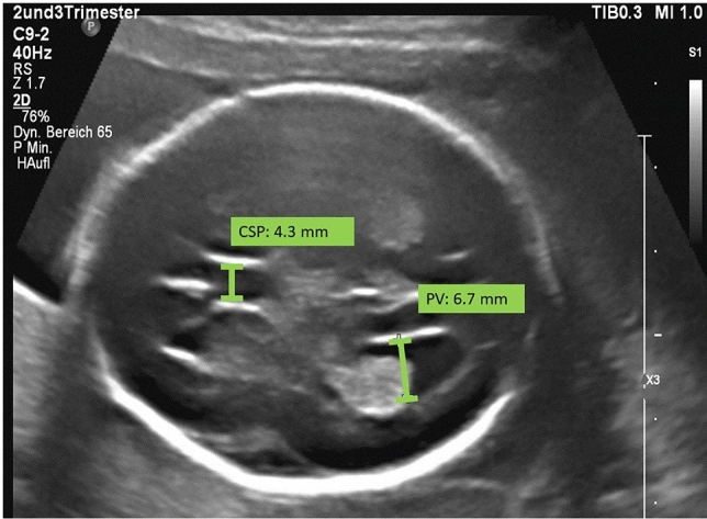
Objective: To compare the fetal brain structures assessed in routine sonographic scans during the second and third trimesters in fetuses with and without congenital heart disease (CHD).
Methods: This is a retrospective cross-sectional single-center study. We measured the head circumference (HC), the transversal diameter of the cerebellum (TCD) and the sizes of the cisterna magna (CM), the cavum septi pellucidi (CSP) and the posterior ventricles (PV) between 20 and 41 weeks of gestation. We compared 160 fetuses with CHD (case group) to 160 fetuses of normal pregnancies (control group). Every patient was matched with a control, considering the gestational age at which the ultrasound was performed. We divided the CHD group into 3 subgroups: retrograde flow in the aortic arch (group 1), right heart anomaly with the antegrade flow in the aortic arch (group 2) and other CHDs with the antegrade flow in the aortic arch (group 3).
Results: The mean width of the PV was larger in fetuses of groups 1 and 3 in comparison to the control group (P < 0.001, P = 0.022; respectively). We found that the APGAR score at 5 min (P < 0.001, P < 0.001; respectively) and gestational age at delivery (P = 0.006, P = 0.001; respectively) were inferior in groups 1 and 3 compared to controls.
Conclusions: Central nervous system biometry is altered in fetuses with CHD. PV is enlarged in CHD fetuses especially with decreased oxygen levels in the aortic arch.
Keywords: Congenital heart disease; Posterior ventricles; Prenatal ultrasound, cavum septi pellucidi.
© 2022. The Author(s).
Conflict of interest statement
All authors declare that they have no conflict of interest.
Figures
References
-
- Masoller N, Sanz-Cortés M, Crispi F, Gómez O, Bennasar M, Egaña-Ugrinovic G, Bargalló N, Martínez JM, Gratacós E. Mid-gestation brain Doppler and head biometry in fetuses with congenital heart disease predict abnormal brain development at birth. Ultrasound ObstetGynecol. 2016;47(1):65–73. doi: 10.1002/uog.14919. - DOI - PubMed
Publication types
MeSH terms
LinkOut - more resources
Full Text Sources
Medical
