Case Reports
. 2022 Apr;9(4):570-576.
doi: 10.1002/acn3.51531. Epub 2022 Mar 16.

Novel CAPN1 missense variants in complex hereditary spastic paraplegia with early-onset psychosis

Affiliations
Case Reports

Novel CAPN1 missense variants in complex hereditary spastic paraplegia with early-onset psychosis

Julian E Alecu et al. Ann Clin Transl Neurol. 2022 Apr.

Abstract

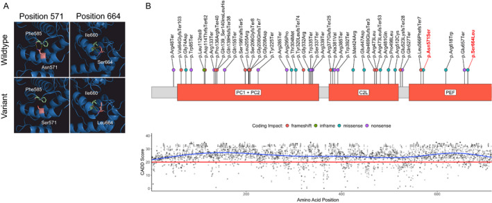
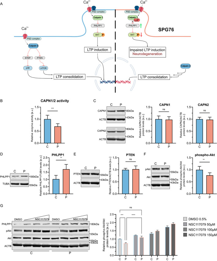
CAPN1-associated hereditary spastic paraplegia (SPG76) is a rare and clinically heterogenous syndrome due to loss of calpain-1 function. Here we illustrate a translational approach to the case of an 18-year-old patient who first presented with psychiatric symptoms followed by spastic gait, intention tremor, and neurogenic bladder dysfunction, consistent with a complex form of HSP. Exome sequencing showed compound-heterozygous missense variants in CAPN1 (NM_001198868.2: c.1712A>G (p.Asn571Ser)/c.1991C>T (p.Ser664Leu)) and a previously reported heterozygous stop-gain variant in RCL1. In silico analyses of the CAPN1 variants predicted a deleterious effect and in vitro functional studies confirmed reduced calpain-1 activity and dysregulated downstream signaling. These findings support a diagnosis of SPG76 and highlight that the psychiatric symptoms can precede the motor symptoms in HSP. Our results also suggest that multiple genes can potentially contribute to complex neuropsychiatric diseases.


Conflict of interest statement

D. E. F. received a speaker honorarium from the Movement Disorder Society, publishing royalties from the Cambridge University Press and reports research funding through a joint research agreement with Astellas Pharmaceuticals Inc.

Figures

Figure 1
Figure 1
Calpain‐1 structure with novel variants, variant distribution, and CADD PHRED scores. (A) The sites of predicted amino acid exchange in the PEF domain in the proband compared to wildtype. Mutated amino acids are colored in light red, interaction partners in light green, and hydrogen bonds in yellow. (B, upper panel) Schematic of the calpain‐1 primary protein structure. Disease‐associated variants identified in the literature are annotated along the protein with colored dots representing coding impacts. Novel variants identified in this report are labeled in red. (B, lower panel) CADD PHRED scores for all possible missense variants aligned to the CAPN1 protein structure. The recommended cut‐off for deleteriousness (20) is depicted as a red line. C2L, C2‐like domain; PC, protease core domains; PEF, large subunit of the penta‐EF‐hand domains. [Colour figure can be viewed at wileyonlinelibrary.com]
Figure 2
Figure 2
Calpain‐1 pathway and results of functional studies. (A) Schematic of the calpain‐1 and calpain‐2 pathways. The left half depicts physiological calpain signaling. The right half shows the impact of the loss of calpain‐1 function and associated dysregulation of downstream signaling in SPG76. (B) Calpain‐1 and calpain‐2 activity are measured in fibroblast lysates using a fluorogenic assay (p = 0.0004). (C) Western blot for calpain‐1 and calpain‐2 (p = 0.67, p = 0.93). (D) Western blot for calpain‐1 substrate PHLPP1 (p = 0.0087). (E) WB for calpain‐2 substrate PTEN (p > 0.99). (F) Western blot for Akt and phospho‐(Ser473)‐Akt (p = 0.004). (G) Western blot of NSC117079‐ or DMSO‐treated fibroblasts for Akt and phospho‐Akt. Whole‐cell fibroblast lysates were used for all WB experiments. Molecular weights are provided in kilodaltons (kDa). C, control; CAPN1, calpain‐1; CAPN2, calpain‐2; P, proband. Values are shown as mean ± SD, (ns = not significant, **p < 0.01, ***p < 0.001, ****p < 0.0001; [B–F] Mann–Whitney U test; [G] two‐way ANOVA with multiple comparisons and Tukey's post hoc analysis). [Colour figure can be viewed at wileyonlinelibrary.com]

References

    1. Shribman S, Reid E, Crosby AH, Houlden H, Warner TT. Hereditary spastic paraplegia: from diagnosis to emerging therapeutic approaches. Lancet Neurol. 2019;18(12):1136‐1146. - PubMed
    1. Erfanian Omidvar M, Torkamandi S, Rezaei S, et al. Genotype‐phenotype associations in hereditary spastic paraplegia: a systematic review and meta‐analysis on 13,570 patients. J Neurol. 2021;268(6):2065‐2082. - PubMed
    1. Saputra L, Kumar KR. Challenges and controversies in the genetic diagnosis of hereditary spastic paraplegia. Curr Neurol Neurosci Rep. 2021;21(4):15. - PMC - PubMed
    1. Gan‐Or Z, Bouslam N, Birouk N, et al. Mutations in CAPN1 cause autosomal‐recessive hereditary spastic paraplegia. Am J Hum Genet. 2016;98(5):1038‐1046. - PMC - PubMed
    1. Brownstein CA, Smith RS, Rodan LH, et al. RCL1 copy number variants are associated with a range of neuropsychiatric phenotypes. Mol Psychiatry. 2021;26(5):1706‐1718. - PMC - PubMed

Publication types