. 2022 Apr 1;149(7):dev200217.
doi: 10.1242/dev.200217. Epub 2022 Mar 31.

The Blimp-1 transcription factor acts in non-neuronal cells to regulate terminal differentiation of the Drosophila eye

Affiliations

The Blimp-1 transcription factor acts in non-neuronal cells to regulate terminal differentiation of the Drosophila eye

Hongsu Wang et al. Development. .

Abstract

The formation of a functional organ such as the eye requires specification of the correct cell types and their terminal differentiation into cells with the appropriate morphologies and functions. Here, we show that the zinc-finger transcription factor Blimp-1 acts in secondary and tertiary pigment cells in the Drosophila retina to promote the formation of a bi-convex corneal lens with normal refractive power, and in cone cells to enable complete extension of the photoreceptor rhabdomeres. Blimp-1 expression depends on the hormone ecdysone, and loss of ecdysone signaling causes similar differentiation defects. Timely termination of Blimp-1 expression is also important, as its overexpression in the eye has deleterious effects. Our transcriptomic analysis revealed that Blimp-1 regulates the expression of many structural and secreted proteins in the retina. Blimp-1 may function in part by repressing another transcription factor; Slow border cells is highly upregulated in the absence of Blimp-1, and its overexpression reproduces many of the effects of removing Blimp-1. This work provides insight into the transcriptional networks and cellular interactions that produce the structures necessary for visual function.

Keywords: Drosophila; Blimp-1; Cone cells; Corneal lens; Pigment cells; Rhabdomeres.


Conflict of interest statement

Competing interests The authors declare no competing or financial interests.

Figures
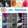
Fig. 1.
Fig. 1.
Blimp-1 is required for normal eye morphology and is transiently expressed in all retinal cell types. (A,B) Scanning electron micrographs of adult eyes: wild type (A) and Blimp-1KG09531 homozygous mutant clones (B). The mutant regions are raised and have a smooth surface. (C,D) Adult eyes with Blimp-1 mutant clones: Blimp-1KG09531 (mutant regions are darker red; C) and Blimp-112 (mutant regions are white; D). (E) Unmarked Blimp-112 mutant clones expressing UAS-Blimp-172.1. (F) Schematic of an ommatidium in the pupal retina showing the photoreceptors (1-8), cone cells (cc), primary (1°), secondary (2°) and tertiary (3°) pigment cells, and bristles (b). (G) Diagram of the Blimp-1 RE transcript, showing untranslated regions in gray, coding regions in blue and the five zinc fingers in white. CRISPR mutants Blimp-112 and Blimp-117 were created by using the two sgRNAs shown to delete the majority of the coding region including the zinc fingers. (H-M) Blimp-1 antibody staining (H″,J″,L″, red in H,J,L) in retinas containing Blimp-117 clones positively labeled with GFP (H‴,J‴,L‴, green in H,J,L) and outlined with dashed lines. Photoreceptors are labeled with anti-Elav staining (H′,J′, blue in H,J) in 24 h APF (H,I) and 30 h APF (J,K) retinas. L, M show 46 h APF retinas stained with anti-Cut (L′, blue in L) to mark cone cells. I, K, M show enlargements of single ommatidia from H, J, L, respectively, at planes in which Blimp-1 staining in each of the cell types is visible. M′ shows the plane that contains photoreceptor nuclei, which do not express Blimp-1 at this stage. CC, cone cells; PC, secondary and tertiary pigment cells; PPC, primary pigment cells; PR, photoreceptors. Scale bars: 100 µm (A-E); 10 µm (H,J,L); 2.5 µm (I,K,M).
Fig. 2.
Fig. 2.
Blimp-1 acts in the higher order pigment cells to maintain bi-convex corneal lens morphology. (A) Horizontal section of adult head with Blimp-117 clones positively labeled with myristoylated Tomato (red). Corneal lenses are stained with an Alexa488-labeled chitin binding domain (CBD, green) and Calcofluor White (blue); in wild-type regions they have a bi-convex shape (blue arrows), whereas in mutant regions they have a plano-convex shape (yellow arrows). (B) Schematic of the normal bi-convex shape of the Drosophila corneal lens in horizontal section. (C) Schematic of the plano-convex lens shape seen in Blimp-1 mutants. W, width; H1, height of external corneal lens; H2, height of internal corneal lens; T, thickness. (D) Schematic of the apical region of an adult ommatidium, showing the positions of cone and primary pigment cells under the central corneal lens and secondary pigment cells at the periphery. (E,F) Plastic sections of adult eyes: control (E) and Blimp-1 RNAi driven in secondary and tertiary pigment cells by 54-GAL4 (F). (G,H) Adult eyes with clones in which Blimp-1 RNAi is driven with 54-GAL4. G shows a horizontal section in which β-gal (red) marks a clone expressing Blimp-1 RNAi with 54-GAL4, stained with CBD (green) and Calcofluor White (blue) to detect chitin. Yellow arrows indicate plano-convex and blue arrows bi-convex lenses. H shows an external eye with a smooth surface in the clones. (I,J) Transmission electron micrographs of ommatidia from a control eye (I) and an eye in which Blimp-1 RNAi is driven with 54-GAL4 (J). Little corneal lens material is deposited above the secondary pigment cells in either case (red arrows). (K) Illustration defining the outer angle of the corneal lens. (L) Quantification of the outer angle of corneal lenses in Blimp-1 mutant or wild-type control clones, n=35 each. (M) Image focal length f′, the parameter that is relevant to fly vision, is calculated using the equations P1=(nl−n)/R1, P2=(n′−nl)/R2, P3=(−t/nl)×P1×P2, Pl=P1+P2+P3, f=n/Pl, f′=n′/Pl. F, object focal point; Fi, image focal point; R1, radius of curvature of the exterior surface facing the air; R2, radius of curvature of the interior surface facing the pseudocone; H and H′, principal points; t, thickness of the lens; f, object focal length; n, refractive index of air; nl, refractive index of lens; n′, refractive index of pseudocone. Equations and refractive indices are those used for wild-type flies (Stavenga, 2003). R2 is a negative number in a bi-convex lens, and R1 is assumed to be infinite in a plano-convex lens. (N) Diagram showing the radius of curvature of a corneal lens surface, which is calculated using the formula R=H2+W2/8H. (O) Calculated image focal length of corneal lenses in Blimp-1 mutant and wild-type control clones, n=20 each. Data are mean±s.d. ****P<0.0001 (Welch's two-tailed t-test). Scale bars: 20 µm (A,E,F,G); 100 µm (H); 10 µm (I, also applies to J).
Fig. 3.
Fig. 3.
Blimp-1 acts in cone cells to generate normal photoreceptor morphology. (A) Horizontal section of adult head in which Blimp-117 mutant clones are negatively marked by the absence of nuclear RFP (A‴, red in A). TrpL staining (A′, green in A) marks photoreceptor rhabdomeres and Elav (A″, blue in A) marks neuronal nuclei. Brackets mark clones in which rhabdomeres fail to extend to the proximal side of the retina and photoreceptor nuclei are misplaced. (B) Schematic of an ommatidium representing loss of Blimp-1 from the cone cells (cc) and primary pigment cells (1°), which express spa-GAL4. 1-8, photoreceptors; 2°, secondary pigment cells; 3°, tertiary pigment cells; b, bristles. (C) Horizontal section of adult head in which Blimp-1 RNAi is driven by spa-GAL4. Photoreceptor rhabdomeres marked by TrpL (C′, green in C) fail to extend and nuclei marked by Elav (C″, magenta in C) are disorganized. (D) Horizontal section of adult head containing clones in which Blimp-1 RNAi and myristoylated Tomato (D‴, red in D) are driven in cone and primary pigment cells with spa-GAL4. Chp (D′, green in D) marks photoreceptor rhabdomeres and Elav (D″, blue in D) marks neuronal nuclei. Brackets mark an RNAi clone, which shows a rhabdomere extension defect and disorganized photoreceptor nuclei. (E) Horizontal section of adult head in which Blimp-117 mutant clones are negatively marked by the absence of nuclear RFP (E‴, red in E). Chp staining (E′, green in E) marks photoreceptor rhabdomeres and Sls (E″, blue in E) marks cone cell feet. Blimp-117 clones show reduced rhabdomere extension and lack Sls staining in the cone cell feet (yellow bracket). Blue bracket indicates wild-type cone cell feet. Inset (E″) shows an enlargement of the indicated region at the border of the clone. (F,G) Quantifications of proximal rhabdomere extension (F) and distal displacement of R8 photoreceptor nuclei (G), defined as a percentage of the length of the retina at that point. Data are mean±s.d. ****P<0.0001 (Welch's two-tailed t-test). n=137 (spa-GAL4/+, F), n=231 (spa>Blimp-1 RNAi, F), n=86 (spa>Blimp-1 RNAi clones, F), n=59 (Blimp-112 clones, F), n=210 (spa-GAL4/+, G), n=213 (spa>Blimp-1 RNAi, G), n=94 (spa>Blimp-1 RNAi clones, G) and n=55 (Blimp-112 clones, G). Scale bars: 50 µm.
Fig. 4.
Fig. 4.
Ecdysone maintains Blimp-1 expression in the pupal retina. (A,B) Horizontal sections of adult eyes stained with CBD (green), Elav (blue) and Chp (A′,B′, red in A,B). (A) w1118 control. (B) EcR RNAi expressed throughout the eye with ey3.5-FLP, Act>CD2>GAL4. Insets in A′ and B′ show enlargements of corneal lenses from sections stained with Calcofluor White. Loss of EcR results in short rhabdomeres, displaced photoreceptor nuclei and flattened corneal lenses. (C-H) Pupal retinas containing clones expressing EcR RNAi with lGMR-GAL4, labeled with GFP (C″,F″, green in C-H) and stained for Blimp-1 (C‴,D′,E′,F‴,G′,H′, magenta in C-E, red in F-H) and Ecad to mark apical cell membranes (C′,F′, blue in F-H) at 30 h APF (C-E) and 41 h APF (F-H). Individual panels are confocal sections showing the nuclei of cone cells (C‴,F‴), photoreceptors and primary pigment cells (D′,G′), and higher order pigment cells (E′,H′). Pigment cell nuclei in wild-type (blue) and knockdown (orange) ommatidia are indicated with arrowheads in E′. Blimp-1 levels are slightly reduced in EcR RNAi clones (outlined with dashed lines) at 30 h APF and strongly reduced at 46 h APF. Scale bars: 50 µm (A,B); 20 µm (C-H).
Fig. 5.
Fig. 5.
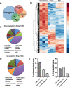
Blimp-1 regulates genes that may function in terminal differentiation. (A) Euler diagram of the number of genes with significant changes in expression level (log2 fold change>1, P-value>0.05, standard deviation/mean<0.5) induced by Blimp-1 overexpression or RNAi. (B) Heat map of the expression levels of Blimp-1-regulated genes in three conditions: Blimp-1 overexpression, lGMR-GAL4/+ control and Blimp-1 RNAi. (C,D) Pie charts showing the major categories of genes with reduced (C) or increased (D) expression in retinas expressing Blimp-1 RNAi. (E) Graphs showing the percentages of genes that are significantly downregulated (left) or upregulated (right) by Blimp-1 RNAi that have an expression peak at 48 h APF or that are stably increasing or decreasing their expression at this stage.
Fig. 6.
Fig. 6.
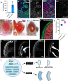
Slbo overexpression causes phenotypes similar to loss of Blimp-1. (A) Graph showing significant upregulation of slbo in RNA-seq data from retinas expressing Blimp-1 RNAi. (B) Confocal sections from a 42 h APF pupal retina in which Blimp-1 mutant clones are marked with GFP (green), stained with anti-β-galactosidase to reveal slbo-lacZ expression (B′,B‴, blue in B,B″) in mutant cone and primary pigment cells (apical section in B,B′) and secondary pigment cells (more basal section in B″,B‴) but not in photoreceptors stained with anti-Elav (red in B,B″). (C-E) Adult eyes: wild type (C), lGMR-GAL4 driving Blimp-1 RNAi (D) and lGMR-GAL4 driving UAS-slbo (E). Both Blimp-1 loss-of-function and slbo overexpression cause a similar glossy eye phenotype. (F) Horizontal section of adult eye expressing UAS-SlboHA with 54-GAL4 in higher order pigment cells, stained with CBD (green), Chp (red) and Elav (blue) shows plano-convex lenses. (G) Quantification of outer lens angles for 54-GAL4/+ control and 54-GAL4; UAS-SlboHA, showing mean±s.d. n=31 (54-GAL4/+) or 36 (54>SlboHA). ****P<0.0001 (Welch's two-tailed t-test). (H,I) Sections of adult eyes stained for Chp (H,I) and Elav (H′,I′). lGMR-GAL4 driving Blimp-1 RNAi (H), lGMR-GAL4 driving UAS-slbo (I). Both manipulations caused shortened rhabdomeres and disorganized photoreceptor nuclei. (J) Model of the Blimp-1 transcriptional network and its major morphological contributions to eye development. Scale bars: 10 µm (B); 100 µm (C-E); 20 µm (F); 50 µm (H-I′).

References

    1. Agawa, Y., Sarhan, M., Kageyama, Y., Akagi, K., Takai, M., Hashiyama, K., Wada, T., Handa, H., Iwamatsu, A., Hirose, S.et al. (2007). Drosophila Blimp-1 is a transient transcriptional repressor that controls timing of the ecdysone-induced developmental pathway. Mol. Cell. Biol. 27, 8739-8747. 10.1128/MCB.01304-07 - DOI - PMC - PubMed
    1. Ahmed, M. I., Elias, S., Mould, A. W., Bikoff, E. K. and Robertson, E. J. (2016). The transcriptional repressor Blimp1 is expressed in rare luminal progenitors and is essential for mammary gland development. Development 143, 1663-1673. 10.1242/dev.136358 - DOI - PMC - PubMed
    1. Akagi, K. and Ueda, H. (2011). Regulatory mechanisms of ecdysone-inducible Blimp-1 encoding a transcriptional repressor that is important for the prepupal development in Drosophila. Dev. Growth Differ. 53, 697-703. 10.1111/j.1440-169X.2011.01276.x - DOI - PubMed
    1. Akagi, K., Sarhan, M., Sultan, A.-R. S., Nishida, H., Koie, A., Nakayama, T. and Ueda, H. (2016). A biological timer in the fat body comprising Blimp-1, betaFtz-f1 and Shade regulates pupation timing in Drosophila melanogaster. Development 143, 2410-2416. 10.1242/dev.133595 - DOI - PubMed
    1. Arnes, L. and Sussel, L. (2015). Epigenetic modifications and long noncoding RNAs influence pancreas development and function. Trends Genet. 31, 290-299. 10.1016/j.tig.2015.02.008 - DOI - PMC - PubMed

Publication types