. 2022 Jul;29(7):2036-2046.
doi: 10.1111/ene.15329. Epub 2022 Apr 4.

Relation of sensorimotor and cognitive cerebellum functional connectivity with brain structural damage in patients with multiple sclerosis and no disability

Collaborators, Affiliations

Relation of sensorimotor and cognitive cerebellum functional connectivity with brain structural damage in patients with multiple sclerosis and no disability

Silvia Tommasin et al. Eur J Neurol. 2022 Jul.

Abstract

Background and purpose: To investigate the relationship between the functional connectivity (FC) of the sensorimotor and cognitive cerebellum and measures of structural damage in patients with multiple sclerosis (MS) and no physical disability.

Methods: We selected 144 relapsing-remitting MS patients with an Expanded Disability Status Scale score of ≤1.5 and 98 healthy controls from the Italian Neuroimaging Network Initiative database. From multimodal 3T magnetic resonance imaging (MRI), including functional MRI at rest, we calculated lesion load, cortical thickness, and white matter, cortical gray matter, and caudate, putamen, thalamic, and cerebellar volumes. Voxel-wise FC of the sensorimotor and cognitive cerebellum was assessed with seed-based analysis, and multiple regression analysis was used to evaluate the relationship between FC and structural damage.

Results: Whole brain, white matter, caudate, putamen, and thalamic volumes were reduced in patients compared to controls, whereas cortical gray matter was not significantly different in patients versus controls. Both the sensorimotor and cognitive cerebellum showed a widespread pattern of increased and decreased FC that were negatively associated with structural measures, indicating that the lower the FC, the greater the tissue loss. Lastly, among multiple structural measures, cortical gray matter and white matter volumes were the best predictors of cerebellar FC alterations.

Conclusions: Increased and decreased cerebellar FC with several brain areas coexist in MS patients with no disability. Our data suggest that white matter loss hampers FC, whereas, in the absence of atrophy, cortical volume represents the framework for FC to increase.

Keywords: cognitive cerebellum; disability; functional magnetic resonance imaging; multiples sclerosis; neural plasticity; sensorimotor cerebellum.


Conflict of interest statement

S.T., V.I. and C.G. have nothing to disclose. M.A.R. received speaker honoraria from Bayer, Biogen Idec, Celgene, Genzyme, Merck Serono, Novartis, Roche, and Teva, and receives research support from the MS Society of Canada and Fondazione Italiana Sclerosi Multipla. N.P. received speaker fees from Biogen and mission support from Genzyme and Novartis. G.T. received funding for travel, speaking honorarium, consulting service, and research projects from Novartis, Genzyme, Teva, Merck, Biogen, Roche, Allergan, Abbvie, Lilly, and Mylan. N.D.S. has received honoraria from Biogen‐Idec, Celgene, Immunic, Merck Serono, Novartis, Roche, Sanofi‐Genzyme, and Teva for consulting services, speaking, and travel support. He serves on advisory boards for Biogen‐Idec, Immunic, Merck Serono, Novartis, Roche, and Sanofi‐Genzyme. He has received research grant support from the Italian Multiple Sclerosis Society. C.P. received consulting and lecture fees from Sanofi‐Aventis, Biogen Idec, Bayer Schering, Merck Serono, and Novartis. He also received research funding from Novartis, Sanofi‐Aventis, Merck Serono, and Bayer Schering. M.F. is Editor‐in‐Chief of the Journal of Neurology; received compensation for consulting services and/or speaking activities from Bayer, Biogen Idec, Merck‐Serono, Novartis, Roche, Sanofi Genzyme, Takeda, and Teva Pharmaceutical Industries; and receives research support from Biogen Idec, Merck‐Serono, Novartis, Roche, Teva Pharmaceutical Industries, Italian Ministry of Health, Fondazione Italiana Sclerosi Multipla, and ARiSLA (Fondazione Italiana di Ricerca per la SLA). P.P. received funding for travel from Novartis, Genzyme, and Bracco and a speaking honorarium from Biogen. She received research support from the Italian Ministry of Foreign Affairs and Fondazione Italiana Sclerosi Multipla.

Figures

FIGURE 1
FIGURE 1
Flowchart of methods. Left side of panel: From a sample of 144 patients with multiple sclerosis (MS) and no disability, we randomly obtained two subgroups of 72 subjects each (MS1 and MS2). For both the sensorimotor cerebellum (smCb) and cognitive cerebellum (cCb), we calculated voxel‐wise maps of altered functional connectivity (FC) in MS1 and MS2 subgroups versus healthy controls (HCs) (alteration masks 1 and 2, increased FC is depicted in yellow and decreased FC in light blue) and intersected the two resulting maps to create masks of significant difference (intersection masks, intersecting areas are shown in red). Then, we compared the intersection masks derived from the two subgroups with the cerebellar FC maps obtained by comparing the whole group of patients and HCs to verify the consistency of FC alterations in patients despite FC variances. Right side of panel: For both smCb and cCb, we calculated FC alterations in 144 patients as compared with 98 HCs and from the areas of significant alterations (alteration map, increased FC is depicted in yellow and decreased FC in light blue), in terms of either increased or decreased FC, we created an alteration mask (shown in green). Within the areas covered by the alteration mask, we calculated voxel‐wise multiple regression between the smCb and cCb and structural measures, such as cortical gray matter, white matter, caudate, putamen, thalamic, and cerebellar inverse volumes, and lesion load. We created an association mask from the areas where FC with either the smCb or cCb was significantly associated with structural measures. Lastly, average smCb or cCb FC within the areas covered by the association mask was associated with the structural measure via stepwise regression to find the best FC alteration predictors [Colour figure can be viewed at wileyonlinelibrary.com]
FIGURE 2
FIGURE 2
Flowchart of sample selection. Out of 1347 records, 1305 were first exams, including three‐dimensional T1‐weighted (3DT1), proton density (PD) or T2‐weighted or dual echo and resting state functional images, of 951 patients (PTNS) with multiple sclerosis (702 relapsing–remitting [RR], 174 secondary progressive [SP], 74 primary progressive [PP]) and 354 healthy controls (HCs). In the RR group, 363 patients had an Expanded Disability Status Scale (EDSS) score of ≤1.5, and 267 underwent a resting‐state acquisition with repetition time (TR) = 3 s and at least 140 volumes. Of these, 144 patients had 3DT1 images and T2 lesion load (T2LL) masks preprocessed and double checked by either Center A or C and were thus included in the following analysis. Among HCs, 199 had TR = 3 s and at least 140 volumes, and 98 had 3DT1 images preprocessed and double checked by either Center A or C and were included in the study [Colour figure can be viewed at wileyonlinelibrary.com]
FIGURE 3
FIGURE 3
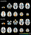
Two‐sample t test. Comparison of resting‐state functional connectivity (FC) of the sensorimotor cerebellum (smCb) (a) and cognitive cerebellum (cCb) (b) between the entire group of patients with multiple sclerosis and healthy controls (HCs). Yellow areas indicate where FC was significantly higher in patients than in HCs, and light blue areas indicate where FC was significantly lower in patients than in HCs. Statistical significance was reached if < 0.05, false discovery rate–corrected. Superimposed onto the result of the two‐sample t test, combined binary masks of smCb and cCb FC obtained by the intersection of MS1 patients versus HCs and MS2 patients versus HCs are shown in red [Colour figure can be viewed at wileyonlinelibrary.com]
FIGURE 4
FIGURE 4
Relation between structural damage and functional connectivity (FC) of the sensorimotor cerebellum. In patients with multiple sclerosis, FC between (a) the sensorimotor cerebellum (smCb) and the frontal lobe, bilateral insulae, primary somatosensory cortex, and precuneus decreases as structural damage increases and (b) the cognitive cerebellum (cCb) and precuneus. Negative associations are shown in blue/light blue. Significant results were reached if < 0.05, false discovery rate–corrected [Colour figure can be viewed at wileyonlinelibrary.com]

References

    1. Timmann D, Drepper J, Frings M, et al. The human cerebellum contributes to motor, emotional and cognitive associative learning. A review. Cortex. 2010;46:845‐857. doi:10.1016/j.cortex.2009.06.009 - DOI - PubMed
    1. Jissendi P, Baudry S, Balériaux D. Diffusion tensor imaging (DTI) and tractography of the cerebellar projections to prefrontal and posterior parietal cortices: a study at 3T. J Neuroradiol. 2008;35:42‐50. doi:10.1016/j.neurad.2007.11.001 - DOI - PubMed
    1. Palesi F, Tournier J‐D, Calamante F, et al. Contralateral cerebello‐thalamo‐cortical pathways with prominent involvement of associative areas in humans in vivo. Brain Struct Funct. 2015;220:3369‐3384. doi:10.1007/s00429-014-0861-2 - DOI - PMC - PubMed
    1. Stoodley CJ, Valera EM, Schmahmann JD. Functional topography of the cerebellum for motor and cognitive tasks: an fMRI study. NeuroImage. 2012;59:1560‐1570. doi:10.1016/j.neuroimage.2011.08.065 - DOI - PMC - PubMed
    1. Mormina E, Petracca M, Bommarito G, et al. Cerebellum and neurodegenerative diseases: beyond conventional magnetic resonance imaging. World J Radiol. 2017;9:371‐388. doi:10.4329/wjr.v9.i10.371 - DOI - PMC - PubMed

Publication types

MeSH terms