. 2022 Mar 18;375(6586):1281-1286.
doi: 10.1126/science.abm5320. Epub 2022 Mar 17.

The histone H3.1 variant regulates TONSOKU-mediated DNA repair during replication

Affiliations

The histone H3.1 variant regulates TONSOKU-mediated DNA repair during replication

Hossein Davarinejad et al. Science. .

Abstract

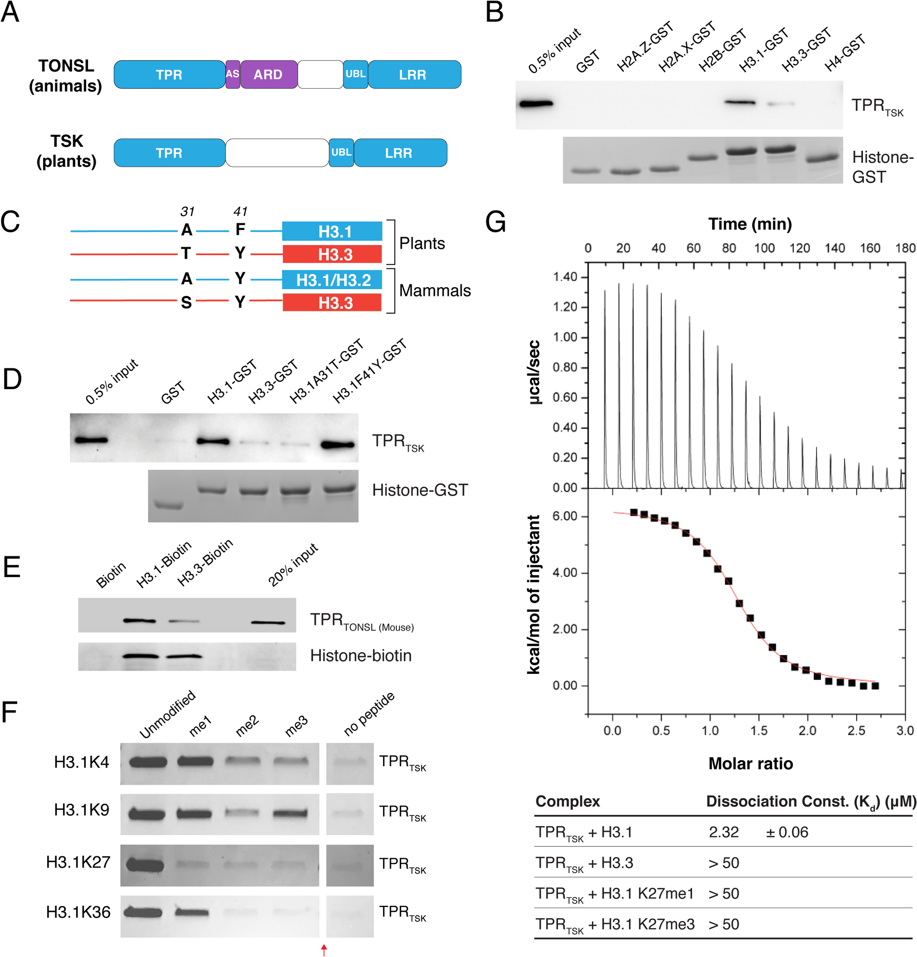
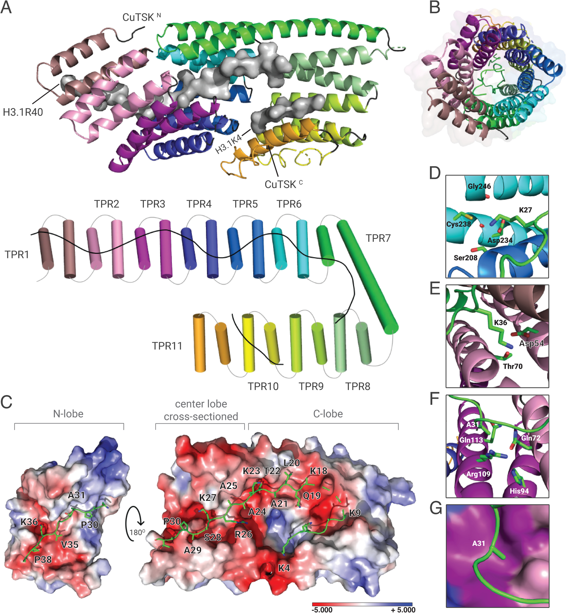
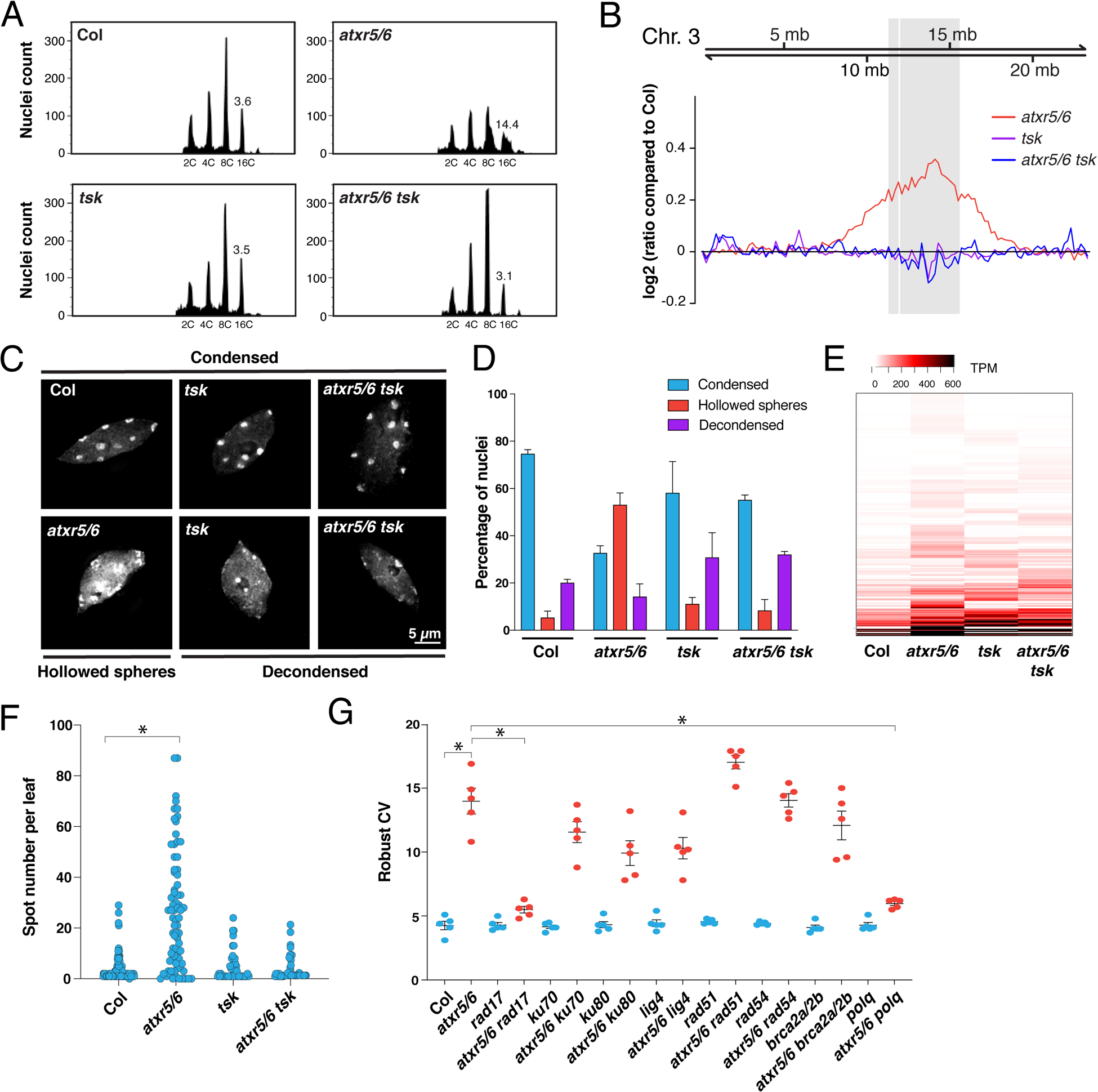
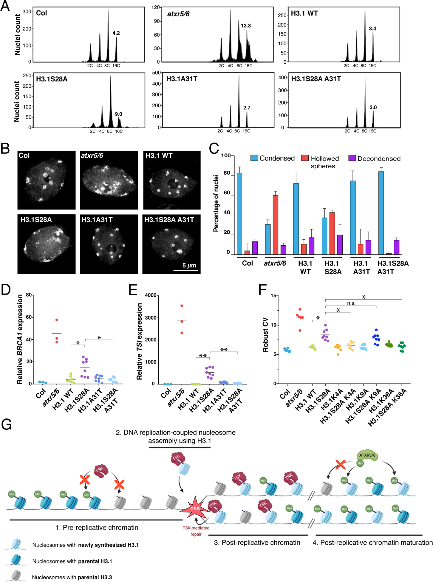
The tail of replication-dependent histone H3.1 varies from that of replication-independent H3.3 at the amino acid located at position 31 in plants and animals, but no function has been assigned to this residue to demonstrate a unique and conserved role for H3.1 during replication. We found that TONSOKU (TSK/TONSL), which rescues broken replication forks, specifically interacts with H3.1 via recognition of alanine 31 by its tetratricopeptide repeat domain. Our results indicate that genomic instability in the absence of ATXR5/ATXR6-catalyzed histone H3 lysine 27 monomethylation in plants depends on H3.1, TSK, and DNA polymerase theta (Pol θ). This work reveals an H3.1-specific function during replication and a common strategy used in multicellular eukaryotes for regulating post-replicative chromatin maturation and TSK, which relies on histone monomethyltransferases and reading of the H3.1 variant.


Conflict of interest statement

Competing interests:

The authors declare that they have no competing interests.

Figures

Figure 1.
Figure 1.. The TPR domain of TSK specifically interacts with the N-terminal tail of the H3.1 variant.
(A) Domain architecture of animal and plant TONSL/TSK. TPR: Tetratricopeptide Repeats, AS: Acidic Sequence, ARD: Ankyrin Repeat Domain, UBL: Ubiquitin-like, LRR: Leucine-Rich Repeats. Conserved domains are shown in blue. (B) Pull-down assay using TPRTSK and GST tagged with the N-terminal tails of histones H2A.Z, H2A.X, H2B, H3.1, H3.3 and H4 from plants. (C) Representation of plant and mammalian H3.1/H3.2 (blue) and H3.3 (red) H3 variants. Thin lines and blocks represent the histone tails and cores, respectively, and numbers indicate amino acid positions in H3. (D) Peptide pull-down assay using plant TPRTSK and GST tagged with the tails of histones H3.1, H3.3, H3.1A31T and H3.1F41Y. (E) Peptide pull-down assay using mouse TPRTONSL and biotin-tagged histones H3.1 and H3.3 (full-length proteins) from mammals. (F) Peptide pull-down assay using plant TPRTSK and methylated peptides at K4, K9, K27 and K36 of H3.1 (a.a. 1–45). The red arrow indicates a gel lane that was removed. (G) ITC assay using plant TPRTSK and different H3 peptides.
Figure 2.
Figure 2.. Crystal structure of plant TPRTSK bound to the H3.1 tail.
(A) The TPR domain is depicted as a cartoon (top) or a cylinder (bottom) with individual TPR motifs as distinct colors. H3.1 is shown as surfaces (top) or line (bottom). (B) Channel view of the TPR solenoid tube showing the space inside the tube where H3.1 is extended (represented as a green line). (C) Surface representation of the TPR domain shown as electrostatic potential gradients contoured from +5.000 kBTe−1 (blue) to −5.000 kBTe−1 (red), where e is the electron, T is temperature and kB is the Boltzmann constant. H3.1 is depicted as sticks. The N-terminal lobe (N-lobe) is rotated 180° along the horizontal axis relative to the center lobe and the C-terminal lobe (C-lobe). The surface of the center lobe is sectioned off to reveal the underlying segment of H3.1. (D, E, and F) Amino acid residues from TPRTSK (3-letter code) interacting with H3.1 residues (1-letter code) in their binding pockets are shown for D) K27, E) K36, and F) A31. (G) Surface representation of the H3.1A31 binding pocket. Surface colors correspond to that of TPR helices shown in panel F.
Figure 3.
Figure 3.. Mutations in TSK suppress heterochromatin amplification of atxr5/6 mutants.
(A) Flow cytometry profiles of Col, atxr5/6, tsk and atxr5/6 tsk leaf nuclei. The numbers below the peaks indicate ploidy levels of the nuclei. The numbers above the 16C peaks indicate the robust coefficient of variation (rCV). (B) Chromosomal view (Chromosome 3 of A. thaliana) of DNA sequencing reads from sorted 16C nuclei. The pericentromeric region is highlighted in gray. (C) Leaf interphase nuclei of Col, atxr5/6, tsk and atxr5/6 tsk stained with DAPI. (D) Quantification of nuclei from experiment shown in panel C. Error bars indicate SEM. (E) Heat map showing the relative expression levels of atxr5/6-induced TEs as measured by TPM (transcripts per million). (F) Average number of blue spots per leaf in Col and atxr5/6 mutants as determined using a GUS reporter for homologous recombination. Error bars represent SEM. Welch’s ANOVA followed by Dunnett’s T3 test: * p < 0.0001. (G) rCV values for 16C nuclei obtained by flow cytometry analyses. Each dot represents an independent biological replicate. Horizontal bars indicate the mean. Error bars represent SEM. Welch’s ANOVA followed by the Dunnett’s T3 test: * p < 0.05.
Figure 4.
Figure 4.. H3.1 is required to mediate genomic instability in atxr5/6 mutants.
(A) Flow cytometry of leaf nuclei. Numbers below the peaks indicate ploidy, and those above indicate rCV. (B) Leaf nuclei of Col, atxr5/6, and first-generation (T1) H3.1 lines stained with DAPI. (C) Quantification from nuclei in B. Error bars indicate SEM. (D, E) RT-qPCR of BRCA1 and TSI. Horizontal bars indicate the mean. Welch’s ANOVA followed by the Dunnett’s T3 test: * p < 0.05, ** p < 0.001. (F) rCV for 16C nuclei obtained by flow cytometry. For Col and atxr5/6, each dot represents a biological replicate. For the H3.1 lines, each dot represents one T1 plant. Horizontal bars indicate the mean. Welch’s ANOVA followed by the Dunnett’s T3 test: * p < 0.05, n.s. = not significantly different. (G) Model depicting the interplay between H3.1, TSK and ATXR5/6 during replication. Step 1. TSK cannot interact with chromatin containing H3.3K27me0 or H3.1K27me1. Step 2. Newly synthesized H3.1 (H3.1K27me0) in complex with TSK are inserted at replication forks. Step 3. DSBs caused by broken replication forks are repaired by TSK. Step 4. Mono-methylation of newly inserted H3.1 (but not H3.3) at K27 by ATXR5/6 prevents binding of TSK.

Comment in

  • One residue-one function.
    Berger F. Berger F. Science. 2022 Mar 18;375(6586):1232-1233. doi: 10.1126/science.abo4219. Epub 2022 Mar 17. Science. 2022. PMID: 35298274

References

    1. Duro E et al., Identification of the MMS22L-TONSL complex that promotes homologous recombination. Mol Cell 40, 632–644 (2010). - PubMed
    1. Huang TH et al., The Histone Chaperones ASF1 and CAF-1 Promote MMS22L-TONSL-Mediated Rad51 Loading onto ssDNA during Homologous Recombination in Human Cells. Mol Cell 69, 879–892 e875 (2018). - PMC - PubMed
    1. O’Connell BC et al., A genome-wide camptothecin sensitivity screen identifies a mammalian MMS22L-NFKBIL2 complex required for genomic stability. Mol Cell 40, 645–657 (2010). - PMC - PubMed
    1. O’Donnell L et al., The MMS22L-TONSL complex mediates recovery from replication stress and homologous recombination. Mol Cell 40, 619–631 (2010). - PMC - PubMed
    1. Piwko W et al., The MMS22L-TONSL heterodimer directly promotes RAD51-dependent recombination upon replication stress. Embo J 35, 2584–2601 (2016). - PMC - PubMed

Publication types

MeSH terms