Review
. 2022 Mar 17;7(3):214-226.
doi: 10.1530/EOR-21-0069.

Understanding shoulder pseudoparalysis: Part I: Definition to diagnosis

Affiliations
Review

Understanding shoulder pseudoparalysis: Part I: Definition to diagnosis

Stefan Bauer et al. EFORT Open Rev. .

Abstract

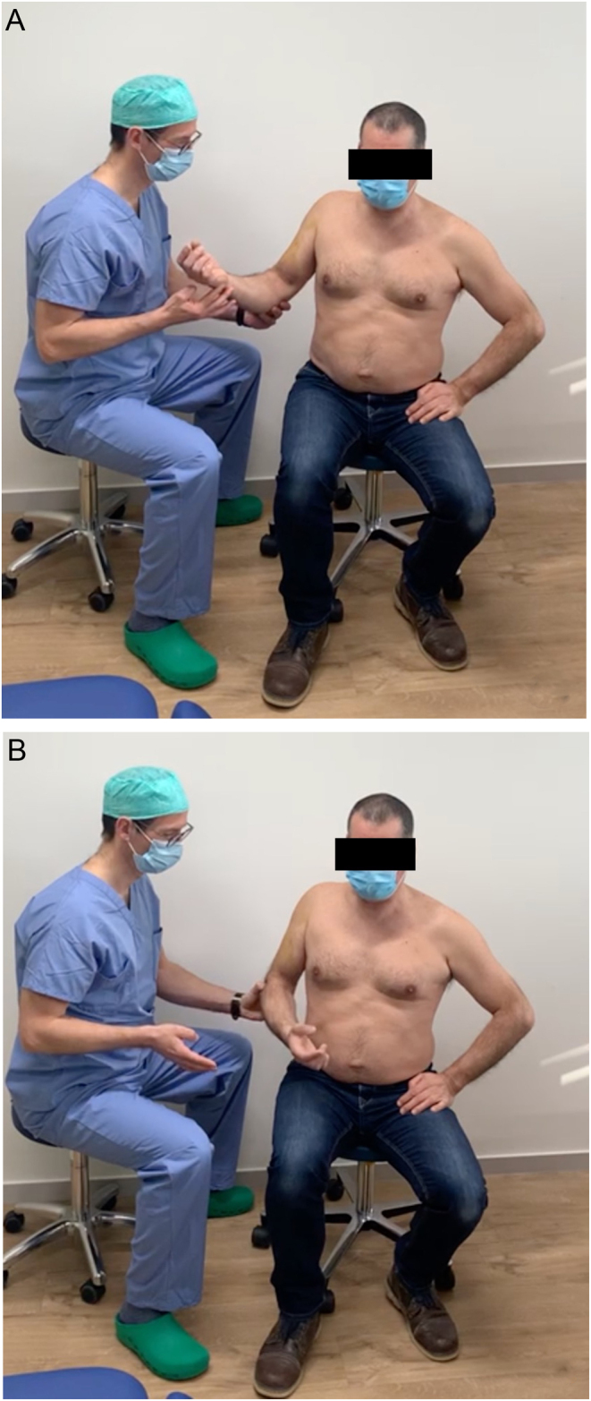
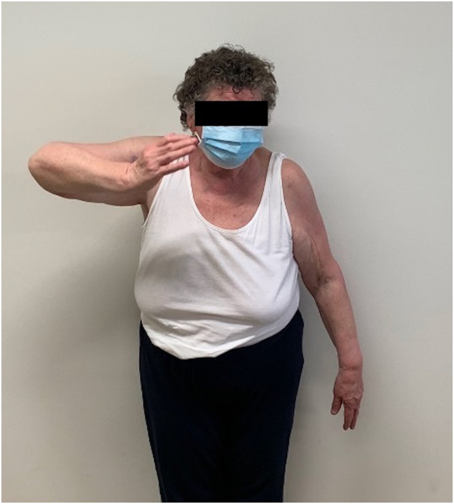
Variable definitions of pseudoparalysis have been used in the literature. Recent systematic reviews and biomechanical studies call for a grading of loss of force couple balance and the use of the terms 'pseudoparesis' and 'pseudoparalysis'. Pain should be excluded as the cause of loss of active function. Key players for loss of force couple balance seem to be the lower subscapularis as an anterior inferior checkrein and the teres minor as a posterior inferior fulcrum. Loss of three out of five muscle-tendon units counting upper and lower subscapularis separately is predictive of pseudoparalysis. Shoulder equator concept: loss of all three posterior, or all three superior, or all three anterior muscle-tendon units is predictive of pseudoparalysis (loss of fulcrum for deltoid force). Every effort should be made to prevent propagation of rotator cuff tears into the subscapularis and posterior rotator cuff (infraspinatus and teres minor) to maintain force couple balance (value of partial cuff repair). Clinical assessment of active forward elevation, active external rotation, and active internal rotation is important to define and grade the severity of loss of force couple balance. Additional features such as patient age, traumatic aetiology, chronicity, fatty infiltration, and stage of cuff tear arthropathy are useful for a specific diagnosis with implications for treatment.

Keywords: arthroscopy; pseudoparalysis; shoulder.


Figures

Figure 1
Figure 1
Clinical photographs of patients attempting AFE. (A) Patient with left shoulder massive cuff tear with AFE <90° and (B) massive cuff tear of the right shoulder with no AFE.
Figure 2
Figure 2
Clinical photographs of a patient being examined for ER1 lag. Passive ER starting position with shoulder in 20° of abduction (A) demonstrating a lag with end position (B).
Figure 3
Figure 3
Clinical photograph of a belly-press test in AIR pseudoparalysis, belly-off sign: wrist flexion of 70° (A) before the elbow is brought forward by examiner passively, which would increase wrist flexion to 90°. Clinically an antero-superior escape of the humeral head is seen (A). Increased ER of the right shoulder due to complete subscapularis tear (B). Radiographic antero-superior subluxation (C). Soft tissue CT sagittal images demonstrate grade 4 fatty infiltration of subscapularis (D).
Figure 4
Figure 4
Clinical photograph of a patient with right shoulder hornblower/dropping sign after RSA.
Figure 5
Figure 5
Illustration demonstrating anterior (A) and posterior (B) views of the right shoulder with horizontal (A) and vertical (B) muscle balance vectors.
Figure 6
Figure 6
Illustration demonstrating schematic representation of a sagittal slice of the humeral head with rotator cuff tendons, as per Collin et al. Horizontal and oblique vertical equators in blue demonstrating the equator concept: type B (complete anterio–superior (AS) cuff loss), type C (complete superior (S) cuff loss), type E (complete postero–superior (PS) cuff loss) (A), and percentage of pseudoparalysis (B). Reproduced with permission.
Figure 7
Figure 7
Clinical photograph of a patient’s right shoulder being examined demonstrating deltoid extension lag test according to Hertel (46).
Figure 8
Figure 8
Clinical photograph of a patient’s right shoulder being examined demonstrating lift-off-lag test (A) and and lift-off test (B).
Figure 9
Figure 9
Plain AP radiograph of a left shoulder with cuff tear arthropathy.
Figure 10
Figure 10
Axial CT with fish backbone sign (A), fish backbone (B), and tangent sign (C). Reproduced with permission from (65).

References

    1. Tokish JM, Alexander TC, Kissenberth MJ, Hawkins RJ. Pseudoparalysis: a systematic review of term definitions, treatment approaches, and outcomes of management techniques. Journal of Shoulder and Elbow Surgery 2017. 26 e177–e187. (10.1016/j.jse.2017.02.024) - DOI - PubMed
    1. Werner CM, Steinmann PA, Gilbart M, Gerber C. Treatment of painful pseudoparesis due to irreparable rotator cuff dysfunction with the delta III reverse-ball-and-socket total shoulder prosthesis. Journal of Bone and Joint Surgery: American Volume 2005. 87 1476–1486. (10.2106/JBJS.D.02342) - DOI - PubMed
    1. Collin P, Matsumura N, Ladermann A, Denard PJ, Walch G. Relationship between massive chronic rotator cuff tear pattern and loss of active shoulder range of motion. Journal of Shoulder and Elbow Surgery 2014. 23 1195–1202. (10.1016/j.jse.2013.11.019) - DOI - PubMed
    1. Burks RT, Tashjian RZ. Should we have a better definition of pseudoparalysis in patients with rotator cuff tears? Arthroscopy 2017. 33 2281–2283. (10.1016/j.arthro.2017.07.024) - DOI - PubMed
    1. Ernstbrunner L, El Nashar R, Favre P, Bouaicha S, Wieser K, Gerber C. Chronic pseudoparalysis needs to be distinguished from pseudoparesis: a structural and biomechanical analysis. American Journal of Sports Medicine 2021. 49 291–297. (10.1177/0363546520969858) - DOI - PubMed