. 2022 Apr 5;34(4):549-563.e8.
doi: 10.1016/j.cmet.2022.02.012. Epub 2022 Mar 16.

Protein tyrosine phosphatase receptor δ serves as the orexigenic asprosin receptor

Affiliations

Protein tyrosine phosphatase receptor δ serves as the orexigenic asprosin receptor

Ila Mishra et al. Cell Metab. .

Abstract

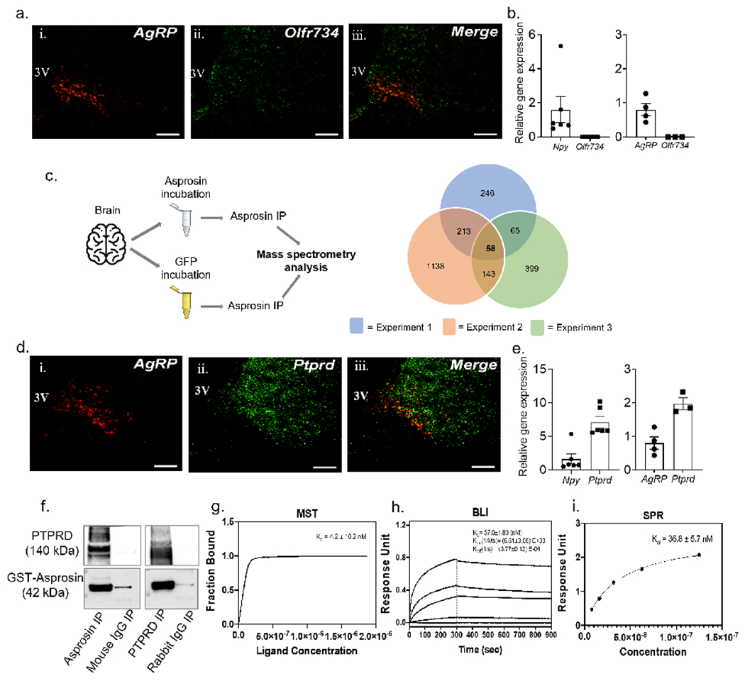
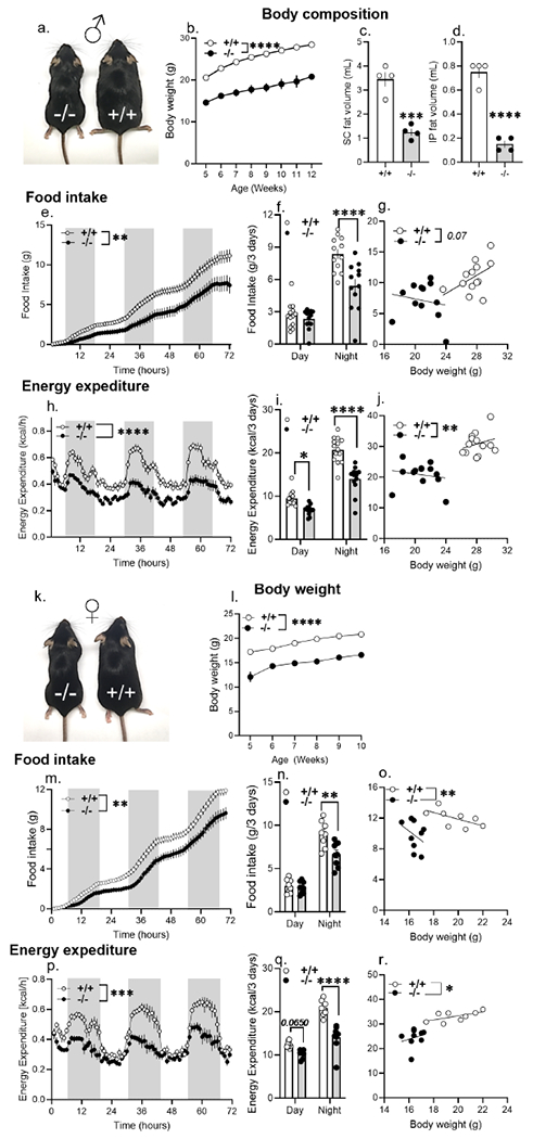
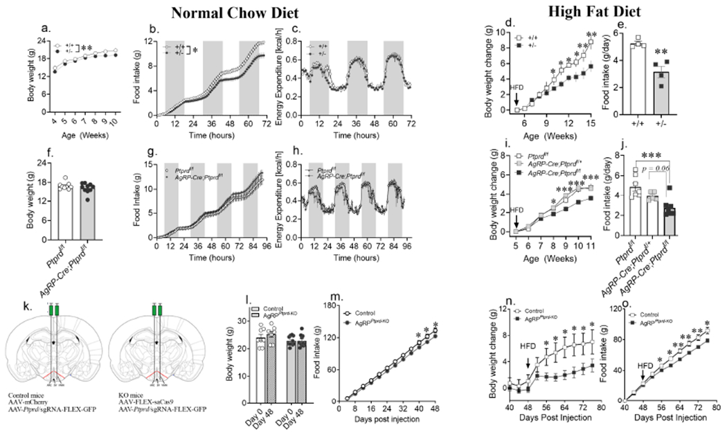
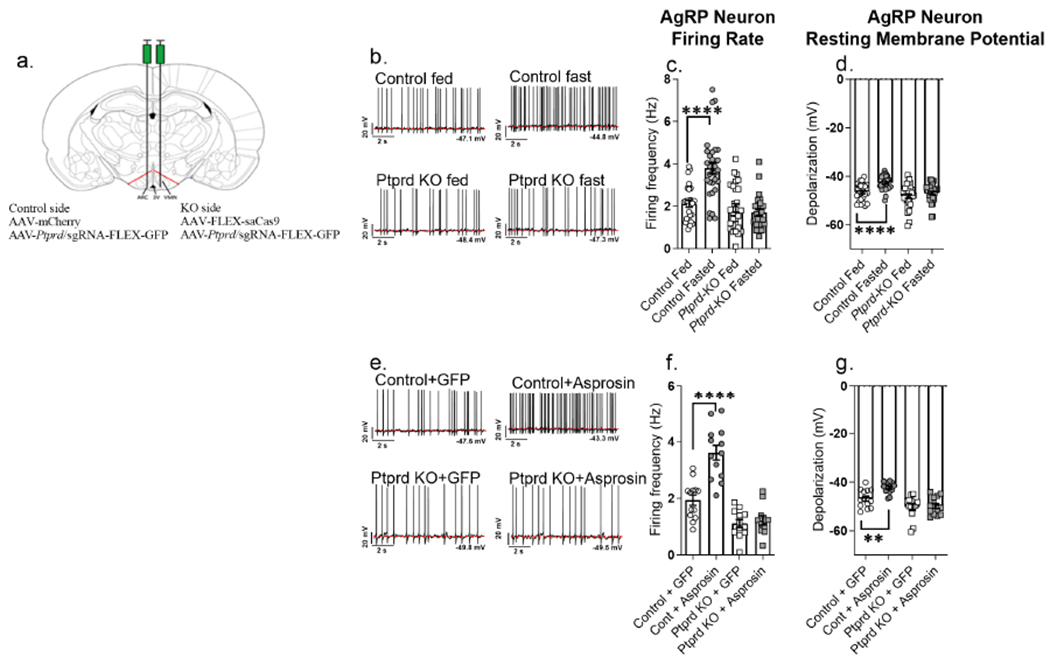
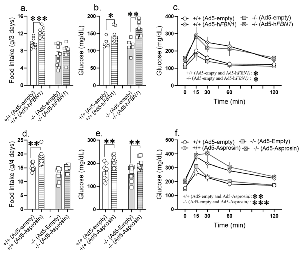
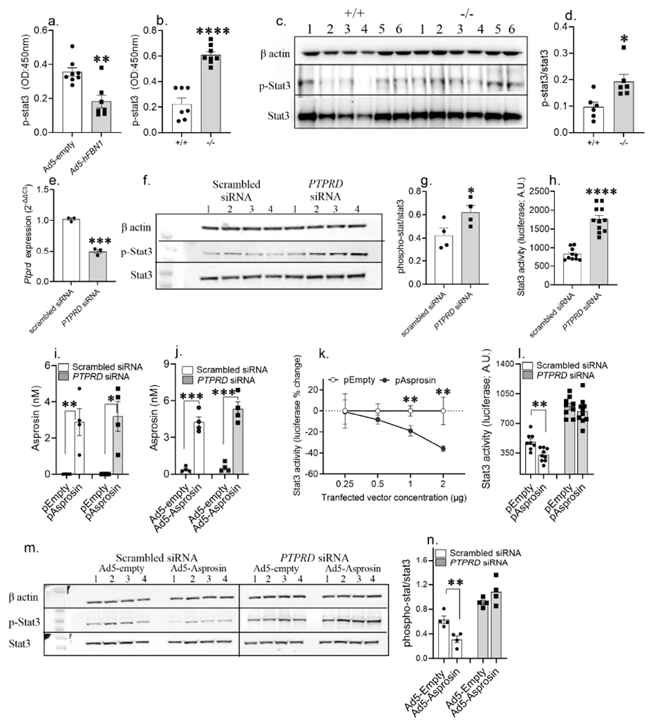
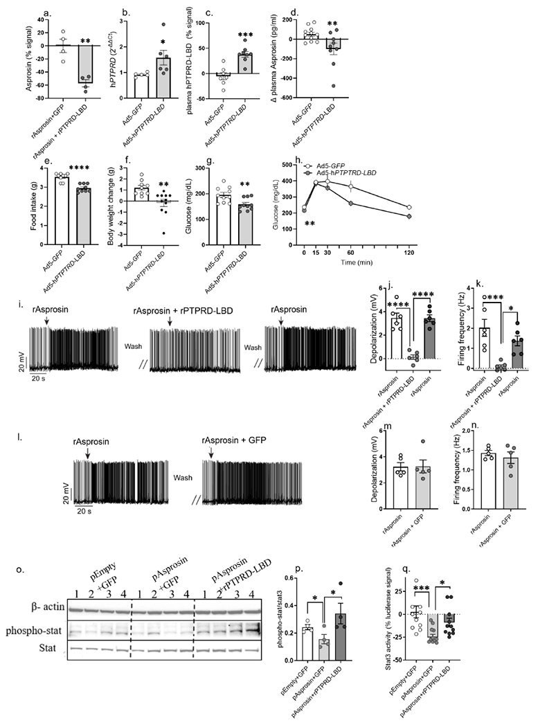
Asprosin is a fasting-induced glucogenic and centrally acting orexigenic hormone. The olfactory receptor Olfr734 is known to be the hepatic receptor for asprosin that mediates its effects on glucose production, but the receptor for asprosin's orexigenic function has been unclear. Here, we have identified protein tyrosine phosphatase receptor δ (Ptprd) as the orexigenic receptor for asprosin. Asprosin functions as a high-affinity Ptprd ligand in hypothalamic AgRP neurons, regulating the activity of this circuit in a cell-autonomous manner. Genetic ablation of Ptprd results in a strong loss of appetite, leanness, and an inability to respond to the orexigenic effects of asprosin. Ablation of Ptprd specifically in AgRP neurons causes resistance to diet-induced obesity. Introduction of the soluble Ptprd ligand-binding domain in the circulation of mice suppresses appetite and blood glucose levels by sequestering plasma asprosin. Identification of Ptprd as the orexigenic asprosin receptor creates a new avenue for the development of anti-obesity therapeutics.

Keywords: AgRP; Ptprd; appetite; asprosin; hypothalamus; metabolism; obesity; receptor.


Conflict of interest statement

Declaration of interests Atul Chopra has been awarded asprosin-related patents and is a co-founder, director, and officer of Vizigen, Inc. and Aceragen, Inc. and holds equity in both companies. The other authors declare no competing interests.

Figures

Figure 1:
Figure 1:. Identification of an asprosin-interacting receptor in the mouse brain
(a) Dual-label immunofluorenscence RNAscope hybridization showing relative overlap between AgRP (red) and Olfr734 (green) expressing cells in the arcuate (ARC) nucleus of wild-type (WT) mice. Scale bars, 100 μm. 3V, 3rd ventricle. (b) qPCR results showing relative mRNA levels of Olfr734 (left) from visually enriched tdTOMATO expressing neurons of AgRP-cre mice and (right) cre-responsive ribotag enriched transcripts from AgRP neurons of AgRP-cre mice. (c) Schematic overview of asprosin immunoprecitation (IP; left) of brain tissue incubated with recombinant asprosin or recombinant GFP before mass spectrometry (MS) analysis, (right) overlap of candidate asprosin-interacting proteins from 3 repeats of IP/MS. 58 candidate proteins, including Ptprd appeared in all 3 repeats. (d) Dual-label immunofluorenscence RNAscope hybridization showing relative overlap between AgRP (red) and Ptprd (green) expressing cells in the ARC nucleus of WT mice. Scale bars, 100 μm. (e) qPCR results showing relative mRNA levels of Ptprd (left) from visually enriched tdTOMATO expressing neurons of AgRP-cre mice and (right) cre-responsive ribotag enriched transcripts from AgRP neurons of AgRP-cre mice. (f) Reciprocal in vitro immunoprecipitation of recombinant GST-asprosin and extracellular domain of his-tagged PTPRD (PTPRD-6his), with mouse IgG and rabbit IgG as negative controls. (g-i) Quantification of the binding affinity between Asprosin and PTPRD by Microscale thermophoresis (k; MST; Kd of 4.2 ±10.2 nM), Bio-layer interferometry (l; BLI; Kd of 57.0 ±1.83 nM), and by Surface plasmon resonance analysis (m; SPR; Kd of 36.8 ±5.7 nM). Data are represented as mean ± SEM.
Figure 2.
Figure 2.. Ptprd ablation phenocopies the body weight and orexigenic deficits associated with genetic asprosin deficiency and pharmacologic asprosin inhibition
(a) A representative photograph of 5-month-old male Ptprd+/+ and Ptprd−/− littermates (n=1 photograph from n = 3 mice per genotype). (b) Weekly body weights of wildtype (Ptprd+/+, n = 25) and Ptprd null (Ptprd−/−, n = 11) male mice from 5- to 12-weeks of age. (c-d) Body composition data using Magnetic Resonance Imaging (MRI) on 5-month-old Ptprd+/+ and Ptprd−/− male mice on normal chow (n = 4/group). (e-g) Cumulative food intake (e), total food intake in the dark and light phase (f), and ANCOVA analysis (g) of total food intake of Ptprd+/+ and Ptprd−/− male mice measured over 3 days using the Promethion metabolic system (n=12 or 13/group). (h-j) Hourly energy expenditure (h) total energy expenditure in the light and dark phase (i), and ANCOVA analysis (j) of energy expenditure of Ptprd+/+ and Ptprd−/− male mice measured over 3 days using the Promethion metabolic system (n = 13/group). (k) A representative photograph of 6-month-old female Ptprd+/+ and Ptprd−/− littermates (n=1 photograph from n = 3 mice per genotype). (l) Weekly body weights of wildtype (Ptprd+/+, n = 16) and Ptprd null (Ptprd−/−, n = 11) female mice from 5- to 10-weeks of age. (m-o) Cumulative food intake (m), total food intake in the dark and light phase (n), and ANCOVA analysis (o) of total food intake of Ptprd+/+ (n = 8) and Ptprd−/− (n = 9) female mice measured over 3 days using Promethion metabolic system. (p-r) Hourly energy expenditure (p) and total energy expenditure in the light and dark phase (q), and ANCOVA analysis (r) of energy expenditure of Ptprd+/+ (n = 8) and Ptprd−/− (n = 9) female mice measured over 3 days using the Promethion metabolic system. *P < 0.05, **P < 0.01, ***P < 0.001 and ****P < 0.0001; ‘effect of genotype’ determined by two-tailed Student’s t-test (c,d,f,i,n,q), Repeated measures two-way ANOVA (b,e,h,l,m,p) or ANCOVA (g,j,o,r). Data are represented as mean ± SEM. See also Figure S1, S2, S3 and S4.
Figure 3.
Figure 3.. Whole-body loss of a single allele and AgRP neuron-specific loss of both alleles of Ptprd is protective against diet-induced obesity in females
(a) Weekly body weights of wildtype (Ptprd+/+; n = 16) and heterozygous Ptprd knockout Ptprd+/−; n = 24) female mice maintained on ad libitum normal chow (NC) diet. (b,c) Cumulative food intake and hourly energy expenditure of 10-week-old Ptprd+/+ and Ptprd+/− female mice on NC over 3 days using the Promethion metabolic system (food intake: Ptprd+/+ n = 8; Ptprd+/− n = 10; Energy expenditure: Ptprd+/− n = 8; Ptprd+/− n = 14). (d,e) Weekly body weight change of Ptprd+/+ and Ptprd+/− females maintained on high fat diet (HFD) from 5-weeks of age, with food intake measured in a randomly selected cohort of 16-week-old female mice from each group (body weight change: Ptprd+/+ n = 9, Ptprd+/− n = 12; food intake: Ptprd+/+ n = 4; Ptprd+/− n = 4). (f) Body weight of 6-week-old Ptprdflox/flox and AgRP-cre; Ptprdflox/flox maintained on ad libitum NC diet (Ptprdflox/flox n = 9, AgRP-cre; Ptprdflox/flox n = 10) (g,h) Cumulative food intake and hourly energy expenditure of 8-week-old Ptprdflox/flox and AgRP-cre; Ptprdflox/flox mice maintained on ad libitum NC over 4 days using the Promethion system (n = 4/group). (i,j) Weekly body weight change of Ptprdflox/flox (n = 9), AgRP-cre; Ptprdflox/+ (n = 4) and AgRP-cre; Ptprdflox/flox (n = 10) females maintained on high fat diet (HFD) from 5 weeks of age, with food intake measured in 14-week-old females from each group (Ptprdflox/flox :n = 8, AgRP-cre; Ptprdflox/+ : n = 5 and AgRP-cre; Ptprdflox/flox: n = 8). (k) Schematic figure showing bilateral stereotactic injection of virus (AAV) containing Ptprd sgRNA with AAV expressing mCherry (control) or Cas9 (AgRPPtprd-KO) in the arcuate (ARC) nucleus of adult AgRP-cre female mice. (l) Body weight of control and AgRPPtprd-KO female mice maintained on NC diet from day 0 to day 48 post stereotactic injection (control: n = 8; AgRPPtprd-KO: n = 11). (m) Cumulative food intake measured every fourth day of control and AgRPPtprd-KO female mice maintained on NC diet (control: n = 6; AgRPPtprd-KO: n = 11). (n,o) Body weight change and cumulative food intake of control and AgRPPtprd-KO female mice subjected to HFD on day 48 post stereotactic injection of AAVs coding cas9 and Ptprd sgRNA (control: n = 6-7; AgRPPtprd-KO: n = 11). *P < 0.05, **P < 0.01, ***P < 0.001 and ****P < 0.0001; by Student’s t-test (e,f,j,l), ‘effect of genotype’ determined by repeated measures two-way ANOVA (a,b,c,g,h) with point-comparison by multiple unpaired t-test (d,i,m,n,o). Circle and square symbol represent mice maintained on NC diet and HFD, respectively. Data are represented as mean ± SEM. See also Figure S5–S7.
Figure 4.
Figure 4.. Deletion of Ptprd renders AgRP neurons unresponsive to asprosin
(a) Schematic figure showing unilateral stereotactic injection of virus (AAV) containing Ptprd sgRNA with AAV expressing mCherry (control) or Cas9 (AgRPPtprd-KO) in the arcuate (ARC) nucleus of adult AgRP-cre male mice (b-d) Representative action potential firing traces, baseline neuronal firing rate and membrane potential of AgRP neurons from AgRP-cre mice subjected to overnight fasting or ad libitum feeding (control + fed: n = 28; control + fasted: n = 31; Ptprd KO + fed: n = 31; Ptprd KO + fasted: n = 28) after unilateral stereotactic injection of Ptprd sgRNA + mCherry expressing AAVs on one side (control side), and Ptprd sgRNA + Cas9 expressing AAVs in the other side (AgRPPtprd-KO; KO side) ARC of hypothalamus (e-g) Representative action potential firing traces, neuronal firing rate and membrane potential of AgRP neurons from AgRP-Cre mice, incubated with recombinant GFP or asprosin for 2 hours (Control+GFP: n = 13, Control+Asprosin: n = 13, Ptprd KO+GFP: n = 13, Ptprd KO+Asprosin: n = 11) after unilateral stereotactic injection of Ptprd sgRNA + mCherry expressing AAVs on one side (control side), and Ptprd sgRNA + Cas9 expressing AAVs in the other side (AgRPPtprd-KO; KO side) of ARC of the hypothalamus. *P < 0.05, **P < 0.01, ***P < 0.001 and ****P < 0.0001; by two-tailed Student’s t-test. Data are represented as mean ± SEM.
Figure 5.
Figure 5.. Ptprd−/− mice are unresponsive to the orexigenic effects of asprosin while responding normally to its glucogenic effects
(a-c) 14-week-old normal chow fed Ptprd+/+ and Ptprd−/− male mice tail-vein transduced with Ad-empty or Ad-hFBN1 (3.6 x 10⁹ pfu/mouse) viruses. (a) Cumulative food intake measured in Promethion metabolic chamber over 3 days; day 8 to day 11 post adenoviral vector transduction (Ad-empty: n = 8 Ptprd+/+ and n = 10 Ptprd−/−; Ad-hFBN1: n = 7 Ptprd+/+ and n = 6 Ptprd−/−. (b,c) ad libitum fed baseline blood glucose levels and glucose tolerance assay in response to intraperitoneal glucose injection (2mg/kg) on day 14 post adenoviral transduction (baseline glucose: Ad-empty: n = 5 Ptprd+/+ and n = 6 Ptprd−/−; Ad-hFBN1: n = 8 Ptprd+/+ and n = 6 Ptprd−/−, GTT: Ad-empty: n = 5 Ptprd+/+ and n = 4 Ptprd−/−; Ad-hFBN1: n = 7 Ptprd+/+ and n = 4 Ptprd−/−). (d-f) 12-week-old normal chow fed Ptprd+/+ and Ptprd−/− male mice tail-vein transduced with Ad-empty or Ad-hAsprosin (5 x 10¹⁰ pfu/mouse) viruses. (d) Cumulative food intake measured in promethion metabolic chamber over 4 days; day 8 to day 12 post adenoviral vector transduction (Ad-empty: n = 8 Ptprd+/+ and n = 8 Ptprd−/−; Ad-hAsprosin: n = 9 Ptprd+/+ and n = 6 Ptprd−/−. (e,f) ad libitum fed baseline blood glucose levels and glucose tolerance assay (GTT) in response to intraperitoneal glucose injection (2mg/kg) on day 15 post adenoviral transduction (Ad-empty: n = 7 Ptprd+/+ and n = 10 Ptprd−/−; Ad-hAsprosin1: n = 7 Ptprd+/+ and n = 7 Ptprd−/−). *P < 0.05, **P < 0.01, ***P < 0.001 and ****P < 0.0001; by two-tailed Student’s t-test (a,b,d,e), and repeated measures two-way ANOVA (c,f). Data are represented as mean ± SEM. See also Figure S8.
Figure 6.
Figure 6.. Asprosin activates Ptprd in a cell autonomous manner
(a) phosphorylated-Stat3 (p-Stat3) levels measured using ELISA in hypothalamic neural lysate of WT normal chow fed lean mice, 15 days post tail-vein transduction with Ad-empty or Ad-hFBN1 (3.6 x 10⁹ pfu/mouse; three technical replicates of n= 7 or 8 biological replicates per group) viruses. (b) p-Stat levels measured using ELISA in hypothalamic neural lysate of normal chow fed lean Ptprd+/+ and Ptprd−/− male mice (three technical replicates of n= 7 or 8 biological replicates per). (c-d) A representative western blot and relative quantification of β-actin, p-Stat3 and Stat3 in hypothalamic neural lysate of normal chow fed Ptprd+/+ and Ptprd−/− male mice (three technical replicates of n = 6 biological replicates per group). (e) PTPRD mRNA levels measured in HEK293T cells 24h post transfection with scrambled (control) and Ptprd siRNA (25nM; three technical replicates of n = 3 biological replicates per treatment). (f-g) A representative western blot and relative quantification of beta actin, p-Stat3 and Stat3 in HEK293T cell lysate 48h post transfection with control and PTPRD siRNA (25nM; three technical replicates of n = 4 biological replicates per treatment). (h) Stat3-response element driven luciferase activity measured in HEK293T cells transduced with 2μg 4xM67 pTATA-TK-Luc plasmid with control or PTPRD siRNA treatment (25nM; three technical replicates of n = 11 biological replicates per treatment). (i,j) Validation of asprosin overexpression in HEK293T cells using IL2-his-asprosin expressing mammalian expression plasmid (empty plasmid as control;i) and Ad5-IL2-his-Asprosin (Ad5-empty as control; j) under Ptprd knockdown condition (non-targeted scrambled siRNA as control; four technical replicates of n =4 biological replicates per group). (k) Percent change in Stat3-response element driven luciferase activity measured in HEK293T cells 72 hours post co-transfection of 2μg 4xM67 pTATA-TK-Luc plasmid with serial dilution of empty or IL2-his-asprosin expressing mammalian expression plasmid (0.25, 0.5, 1 and 2 μg plasmid; two technical replicates of n = 5 or 6 biological replicates per treatment). (l) Stat3-response element driven luciferase activity measured in HEK293T cells 72 hours post co-transfection of 2μg 4xM67 pTATA-TK-Luc plasmid with 1 pg empty or asprosin expressing plasmid under conditions of control or PTPRD knockdown (25nM; three technical replicates of n = 9 or 10 biological replicates pertreatment). (m,n) A representative western blot and relative quantification of β-actin, p-Stat3 and Stat3 of HEK293T cell lysate, 72 h post co-treatment of control and PTPRD siRNA (25 nM) with control or asprosin expressing Ad5 vectors (100vp/cell; three technical replicates of n = 4 biological replicates per treatment). *P < 0.05, **P < 0.01, ***P < 0.001 and ****P < 0.0001; by two-tailed Student’s t-test. Data are represented as mean ± SEM. See also Figure S9.
Figure 7.
Figure 7.. Ectopic introduction of the PTPRD ligand-binding-domain (PTPRD-LBD) into the circulation functions as an “asprosin-trap” to ameliorate appetite, body weight and blood glucose in obese mice
(a) Asprosin detected by sandwich ELISA in 5nM recombinant asprosin (rAsprosin) preincubated with 500ng GFP or recombinant PTPRD-ligand binding domain (rPTPRD-LBD) for one hour in phosphate buffer saline (two technical replicates of n = 4 biological replicates per treatment). Asprosin levels plotted relative to signal detected in rAsprosin+GFP. (b-c) Adenoviral vector (Ad5) mediated overexpression of the IL2-tagged PTPRD ligand-binding-domain (Ptprd-LBD) detected at mRNA level in hepatic tissue (b) and as protein in plasma (c) of 18-week-old DIO mice, 15 days post tail vein injection of Ad5-GFP or Ad5-hPTPRD-LBD (1 x 10¹¹ vp/mouse) viruses. (d) Plasma asprosin levels of 18-week-old DIO mice, 4 days post tail vein injection of Ad5-GFP or Ad5-hPTPRD-LBD (2 x 10¹¹ vp/mouse) viruses. Plasma asprosin plotted relative to day 0 asprosin levels for each mouse (one technical replicate of n = 11 Ad5-GFP and 10 Ad5-hPTPRD-LBD biological replicates). (e-h) Food intake, body weight change, baseline glucose and glucose tolerance test (GTT) measured in 18-week-old male DIO mice 12-14 days post tail-vein-injection of Ad5-GFP or Ad5-hPTPRD-LBD (1 x 10¹¹ vp/mouse) viruses (body weight change, food intake: Ad5-GFP: n = 9; Ad5-hPTPRD-LBD: n = 10; baseline glucose: n = 10/treatment; GTT: Ad5-GFP: n = 8; Ad5h-PTPRD-LBD: n = 7). (i-k) Representative action potential firing traces, depolarization and firing frequency of AgRP neurons after recombinant asprosin (rAsprosin), rAsprosin + recombinant PTPRD-LBD (rPTPRD-LBD), and rAsprosin treatment (three technical replicates of n = 6 biological replicates per treatment). (l-n) Representative action potential firing traces, depolarization and firing frequency of AgRP neurons after rAsprosin and rAsprosin + GFP treatment, to test the effect of irrelevant protein (GFP) treatment (three technical replicates of n = 5 biological replicates per treatment). (o,p) A representative western blot and relative quantification of β-actin, p-Stat3 and Stat3 in 10μg cell lysate from HEK293T cells transfected with empty-backbone (pEmpty) or asprosin coding (pAsprosin) mammalian expression plasmid (2 μg) for 48 hours, followed by 8 hours of treatment with rGFP or rPTPRD-LBD (1 μg; three technical replicates of n = 4 biological replicates per treatment). (q) Percent change in Stat3-response element driven luciferase activity measured in HEK293T cells treated with GFP or rPTPRD-LBD (1 μg) for 12 hours, 72 hours post co-transfection of 2μg 4xM67 pTATA-TK-Luc plasmid with 1 μg pEmpty or pAsprosin (pEmpty+ GFP: n = 11; pAsprosin+ GFP: n = 12; pAsprosin+ rPTPRD-LBD: n = 11). *P < 0.05, **P < 0.01, ***P < 0.001 and ****P < 0.0001; by two-tailed Student’s t-test and 2-way ANOVA (h). Data are represented as mean ± SEM.See also Figure S10.

References

    1. Alan M, Gurlek B, Yilmaz A, Aksit M, Aslanipour B, Gulhan I, Mehmet C, and Taner CE (2018). Asprosin: a novel peptide hormone related to insulin resistance in women with polycystic ovary syndrome. Gynecol Endocrinol 35, 1–4. - PubMed
    1. Azevedo EP, Ivan VJ, Friedman JM, and Stem SA (2021). Higher-Order Inputs Involved in Appetite Control. Biol Psychiatry S0006-3223, 01468–2. - PMC - PubMed
    1. Baykus Y, Yavuzkir S, Ustebay S, Ugur K, Deniz R, and Aydin S (2019). Asprosin in umbilical cord of newborns and maternal blood of gestational diabetes, preeclampsia, severe preeclampsia, intrauterine growth retardation and macrosemic fetus. Peptides 120, 170132. - PubMed
    1. Bienvenu T, Lebrun N, Clarke J, Duriez P, Gorwood P, and Ramoz N (2020). De novo deleterious variants that may alter the dopaminergic reward pathway are associated with anorexia nervosa. Eat Weight Disord - Stud Anorexia Bulimia Obes 25, 1643–1650. - PubMed
    1. Böhmer F-D, and Friedrich K (2014). Protein tyrosine phosphatases as wardens of STAT signaling. Jak-Stat 3, e28087. - PMC - PubMed

Publication types