. 1986 Oct;147(4):657-63.
doi: 10.2214/ajr.147.4.657.

Vascular complications after liver transplantation: a 5-year experience

Vascular complications after liver transplantation: a 5-year experience

P Wozney et al. AJR Am J Roentgenol. 1986 Oct.

Abstract

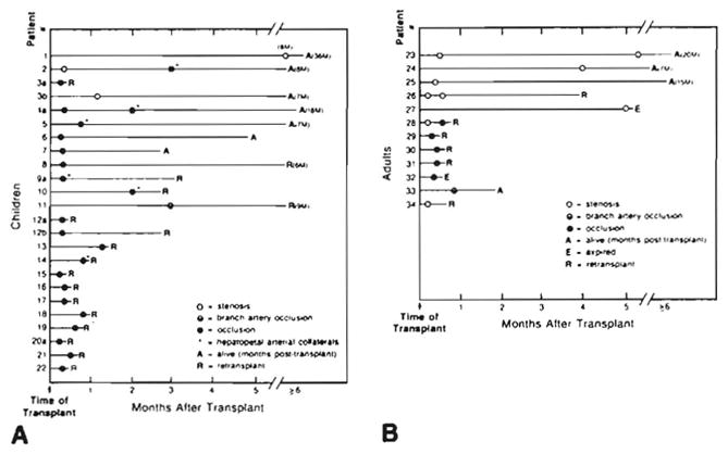
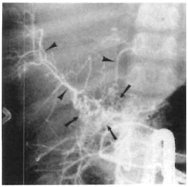
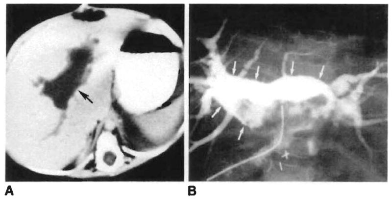
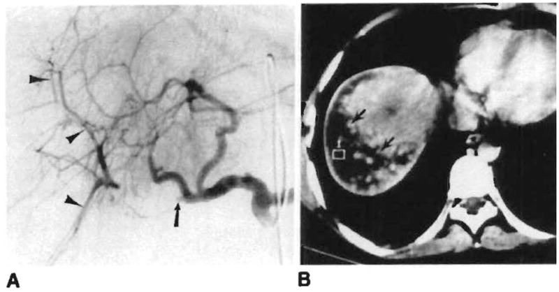
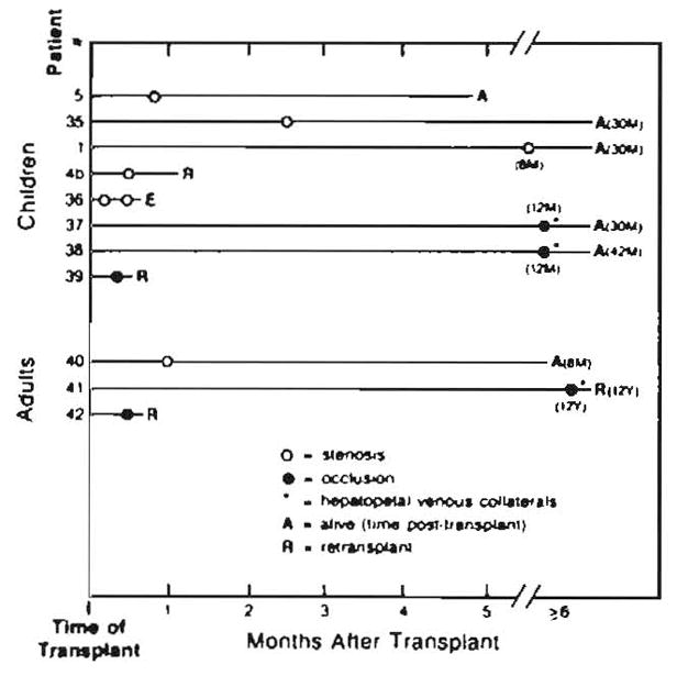
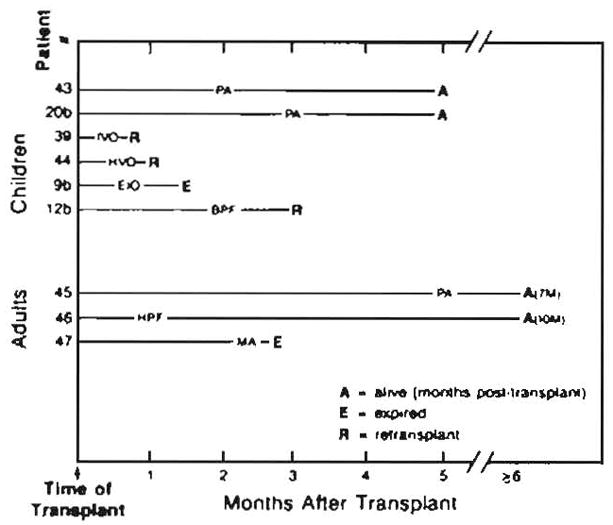
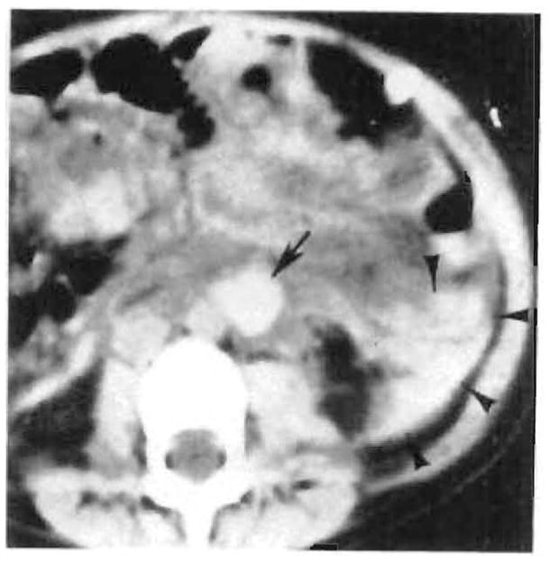
During the past 5 years, 104 angiographic studies were performed in 87 patients (45 children and 42 adults) with 92 transplanted livers for evaluation of possible vascular complications. Seventy percent of the studies were abnormal. Hepatic artery thrombosis was the most common complication (seen in 42% of children studied, compared with only 12% of adults) and was a major complication that frequently resulted in graft failure, usually necessitating retransplantation. In six children, reconstitution of the intrahepatic arteries by collaterals was seen. Three survived without retransplant. Arterial stenosis at the anastomosis or in the donor hepatic artery was observed in 11% of patients. Portal vein thrombosis or stenosis occurred in 13% of patients. Two children and one adult with portal vein thrombosis demonstrated hepatopetal collaterals that reconstituted the intrahepatic portal vessels. Uncommon complications included anastomotic and donor hepatic artery pseudoaneurysms, a hepatic artery-dissecting aneurysm, pancreaticoduodenal mycotic aneurysms, hepatic artery-portal vein fistula, biliary-portal vein fistula, hepatic vein occlusion, and inferior vena cava thrombosis.


Figures

Fig. 1
Fig. 1
Hepatic artery complications in liver transplants. A. Children. B. Adults. Placement of circles indicates time of angiographic study after transplant. Patients are numbered consecutively, with letters indicating separate transplanted livers. Numbers refer to case numbers in Results and Discussion sections. Numbers followed by M are number of months (e.g., 8M = 8 months).
Fig. 2
Fig. 2
Collateral circulation after hepatic artery thrombosis. Extensive arterial collaterals (arrows), which reconstitute intrahepatic arteries (arrowheads), are demonstrated on selective superior mesenteric arteriogram 2 months after transplant
Fig. 3
Fig. 3
Biliary–portal vein fistula in a 1-year-old girl with hepatic artery thrombosis and recurrent intermittent sepsis. A, Large intrahepatic fluid collection (arrow) is seen on CT 2 months after transplant. B, A biloma (arrows) communicating with intrahepatic portal veins is demonstrated after percutaneous drainage.
Fig. 4
Fig. 4
Branch hepatic artery occlusion. A. Occlusion of proximal right hepatic artery (arrow) is seen on celiac arteriogram 3 weeks after transplant. The peripheral branches (arrowheads) are reconstituted by intrahepatic collaterals. B, Probable right hepatic lobe infarct (arrows) seen on abdominal CT performed the same day.
Fig. 5
Fig. 5
Portal vein complications in liver transplants. Placement of circles indicates time of angiographic study after transplant. Numbers followed by M or Y are number of months or years (e.g., 30M = 30 months).
Fig. 6
Fig. 6
Miscellaneous vascular complications in liver transplants. PA = pseudoaneurysm, IVO = inferior vena cava occlusion, HVO = hepatic vein occlusion, EIO = external iliac artery occlusion, BPF = biliary–portal vein fistula, HPF = hepatic artery—portal vein fistula, MA = mycotic aneurysms. Numbers followed by M are number of months (e.g., 7M = 7 months).
Fig. 7
Fig. 7
Pseudoaneurysm (curved arrow) with large right-hepatic-lobe hematoma (straight arrows) seen on hepatic arteriogram 7 weeks after transplant
Fig. 8
Fig. 8
Large aneurysm (curved arrow) just beyond proximal anastomosis (straight arrow) of aortic graft with preservation of distal arterial flow (small arrows) demonstrated on angiogram 3 months after transplant. Surgery showed this to be a dissecting aneurysm starting at proximal anastomosis of graft.
Fig. 9
Fig. 9
Paraaortic (arrow) and retroperitoneal (arrowheads) hematoma originating from proximal anastomosis of aortic graft seen on nonenhanced abdominal CT scan 1 week after transplant. Angiography was normal.

Similar articles

Cited by

References

    1. Starzl TE, Iwatsuki S, Shaw BW, Jr, Gordon RD. Orthotopic liver transplantation in 1984. Transplant Proc. 1985;17:250–258. - PubMed
    1. Iwatsuki S, Shaw BW, Jr, Starzl TE. Five-year survival after liver transplantation. Transplant Proc. 1985;17:259–263. - PMC - PubMed
    1. Iwatsuki S, Shaw BW, Jr, Starzl TE. Current status of hepatic transplantation. Semin Liver Dis. 1983;3:173–180. - PMC - PubMed
    1. Starzl TE, Iwatsuki S, Shaw BW, Jr, et al. Analysis of liver transplantation. Hepatology. 1984;4:47S–49S. - PMC - PubMed
    1. Shaw BW, Jr, Gordon RD, Iwatsuki S, Starzl TE. Hepatic retransplantation. Transplant Proc. 1985;17:264–271. - PMC - PubMed