Review
. 2021 Mar 22;9(2):111-141.
doi: 10.5599/admet.942. eCollection 2021.

Organ-on-a-Chip systems for new drugs development

Affiliations
Review

Organ-on-a-Chip systems for new drugs development

Ronny Vargas et al. ADMET DMPK. .

Abstract

Research on alternatives to the use of animal models and cell cultures has led to the creation of organ-on-a-chip systems, in which organs and their physiological reactions to the presence of external stimuli are simulated. These systems could even replace the use of human beings as subjects for the study of drugs in clinical phases and have an impact on personalized therapies. Organ-on-a-chip technology present higher potential than traditional cell cultures for an appropriate prediction of functional impairments, appearance of adverse effects, the pharmacokinetic and toxicological profile and the efficacy of a drug. This potential is given by the possibility of placing different cell lines in a three-dimensional-arranged polymer piece and simulating and controlling specific conditions. Thus, the normal functioning of an organ, tissue, barrier, or physiological phenomenon can be simulated, as well as the interrelation between different systems. Furthermore, this alternative allows the study of physiological and pathophysiological processes. Its design combines different disciplines such as materials engineering, cell cultures, microfluidics and physiology, among others. This work presents the main considerations of OoC systems, the materials, methods and cell lines used for their design, and the conditions required for their proper functioning. Examples of applications and main challenges for the development of more robust systems are shown. This non-systematic review is intended to be a reference framework that facilitates research focused on the development of new OoC systems, as well as their use as alternatives in pharmacological, pharmacokinetic and toxicological studies.

Keywords: Cell Culture; Drug Discovery; Lab-On-A-Chip Devices; Organoids; Preclinical models; Tissue Engineering.


Conflict of interest statement

Conflict of interest : The authors have no conflicts of interest to declare.

Figures

Figure 1.
Figure 1.
Examples of OoC devices. A) Three-layer chip with a cell culture receptacle on the bottom plate, and multiple channel connectors for feed and signal control on the top plate. Adapted from [46] with permission from The Royal Society of Chemistry. Copyright (2018) B) A Two-channel OoC for the simulation of an epithelial tissue. Reproduced from [47] under terms of the Creative Commons Attribution License. C) OoC model for the simulation of the blood-brain barrier with gel suspended astrocytes and neurons. Reproduced from [48] with permission from The Royal Society of Chemistry. Copyright (2017).
Figure 2.
Figure 2.
Organ interaction in OoC. A) Schematic representation of miniaturized digestive system. B) Heart–liver body-on-a-chip with a liver module, cardiomyocytes and a skin module on a single chip. C) Body-on-a-chip simulation with gastrointestinal tract and liver tissue with two coupled chips. Adapted respectively from [69], [67], and [68] with permission from The Royal Society of Chemistry. Copyright (2019, 2020 & 2014)
Figure 3.
Figure 3.
Diagram of iPSC obtainment from i) Fibroblasts, keratinocytes or melanocytes obtained in skin biopsy, ii) CD34+ from blood samples, iii) CD133+ from umbilical cord or iv) multipotent cell from adipose tissue; iPSC reprogrammed towards pluripotent cells through different induction mechanisms such as: v) microRNA delivery, vi) viral transfection, vii) integration vectors or viii) protein transfection. Adapted from [40] under terms of the Creative Commons Attribution License. Copyright (2017).
Figure 4.
Figure 4.
Heart-on-a-chip for the evaluation of physiological parameters. A) Device diagram and structure. Graphs B) and C) respectively show the effect of two different isoprotenol concentrations on the heart rate and the magnitude of contractile displacement, compared to no treatment values. Adapted from [82] under terms of the Creative Commons Attribution License. Copyright (2020).
Figure 5.
Figure 5.
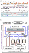
Kidney-on-a-chip device for drug screening and nephrotoxicity assessment. A) Schematic representation of the chip’s two chambers. B) Diagram of the complete device including: the chip and the temperature and the flow control device. Adapted from [97] under terms of the Creative Commons Attribution License. Copyright (2020).
Figure 6.
Figure 6.
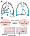
Mimicking of breathing mechanics. Schematic representation of A) the movement of the diaphragm leading to the lungs expansion during inhalation and B) the imitation of this motion by a mechanical microdiaphragm used in lung-on-a-chip devices. Reproduced from [100] under terms of the Creative Commons Attribution License. Copyright (2019)
Figure 7.
Figure 7.
Gut-on-a-chip (GOC) formed by two channels separated by a thin membrane with perfusion vasculature on the lower channel. Adapted from [105] under terms of the Creative Commons Attribution License. Copyright (2020).
Figure 8.
Figure 8.
Schematic examples of liver-on-a-chip devices. A) Multichannel OoC. Representation of a i) microfluidic device with several channels of different compositions for the formation of encapsulated hepatic plate structures, and a ii) system of syringe pumps supplying the microfluidic channels of the OoC with solutions. Adapted from [43] under terms of the Creative Commons Attribution License. Copyright (2020). B) Diagram of a pressure-driven flow control system of a 3D liver bioreactor for hepatotoxicity testing under perfusion conditions. Adapted from [107] under terms of the Creative Commons Attribution License. Copyright (2018).
Figure 9.
Figure 9.
BBB-on-a-chip with microelectrodes incorporated. A) Diagram of the main channel. B) Diagram of the electrodes and the chip structure. C) Photograph of the integrated system. Adapted from [123] under terms of the Creative Commons Attribution License. Copyright (2020).
Figure 10.
Figure 10.
Microfluidic device allowing cell migration. A) Simulation of an in vivo scenario with the chip construction. B) Schematic of the device’s manufacture by soft lithography. Reproduced from [131] with permission from The Royal Society of Chemistry. Copyright (2015).
Figure 11.
Figure 11.
Pancreatic-cancer-on-a-chip. A) i: image of a pancreatic duct (seeded with pancreatic cancer cells) and a blood vessel (seeded with endothelial cells) nested within a collagen matrix that are shown on the right image; ii: representation of the cross section of the right image. B) Invasion distance that cancer cells traverse within the collagen matrix as a function of time, with and without the addition of human umbilical vein endothelial cells (HUVECs). C) and D) Invasion of tumoral cells (green) into the blood vessel (red). E) i: invasion of the blood vessel by cancer cells, inducing to the apoptosis (marked in white in ii) of endothelial cells. Modified from [125] under terms of the Creative Commons Attribution License. Copyright (2019).
Figure 12.
Figure 12.
Biosensors and sensors on BBB-on-a-chip transducing signals to a PC or mobile interface. Reproduced from [146] under terms of the Creative Commons Attribution License. Copyright (2019).

Similar articles

Cited by

References

    1. Mittal R., Woo F.W., Castro C.S., Cohen M.A., Karanxha J., Mittal J., Chhibber T., Jhaveri V.M.. Organ-on-chip models: Implications in drug discovery and clinical applications. Journal of Cellular Physiology 234 (2019) 8352-8380. https://doi.org/10.1002/jcp.27729 10.1002/jcp.27729. - DOI - PubMed
    1. Jodat Y.A., Kang M.G., Kiaee K., Kim G.J., Martinez A.F.H., Rosenkranz A., Bae H., Shin S.R.. Human-Derived Organ-on-a-Chip for Personalized Drug Development. Current Pharmaceutical Design 24 (2019) 5471-5486. https://doi.org/10.2174/1381612825666190308150055 10.2174/1381612825666190308150055. - DOI - PMC - PubMed
    1. Lin A., Sved Skottvoll F., Rayner S., Pedersen-Bjergaard S., Sullivan G., Krauss S., Ray Wilson S., Harrison S.. 3D cell culture models and organ-on-a-chip: Meet separation science and mass spectrometry. Electrophoresis 41 (2020) 56-64. https://doi.org/10.1002/elps.201900170 10.1002/elps.201900170. - DOI - PubMed
    1. Kim J.A., Hong S., Rhee W.J.. Microfluidic three-dimensional cell culture of stem cells for high-throughput analysis. World J Stem Cells 11 (2019) 803-816. https://doi.org/10.4252/wjsc.v11.i10.803 10.4252/wjsc.v11.i10.803. - DOI - PMC - PubMed
    1. Miranda C.C., Fernandes T.G., Diogo M.M., Cabral J.M.S.. Towards multi-organoid systems for drug screening applications. Bioengineering 5 (2018) 1-17. https://doi.org/10.3390/bioengineering5030049. 10.3390/bioengineering5030049 - DOI - PMC - PubMed

LinkOut - more resources