. 2022 Mar;46(1):166-177.
doi: 10.1007/s12639-021-01431-9. Epub 2021 Aug 10.

Remarkable histopathological improvement of experimental toxoplasmosis after receiving spiramycin-chitosan nanoparticles formulation

Affiliations

Remarkable histopathological improvement of experimental toxoplasmosis after receiving spiramycin-chitosan nanoparticles formulation

Amal Farahat Allam et al. J Parasit Dis. 2022 Mar.

Abstract

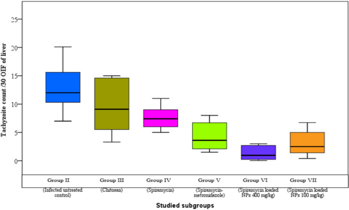
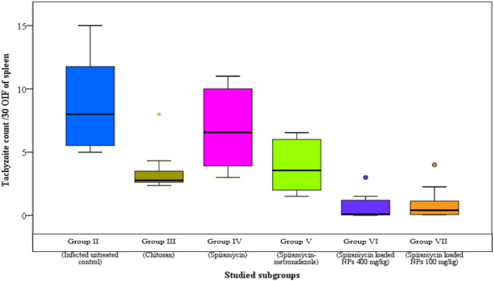
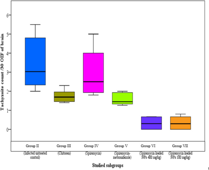
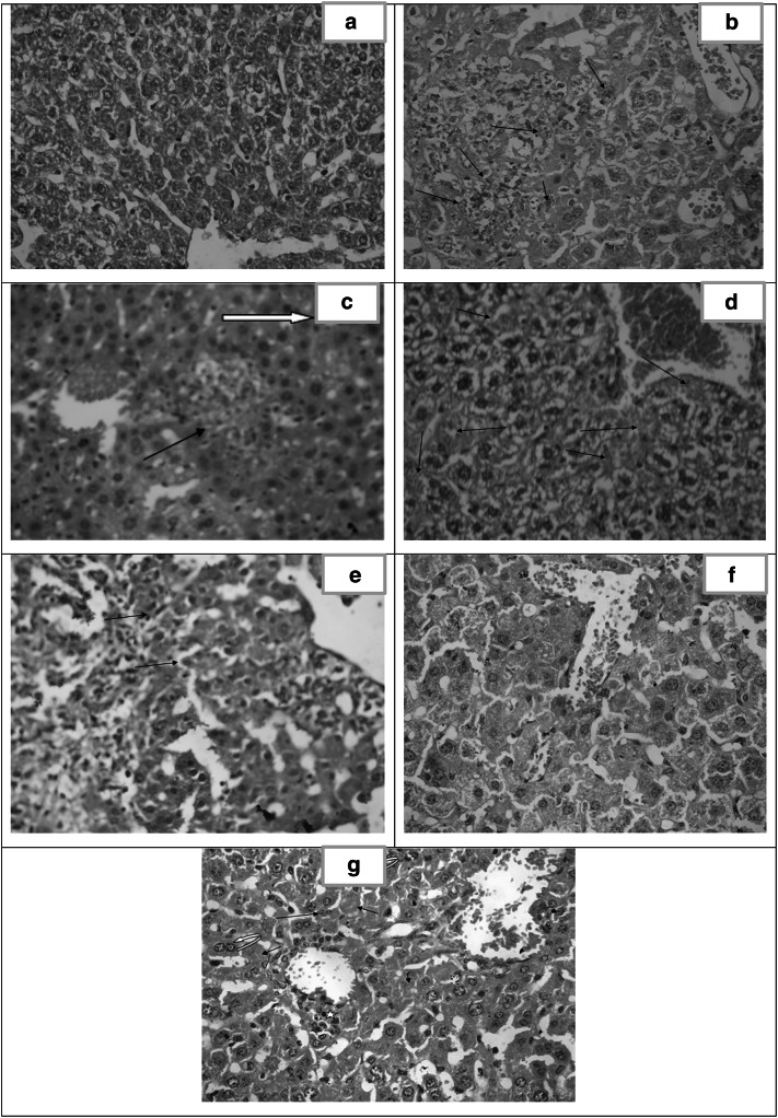
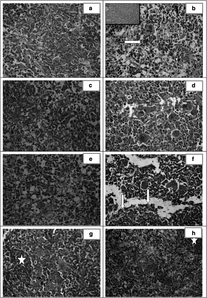
The present study investigated the anti-Toxoplasma effect of chitosan nanoparticles [CS NPs], spiramycin, spiramycin co-administered with metronidazole and spiramycin-CS NPs formulation on the parasite burden and histopathological changes in the liver, spleen and brain in experimentally infected mice. Seventy male Swiss albino mice were classified into seven equal groups: healthy control (I), infected untreated control (II), infected group receiving CS NPs (III), spiramycin administered infected group (IV), infected group receiving spiramycin-metronidazole (V), infected receiving 400 mg/kg spiramycin-CS NPs (VI) and infected treated with spiramycin-loaded CS NPs 100 mg/kg (VII). All groups were inoculated intraperitoneally with 2500 T. gondii tachyzoites RH strain except the healthy control group. All groups were sacrificed on the 8th day after infection. Density of the parasite and histopathological examination of the liver, spleen and brain of all treated mice revealed reduction in the mean tachyzoites count as well as decreased inflammation, congestion and necrosis within tissue sections. Spiramycin-loaded NPs displayed the highest significant reduction in the pathological insult tailed by spiramycin-metronidazole and CS NPs. In conclusion, spiramycin-loaded CS NPs showed a promising synergistic combination in the treatment of the histopathology caused by toxoplasmosis.

Keywords: Chitosan nanoparticles; Spiramycin; Spiramycin-chitosan nanoparticles formulation; Spiramycin-metronidazole; Toxoplasma gondii.


Conflict of interest statement

Conflict of interestThe authors declare that there is no conflict of interest.

Figures

Fig. 1
Fig. 1
Tachyzoites count in liver impression smears of the different studied groups
Fig. 2
Fig. 2
Tachyzoites count in spleen impression smears of the different studied groups
Fig. 3
Fig. 3
Tachyzoites count in brain impression smears of the different studied groups
Fig. 4
Fig. 4
Histopathological study of the liver. a Liver section of healthy control (group I) showing normal architecture. [H&E X400]. b Liver section of infected untreated control (group II) showing focal necrosis with acute and chronic mononuclear inflammatory infiltrate. Tachyzoites are seen among the hepatocytes [arrows]. Marked dilated congested central vein and the sinusoids are observed. [H&E X400]. c Liver section of CS NPs treated group (III) showing marked dilated congested central vein and sinusoids. Foci of hepatocellular necrosis and inflammatory cell infiltration(arrow). Pseudocysts with necrotic tachyzoites are observed [white arrow]. [H&E X400]. d Liver section of spiramycin treated group (IV) showing markedly dilated congested sinusoid. Dispersed necrotic hepatocytes [arrows] and widespread vacuolar degeneration. [H&E X400]. e Liver section of spiramycin-metronidazole treated group (V) showing dilated sinuses, moderate portal inflammatory infiltrate with fibrosis and interface hepatitis [arrows]. [H&E X400]. f Liver section of spiramycin-CS NPs 400 mg/kg treated group (VI) showing dilated congested sinusoid with near normal architecture. [H&E X400]. g Liver section of spiramycin-CS NPs 100 mg/kg treated group (VII) showing dilatation and congestion of portal and central veins. Minimal perivascular inflammatory infiltrate formed mainly of macrophages [star]. Pseudocysts with necrotic tachyzoites are identified (arrows) as well as binucleated regenerating hepatocytes [curved arrows]. [H&E X400]
Fig. 5
Fig. 5
Histopathological study of the spleen. a Spleen section of healthy control (group I) showing normal architecture. [H&E X400]. b and c Spleen section of infected untreated control (group II) showing tachyzoites [explosion shapes], marked disorganized splenic architecture with areas of necrosis [inset and red arrow] and presence of extramedullary hematopoiesis [megakaryocytes: black arrows, normoblasts: white arrow and myeloid cells: star]. [H&E X400]. d Spleen section of CS NPs treated group (III) showing disorganized architecture with extramedullary hematopoiesis. [(H&E X400]. e Spleen section of spiramycin treated group (IV) showing severe disorganized architecture with necrotic tachyzoites [arrows] and apoptotic cells. [H&E X400]. f Spleen section of spiramycin-metronidazole treated group (V) showing moderate disorganized architecture with extramedullary hematopoiesis and excess eosinophils [white arrows]. [H&E X400]. g Spleen section of spiramycin-CS NPs 400 mg/kg treated group (VI) showing mild disorganized architecture with extramedullary hematopoiesis and frequent necrotic tachyzoites [arrows]. Regenerating white pulp is evident [star]. [H&E X400]. h Spleen section of spiramycin-CS NPs 100 mg/kg treated group (VII) showing moderate disorganized spleen architecture with frequent epithelioid histiocytes loaded with necrotic tachyzoites [arrows]. Regenerating white pulp is noticed [star]. [H&E X400]
Fig. 6
Fig. 6
Histopathological study of the brain. a Section in brain of healthy control (group I) showing normal cortex architecture. [H&E X400]. b Section of cerebral cortex of infected untreated control (group II) showing gliosis, apoptosis [white arrows] and perivascular inflammatory infiltrate [arrow]. [H&E X400]. c Brain section of CS NPs treated group (III) showing astrocyte proliferation [gliosis], apoptosis [white arrow] and dilated sinuses [arrows]. [H&E X40]. d Brain section of spiramycin treated group (IV) showing gliotic nodule [star], dead neurons [red arrows] together with perivascular and parenchymal inflammatory infiltrate [black arrows]. [H&E X400]. e Brain section of spiramycin-metronidazole treated group (V) showing mild congested blood vessels, moderate gliosis and mineralized bodies of necrotic neurons so called ferrugination [arrows]. [H&E X400]. f Brain section of spiramycin-CS NPs 400 mg/kg treated group (VI) showing mild dilation of blood vessels (BV) with absence of tachyzoites and near normal architecture. [H&E X 400]. g Brain section of spiramycin-CS NPs 100 mg/kg treated group (VII) showing moderate gliosis, dispersed apoptotic cells [arrows] and extensive vacuolation in the cortex [spongiosis]. [H&E X 400]

References

    1. Agnihotri SA, Mallikarjuna NN, Aminabhavi TM. Recent advances on chitosan based micro- and nanoparticles in drug delivery. J Control Release. 2004;100(1):5–28. doi: 10.1016/j.jconrel.2004.08.010. - DOI - PubMed
    1. Ahmed SA, El-Mahallawy HS, Karanis P. Inhibitory activity of chitosan nanoparticles against Cryptosporidium parvum oocysts. Parasitol Res. 2019;118(7):2053–2063. doi: 10.1007/s00436-019-06364-0. - DOI - PubMed
    1. Allam AF, Shehab AY, Mogahed NMFH, Farag HF, Elsayed Y, El-Latif NF. Effect of nitazoxanide and spiramycin metronidazole combination in acute experimental toxoplasmosis. Heliyon. 2020;6(4):e03661. doi: 10.1016/j.heliyon.2020.e03661. - DOI - PMC - PubMed
    1. Araujo FG, Prockocimer P, Lin L, Remington SJ. Activity of clarithromycin alone or in combination with other drugs for treatment of murine toxoplasmosis. Antimicrob Agents Chemother. 1992;36:2454–2457. doi: 10.1128/aac.36.11.2454. - DOI - PMC - PubMed
    1. Asgari Q, Keshavarz H, Shojaee S, Motazedian MH, Mohebali M, Miri R, Mehrabani D, Rezaeian M. In vitro and in vivo potential of RH strain of Toxoplasma gondii [Type I] in tissue cyst forming. Iran J Parasitol. 2013;8(3):367–375. - PMC - PubMed

LinkOut - more resources