Case Reports
. 2022 Mar 9;7(6):20210019.
doi: 10.1259/bjrcr.20210019. eCollection 2022 Mar.

Aortic intramural hematoma and classic aortic dissection: two sides of the same coin within the acute aortic syndrome for an interventional radiologist

Affiliations
Case Reports

Aortic intramural hematoma and classic aortic dissection: two sides of the same coin within the acute aortic syndrome for an interventional radiologist

Pietro Pitrone et al. BJR Case Rep. .

Abstract

Management of acute type B aortic intramural haematoma (AIH) still represents a challenging issue. Although most resolve spontaneously or with conservative therapy, several cases of AIH may complicate into classic aortic dissection with subsequent risk of aortic rupture and visceral malperfusion, thus needing urgent or preemptive thoracic endovascular aneurysm repair (TEVAR). Despite the long-term aorta-related survival, TEVAR might lead to graft obstruction, migration, infection, stroke/paraplegia, visceral ischemia, endoleak and, last but not least, retrograde aortic dissection (AD), frequent in the acute phase and associated with a high mortality risk. In order to highlight such a close relationship between AIH and AD and the possibility to perform endovascular treatment, we report the experience of an adult female patient with an aortic intramural haematoma evolving into a classic aortic dissection. Despite successful thoracic endovascular aneurysm repair (TEVAR), our patient developed an aortic dissection type A at one month with subsequent indication for cardiac surgery still representing the elective approach in case of pathologies including the ascending aorta. Thus, the aim of our discussion is to create a debate on the most appropriate management for the treatment of descending AIH.


Figures

Figure 1.
Figure 1.
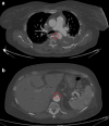
(day 1): (a) Non-enhanced CT axial scan at the level of the origin of the left pulmonary artery: a 11-mm crescent-shaped hyper attenuating (60 HU) thickening within the wall of the descending aorta on its lateral aspect is seen. (day 4, b) Contrast-enhanced CT coronal scan at the level of thoracic aorta: we appreciate the approximate extension of the above-described aortic thickening, that is, from the left subclavian artery to the celiac trunk. (c) Contrast-enhanced axial CT scan at the level of the upper abdomen: a small bulge of contrast is seen on the anterior aspect of the abdominal aorta, likely representing the “exit tear” of an aortic dissection whose thrombosis has led to the development of the AIH. (day 4, d) Contrast-enhanced CT axial scan at the level of the origin of the left pulmonary artery: a focal blush within a thrombotic lesion (7 × 3 mm, with a millimetric “peduncle”) on the medial aspect of the descending aorta is seen, likely representing the “entry tear” of the above-mentioned aortic dissection.
Figure 2.
Figure 2.
(day 8): Contrast-enhanced CT axial images at the level of the bifurcation the pulmonary trunk (a) and the upper abdomen (b): enlargement of both the entry (a) and the exit tears (b) is seen, indicating impending aortic dissection or rupture.
Figure 3.
Figure 3.
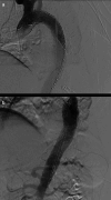
(day 8): Digital subtraction angiography images at the level of the cranial (a) and caudal (b) segments of the thoracic aorta (a): optimal positioning of a Valiant Navion 34 × 174 mm stent graft (Medtronic Vascular, Santa Rosa, Calif) is documented, with the “free-flow” portion at the origin of the left subclavian artery (with caudal aortic diameter measuring 29 mm).
Figure 4.
Figure 4.
(day 42, a, b) Contrast-enhanced CT axial scans at the level of the origin of the left pulmonary artery: an intimal-medial flap cranially to the stent graft extending to the origin of the right coronary artery is seen and is consistent with “retrograde aortic dissection type A”. (day 52, (c) Contrast-enhanced CT axial scan at the level of the aortic arch following cardiac surgery: a small bulge of contrast on the antero medial aspect of the ascending aorta is seen, likely representing the “entry tear” of above-mentioned retrograde dissection type A.

References

    1. Ferrera C, Vilacosta I, Cabeza B, Cobiella J, Martínez I, Saiz-Pardo Sanz M, et al. . Diagnosing aortic intramural hematoma: current perspectives. Vasc Health Risk Manag 2020; 16: 203–13. doi: 10.2147/VHRM.S193967 - DOI - PMC - PubMed
    1. Sahu KK, Lal A, Mishra AK. Additional insights regarding aortic intramural hematoma. Can J Anaesth 2020; 67: 382–3. doi: 10.1007/s12630-019-01490-w - DOI - PubMed
    1. Mendoza D, Terpenning S, Verma S, Stillman AE, Leshnower BG, Little BP. Retrospective comparative analysis of computed tomography findings of acute and chronic aortic dissections and intramural hematomas. J Thorac Imaging 2019; 34: 400–3. doi: 10.1097/RTI.0000000000000401 - DOI - PubMed
    1. Razek AAKA, Gaballa G, Megahed AS, Elmogy E. Time resolved imaging of contrast kinetics (tricks) Mr angiography of arteriovenous malformations of head and neck. Eur J Radiol 2013; 82: 1885–91. doi: 10.1016/j.ejrad.2013.07.007 - DOI - PubMed
    1. Razek AAKA, Saad E, Soliman N, Elatta HA. Assessment of vascular disorders of the upper extremity with contrast-enhanced magnetic resonance angiography: pictorial review. Jpn J Radiol 2010; 28: 87–94. doi: 10.1007/s11604-009-0394-4 - DOI - PubMed

Publication types