. 2022 Jun;53(6):1964-1974.
doi: 10.1161/STROKEAHA.121.036307. Epub 2022 Mar 18.

Cerebral Autosomal Dominant Arteriopathy With Subcortical Infarcts and Leukoencephalopathy Family Members With a Pathogenic NOTCH3 Variant Can Have a Normal Brain Magnetic Resonance Imaging and Skin Biopsy Beyond Age 50 Years

Affiliations

Cerebral Autosomal Dominant Arteriopathy With Subcortical Infarcts and Leukoencephalopathy Family Members With a Pathogenic NOTCH3 Variant Can Have a Normal Brain Magnetic Resonance Imaging and Skin Biopsy Beyond Age 50 Years

Remco J Hack et al. Stroke. 2022 Jun.

Abstract

Background: To determine whether extremely mild small vessel disease (SVD) phenotypes can occur in NOTCH3 variant carriers from Cerebral Autosomal Dominant Arteriopathy with Subcortical Infarcts and Leukoencephalopathy (CADASIL) pedigrees using clinical, genetic, neuroimaging, and skin biopsy findings.

Methods: Individuals from CADASIL pedigrees fulfilling criteria for extremely mild NOTCH3-associated SVD (mSVDNOTCH3) were selected from the cross-sectional Dutch CADASIL cohort (n=200), enrolled between 2017 and 2020. Brain magnetic resonance imaging were quantitatively assessed for SVD imaging markers. Immunohistochemistry and electron microscopy was used to quantitatively assess and compare NOTCH3 ectodomain (NOTCH3ECD) aggregation and granular osmiophilic material deposits in the skin vasculature of mSVDNOTCH3 cases and symptomatic CADASIL patients.

Results: Seven cases were identified that fulfilled the mSVDNOTCH3 criteria, with a mean age of 56.6 years (range, 50-72). All of these individuals harbored a NOTCH3 variant located in one of EGFr domains 7-34 and had a normal brain magnetic resonance imaging, except the oldest individual, aged 72, who had beginning confluence of WMH (Fazekas score 2) and 1 cerebral microbleed. mSVDNOTCH3 cases had very low levels of NOTCH3ECD aggregation in skin vasculature, which was significantly less than in symptomatic EGFr 7-34 CADASIL patients (P=0.01). Six mSVDNOTCH3 cases had absence of granular osmiophilic material deposits.

Conclusions: Our findings demonstrate that extremely mild SVD phenotypes can occur in individuals from CADASIL pedigrees harboring NOTCH3 EGFr 7-34 variants with normal brain magnetic resonance imaging up to age 58 years. Our study has important implications for CADASIL diagnosis, disease prediction, and the counseling of individuals from EGFr 7-34 CADASIL pedigrees.

Keywords: biopsy; brain; capillaries; vascular dementia; white matter.


Figures
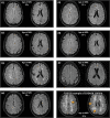
Figure 1.
Figure 1.
Brain magnetic resonance imaging (MRIs) of the 7 individuals with a NOTCH3cys variant and an extremely mild small vessel disease (SVD) phenotype. Brain MRI FLAIR images of 7 individuals with minimal small vessel disease (mSVDNOTCH3 case No. 1–7) showing absence of lacunes and only punctate foci of white matter hyperintensities (Fazekas deep white matter [DWM] 1), except for mSVDNOTCH3 case No. 3, who has beginning white matter hyperintensities (WMH) confluency (Fazekas DWM 2). For comparison, a brain MRI of a typical Cerebral Autosomal Dominant Arteriopathy with Subcortical Infarcts and Leukoencephalopathy (CADASIL) patient with a NOTCH3cys variant located in EGFr (epidermal growth-factor-like repeat) domains 7-34 is shown, who has large confluent WMH (Fazekas DWM 3) and multiple lacunes (indicated by arrows).
Figure 2.
Figure 2.
Pedigrees of 3 individuals with extremely mild small vessel disease. A and B, High intrafamilial variability in disease expression between family members, ranging from a normal brain magnetic resonance imaging (MRI) at age 50 (mSVDNOTCH3 case No. 1 and 2) to a classical severe Cerebral Autosomal Dominant Arteriopathy with Subcortical Infarcts and Leukoencephalopathy (CADASIL) phenotype before age 50 y (A-II-2 and B-II-1). A, Brother of mSVDNOTCH3 case No. 1 (A-II-2) has a classical severe CADASIL phenotype with stroke and cognitive deficits before age 50, with on brain MRI extensive confluent white matter hyperintensities (WMHs), 11 lacunes and 7 cerebral microbleed (CMB). B, mSVDNOTCH3 case No. 2 (B-II-2) and her sister (B-II-3) have a much milder disease expression than their brother who has a classical severe CADASIL phenotype with severe cognitive deficits, apathy, and mood disturbances before age 50 with on brain MRI extensive confluent WMHs, 3 lacunes and 2 CMB. C, In another family, the disease expression is predominantly mild, most individuals are above age 60 y and asymptomatic with only minimal small vessel disease markers on brain MRI, except for one individual (C-II-2) aged 65–70 y, who has severe mood disturbances, history of TIA and frequent attacks of dizziness with 3 lacunes and 4 CMB, but a relatively low burden of WMH on brain MRI for a CADASIL patient of his age.
Figure 3.
Figure 3.
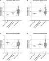
Magnetic resonance imaging (MRI) small vessel disease (SVD) burden in extremely mild small vessel disease cases, compared with their first-degree relatives with a NOTCH3cys variant and other individuals in the Dutch Cerebral Autosomal Dominant Arteriopathy with Subcortical Infarcts and Leukoencephalopathy (CADASIL) cohort with a NOTCH3cys EGFr (epidermal growth-factor-like repeat) 7-34 variant. A–D, Scatter plots showing standardized scores (corrected for age) of SVD imaging markers, based on all individuals with a NOTCH3cys EGFr 7-34 variant in the Dutch CADASIL cohort, including mSVDNOTCH3 cases* and their first-degree relatives†: mSVDNOTCH3 cases have a lower burden of SVD imaging markers than their first-degree relatives (nWMH volume P=0.009; nLacune volume P=0.009; brain parenchymal fraction [BPF] P=0.09; cerebral microbleed [CMB] count P=0.18). The fact that the mSVDNOTCH3 cases did not always have the lowest standardized scores is due to age differences. There was a high variability in burden of SVD imaging markers in symptomatic first-degree relatives, which was similar to the variability seen in the CADASIL EGFr 7-34 cohort. There was no significant difference in standardized scores of SVD imaging markers between first-degree relatives and the complete CADASIL EGFr 7-34 cohort: nWMH volume (P=0.09); nLacune volume (P=0.55); BPF (P=0.29); and CMB count (P=0.55). * The nWMH volume and BPF could not be quantified for mSVDNOTCH3 case No. 7, because 3D-FLAIR and 3D-T1 MRI sequences were not available. †Only first-degree relatives harboring a heterozygous cysteine altering missense NOTCH3 variant (NOTCH3cys) variant with a brain MRI performed on our 3T MRI scanner were included.
Figure 4.
Figure 4.
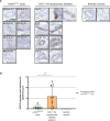
NOTCH3-immunohistochemistry of skin vessels in extremely mild small vessel disease cases compared with symptomatic Cerebral Autosomal Dominant Arteriopathy with Subcortical Infarcts and Leukoencephalopathy (CADASIL) patients with an EGFr (epidermal growth-factor-like repeat) 7-34 variant and healthy controls. A, Representative images of NOTCH3-immunohistochemistry of skin biopsies of 7 extremely mild NOTCH3-associated small vessel disease (mSVDNOTCH3) cases and 9 symptomatic CADASIL patients with a cysteine altering missense NOTCH3 (NOTCH3cys) variant located in EGFr 7-34, of which 4 individuals are first-degree relatives of the mSVDNOTCH3 cases (A-II-2, B-I-1, B-II-1, and C-II-2), and 2 healthy controls. NOTCH3 staining of mSVDNOTCH3 cases did not show the intense granular NOTCH3 staining typically seen in CADASIL, except for in mSVDNOTCH3 case No. 6. B, The NOTCH3 score was significantly lower in mSVDNOTCH3 cases than in symptomatic CADASIL patients: median 1.0 (interquartile range [IQR], 1.7 [range, 0.0–5.2]) vs median 10.3 (IQR, 19.4 [range, 0.0–25.7]; P=0.010). Bar represents 50 µm. *P<0.05.

Comment in

  • Clinical patterns in CADASIL.
    Gailani G, Robertson NP. Gailani G, et al. J Neurol. 2022 Aug;269(8):4575-4577. doi: 10.1007/s00415-022-11261-1. Epub 2022 Jul 14. J Neurol. 2022. PMID: 35833984 Free PMC article. No abstract available.

References

    1. Chabriat H, Joutel A, Dichgans M, Tournier-Lasserve E, Bousser MG. Cadasil. Lancet Neurol. 2009;8:643–653. doi: 10.1016/S1474-4422(09)70127-9 - PubMed
    1. Rutten JW, Hack RJ, Duering M, Gravesteijn G, Dauwerse JG, Overzier M, van den Akker EB, Slagboom E, Holstege H, Nho K, et al. . Broad phenotype of cysteine-altering NOTCH3 variants in UK Biobank: CADASIL to nonpenetrance. Neurology. 2020;95:e1835–e1843. doi: 10.1212/WNL.0000000000010525 - PMC - PubMed
    1. Hack RJ, Rutten JW, Person TN, Li J, Khan A, Griessenauer CJ, Abedi V, Lesnik Oberstein SAJ, Zand R; Regeneron Genetics Center. Cysteine-Altering NOTCH3 variants are a risk factor for stroke in the elderly population. Stroke. 2020;51:3562–3569. doi: 10.1161/STROKEAHA.120.030343 - PMC - PubMed
    1. Cho BPH, Nannoni S, Harshfield EL, Tozer D, Gräf S, Bell S, Markus HS. NOTCH3 variants are more common than expected in the general population and associated with stroke and vascular dementia: an analysis of 200 000 participants. J Neurol Neurosurg Psychiatry. 2021;92:694–701. doi: 10.1136/jnnp-2020-325838 - PMC - PubMed
    1. Opherk C, Peters N, Herzog J, Luedtke R, Dichgans M. Long-term prognosis and causes of death in CADASIL: a retrospective study in 411 patients. Brain. 2004;127(Pt 11):2533–2539. doi: 10.1093/brain/awh282 - PubMed

Publication types