. 2022 Oct 6;60(4):2102411.
doi: 10.1183/13993003.02411-2021. Print 2022 Oct.

Clinical, radiological and pathological findings in patients with persistent lung disease following SARS-CoV-2 infection

Affiliations

Clinical, radiological and pathological findings in patients with persistent lung disease following SARS-CoV-2 infection

Claudia Ravaglia et al. Eur Respir J. .

Abstract

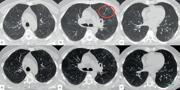
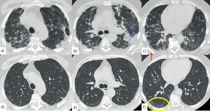
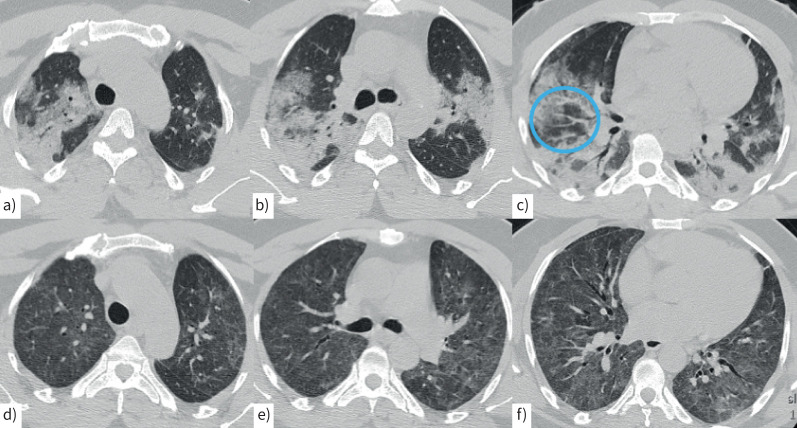
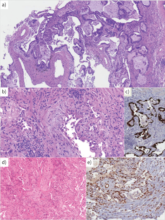
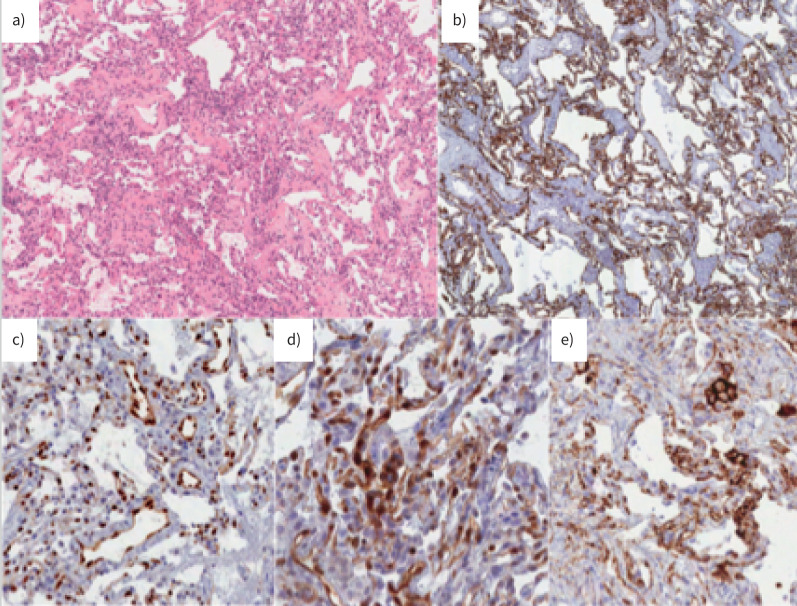
Some patients experience pulmonary sequelae after SARS-CoV-2 infection, ranging from self-limited abnormalities to major lung diseases. Morphological analysis of lung tissue may help our understanding of pathogenic mechanisms and help to provide consistent personalised management. The aim of this study was to ascertain morphological and immunomolecular features of lung tissue. Transbronchial lung cryobiopsy was carried out in patients with persistent symptoms and computed tomography suggestive of residual lung disease after recovery from SARS-CoV-2 infection. 164 patients were referred for suspected pulmonary sequelae after COVID-19; 10 patients with >5% parenchymal lung disease underwent lung biopsy. The histological pattern of lung disease was not homogeneous and three different case clusters could be identified, which was mirrored by their clinical and radiological features. Cluster 1 ("chronic fibrosing") was characterised by post-infection progression of pre-existing interstitial pneumonias. Cluster 2 ("acute/subacute injury") was characterised by different types and grades of lung injury, ranging from organising pneumonia and fibrosing nonspecific interstitial pneumonia to diffuse alveolar damage. Cluster 3 ("vascular changes") was characterised by diffuse vascular increase, dilatation and distortion (capillaries and venules) within otherwise normal parenchyma. Clusters 2 and 3 had immunophenotypical changes similar to those observed in early/mild COVID-19 pneumonias (abnormal expression of STAT3 in hyperplastic pneumocytes and PD-L1, IDO and STAT3 in endothelial cells). This is the first study correlating histological/immunohistochemical patterns with clinical and radiological pictures of patients with post-COVID lung disease. Different phenotypes with potentially different underlying pathogenic mechanisms have been identified.


Conflict of interest statement

Conflict of interest: The authors report no competing interests.

Figures

FIGURE 1
FIGURE 1
Computed tomography (CT) scan in a 60-year-old male smoker. a–c) During acute infection, mild peripheral ground-glass attenuation is present in both upper lobes, mainly in the left hemithorax (red circle). Scattered areas of cystic changes suggestive of airspace enlargement with associated fibrosis (AEF) with mild architectural distortion are present bilaterally. d–f) A CT scan performed 4 months later shows a reduction in ground-glass attenuation, with residual ground-glass attenuation and areas of AEF (f, green arrow).
FIGURE 2
FIGURE 2
Computed tomography (CT) scan in a 62-year-old man with a–c) acute COVID infection characterised by bilateral, peripheral consolidations (b, blue arrow) and perilobular pattern (c, red arrow). d–f) A CT scan performed 2 months later shows mild peripheral reticulation and minimal perilobular pattern, mainly in the right lower lobe (f, yellow circle).
FIGURE 3
FIGURE 3
Computed tomography (CT) scan in a 50-year-old man with a–c) COVID-related acute pneumonia with extensive, peribronchovascular consolidations in both lungs and lobular sparing in both lower lobes. Moderate ground-glass attenuation is present in both upper lobes associated with vessel enlargement (c, blue circle). d–f) A CT scan performed 2 months later shows mild, diffuse ground-glass attenuation in both lungs associated with central bronchiectasis in the middle lobe and in both lower lobes.
FIGURE 4
FIGURE 4
a) Cluster 1. A case with a usual interstitial pneumonia pattern: architectural distortion, spatial and temporal heterogeneity of scarring modifications and microscopic honeycombing. Haematoxylin and eosin (H&E) staining ×10 magnification. b) Cluster 2. A case with morphological evidence of ongoing interstitial fibrosis and extended alveolar epithelial type II cell hyperplasia as observed in diffuse alveolar damage, proliferative phase. H&E staining. c) Cluster 2. Cytokeratin 7 immunostaining of epithelial cells, at low magnification, showing the severe effacement of parenchymal structure. d) A case with organising pneumonia pattern. H&E staining. e) Most epithelial cells and endothelial cells express nuclear phosphorylated signal transducer and activator of transcription 3. b–e ×20 magnification.
FIGURE 5
FIGURE 5
Cluster 3. A case with diffuse vascular increase, dilatation and distortion (both capillaries and venules) within an otherwise normal parenchyma. a) Haematoxylin and eosin staining ×10 magnification, b) cytokeratin 7 immunostaining, c) diffuse and strong endothelial expression of phosphorylated signal transducer and activator of transcription 3, d) indoleamine 2, 3-dioxygenase and e) programmed cell death 1 ligand 1. b–e ×20 magnification.

References

    1. Ghosn J, Piroth L, Epaulard O, et al. Persistent COVID-19 symptoms are highly prevalent 6 months after hospitalization: results from a large prospective cohort. Clin Microbiol Infect 2021; 27: 1041. doi: 10.1016/j.cmi.2021.03.012 - DOI - PMC - PubMed
    1. Wells AU, Devaraj A, Desai SR. Interstitial lung disease after COVID-19 infection: a catalog of uncertainties. Radiology 2021; 299: E216–E218. doi: 10.1148/radiol.2021204482 - DOI - PMC - PubMed
    1. Han X, Fan Y, Alwalid O, et al. Six-month follow-up chest CT findings after severe COVID-19 pneumonia. Radiology 2021; 299: E177–E186. doi: 10.1148/radiol.2021203153 - DOI - PMC - PubMed
    1. Darcis G, Bouquegneau A, Maes N, et al. Long-term clinical follow up of patients suffering from moderate to severe COVID-19 infection: a monocentric prospective observational cohort study. Int J Infect Dis 2021; 109: 209–216. doi: 10.1016/j.ijid.2021.07.016 - DOI - PMC - PubMed
    1. Nehme M, Braillard O, Chappuis F, et al. Prevalence of symptoms more than seven months after diagnosis of symptomatic COVID-19 in an outpatient setting. Ann Intern Med 2021; 174: 1252–1260. doi: 10.7326/M21-0878 - DOI - PMC - PubMed

Publication types