. 2022 Mar 17;5(1):235.
doi: 10.1038/s42003-022-03180-8.

Characterisation of PDGF-BB:PDGFRβ signalling pathways in human brain pericytes: evidence of disruption in Alzheimer's disease

Affiliations

Characterisation of PDGF-BB:PDGFRβ signalling pathways in human brain pericytes: evidence of disruption in Alzheimer's disease

Leon C D Smyth et al. Commun Biol. .

Abstract

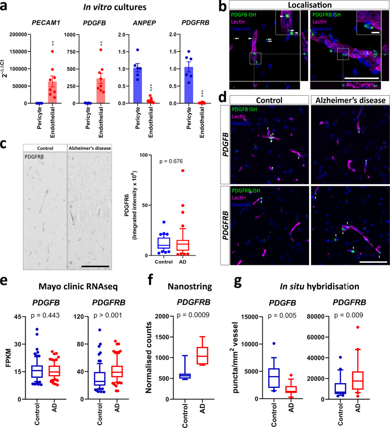
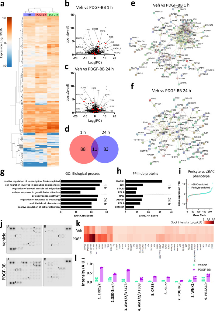
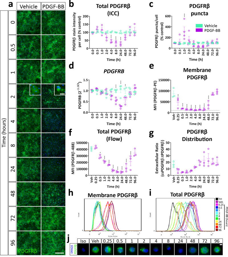
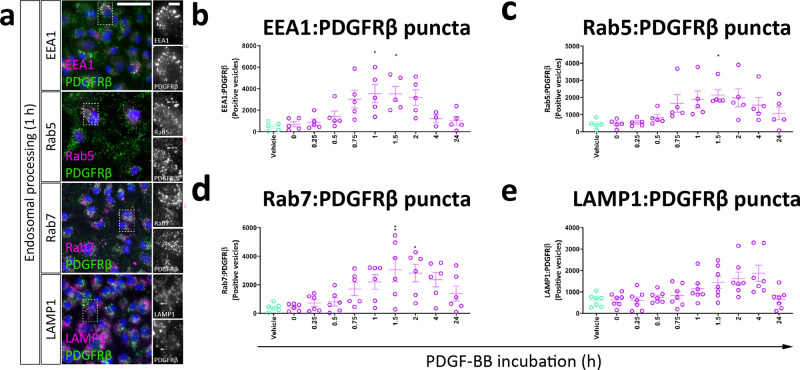
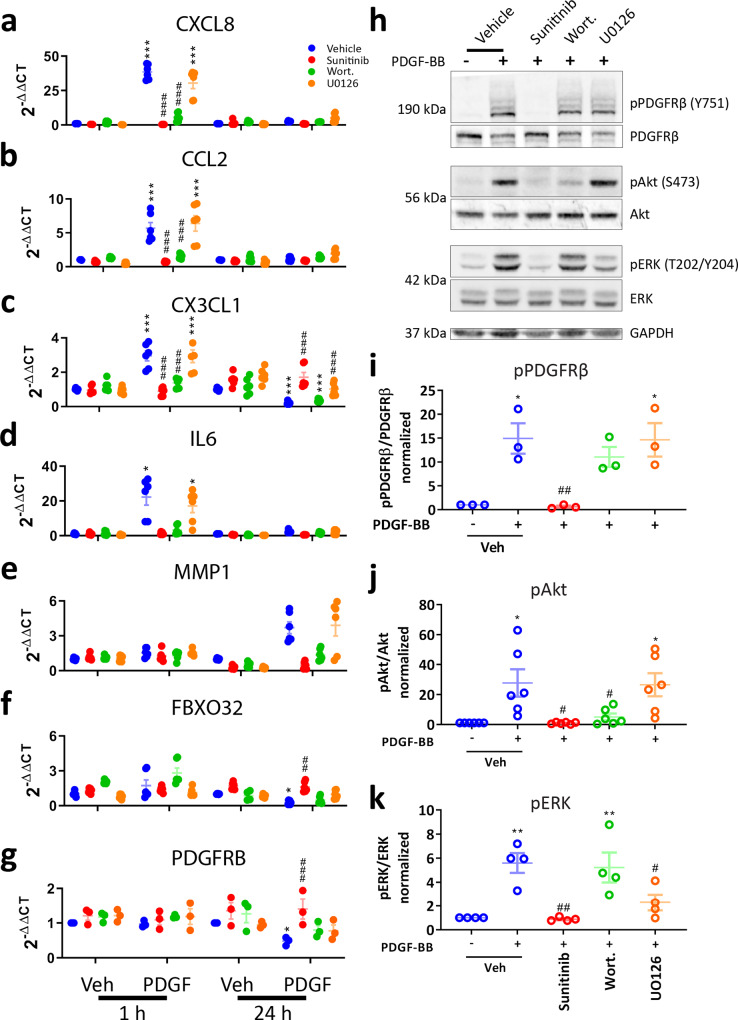
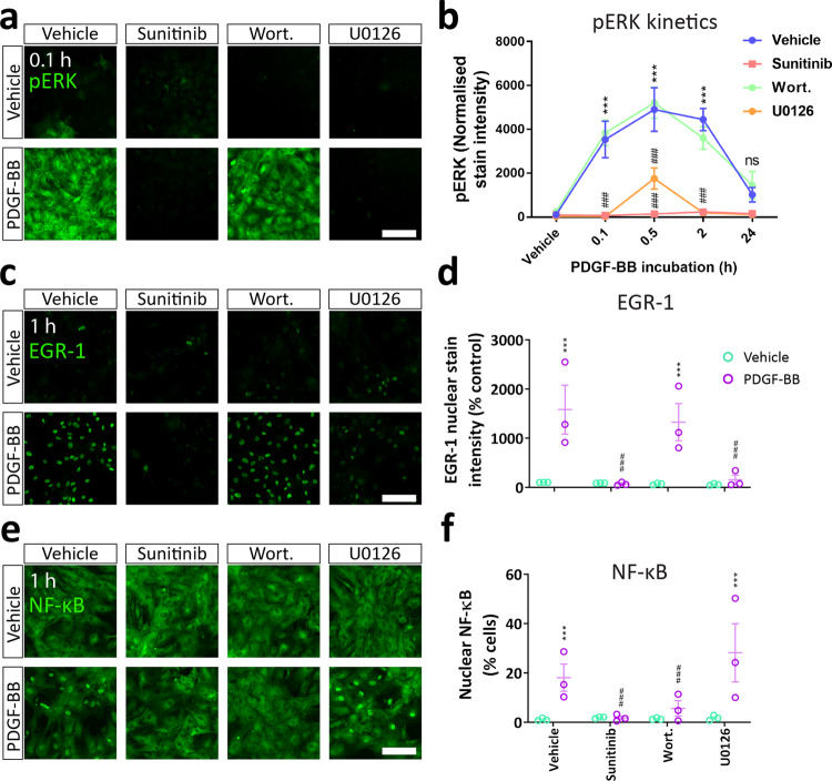
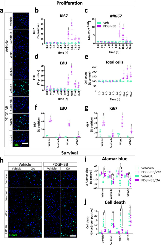
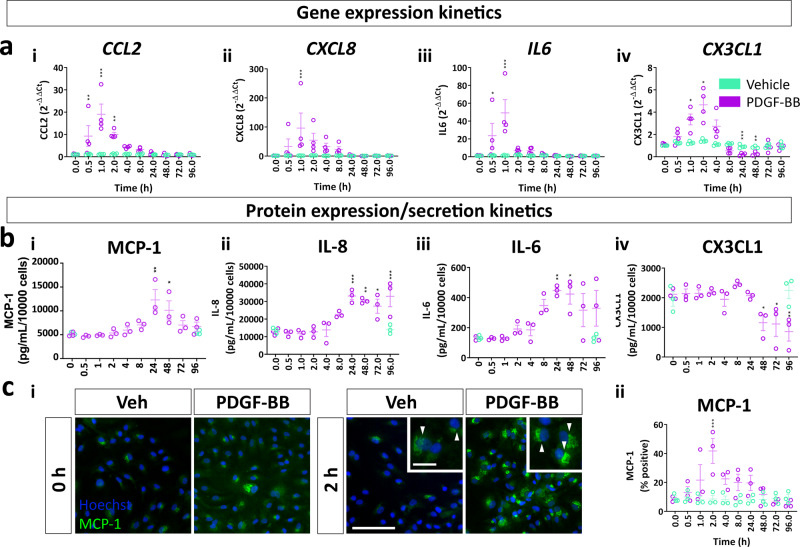
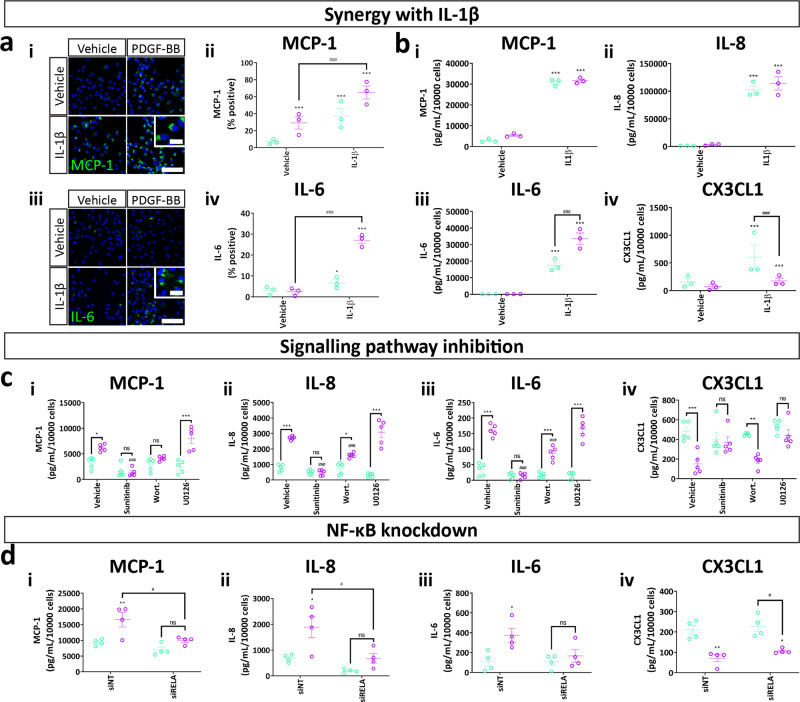
Platelet-derived growth factor-BB (PDGF-BB):PDGF receptor-β (PDGFRβ) signalling in brain pericytes is critical to the development, maintenance and function of a healthy blood-brain barrier (BBB). Furthermore, BBB impairment and pericyte loss in Alzheimer's disease (AD) is well documented. We found that PDGF-BB:PDGFRβ signalling components were altered in human AD brains, with a marked reduction in vascular PDGFB. We hypothesised that reduced PDGF-BB:PDGFRβ signalling in pericytes may impact on the BBB. We therefore tested the effects of PDGF-BB on primary human brain pericytes in vitro to define pathways related to BBB function. Using pharmacological inhibitors, we dissected distinct aspects of the PDGF-BB response that are controlled by extracellular signal-regulated kinase (ERK) and Akt pathways. PDGF-BB promotes the proliferation of pericytes and protection from apoptosis through ERK signalling. In contrast, PDGF-BB:PDGFRβ signalling through Akt augments pericyte-derived inflammatory secretions. It may therefore be possible to supplement PDGF-BB signalling to stabilise the cerebrovasculature in AD.


Conflict of interest statement

The authors declare no competing interests.

Figures

Fig. 1
Fig. 1. PDGF-BB:PDGFRβ signalling components are altered in the AD brain.
Primary human brain pericytes and endothelial cells were isolated and PDGFRβ signalling components were measured by qPCR. a qPCR of lineage markers (PECAM1 and ANPEP) and PDGF-BB:PDGFRβ signalling components in isolated human brain endothelial cells and pericytes. N = 6–8. **p < 0.01, ***p < 0.001, unpaired t test. Tissue microarray blocks containing control and AD cores were stained with PDGFRβ (n = 41–42), or UEA lectin to label blood vessels and RNAscope probes against PDGFB and PDGFRB (n = 14–28). b Confocal images of PDGFB and PDGFRB probes relative to lectin in epilepsy biopsy cortex. Scale bar = 100 μm, inset = 10 μm. c Representative immunohistochemical staining for PDGFRβ in control and AD cortex, and quantification of staining intensity. Scale bar = 100 μm. d Representative images of PDGFB and PDGFRB ISH in AD and control cortex. Scale bar = 100 μm. e Expression of PDGFB and PDGFRB in bulk RNAseq data of control and AD temporal cortex, derived from Allen et al. 51. n = 80, unpaired t test. f PDGFRB expression in frozen temporal cortex tissue from control and AD brains, detected by Nanostring. n = 9, unpaired t test. g Quantification of PDGFB and PDGFRB puncta associated with lectin-positive blood vessels. n = 14–21, unpaired t test.
Fig. 2
Fig. 2. PDGF-BB activates a biphasic response in brain pericytes.
Pericytes derived from neurologically normal post-mortem patients were treated with vehicle or PDGF-BB (10 ng/mL) for 1 or 24 h and gene expression analysed by RNAseq. a Dendrogram of PDGF-BB response in pericytes. Volcano plots comparing b 1 h and c 24 h PDGF-BB treatment to vehicle, with hits highlighted. d Venn diagram of differentially expressed gene lists at 1 and 24 h of PDGF-BB treatment. STRING network analysis of hits from e 1 and f 24 h of PDGF-BB treatment. g Gene ontology analysis and h interaction hub analysis of differentially expressed gene lists. i Expression of pericyte and vascular smooth muscle cell lineage markers identified by Vanlandewijck et al., ranked by fold change. Pericytes derived from the middle temporal gyrus of epilepsy patients were serum starved overnight, then treated with either PDGF-BB (100 ng/mL) or vehicle and lysed. Phosphorylation was detected in lysates using an antibody array. j Representative images, k heatmap of all phosphorylation sites and l levels of selected phosphorylation events in vehicle or PDGF-BB-treated pericytes.
Fig. 3
Fig. 3. PDGF-BB treatment causes a transient loss of PDGFRβ expression due to internalisation in pericytes.
Brain pericytes were treated with PDGF-BB (10 ng/mL) or vehicle for the specified time, then stained for cell-surface PDGFRβ for flow cytometry, fixed and stained for total PDGFRβ expression or RNA extracted for qPCR. a Representative images and quantification of b total PDGFRβ immunostaining and c PDGFRβ puncta formation in pericytes treated with vehicle or PDGF-BB. Scale bar = 500 μm, inset = 25 μm. n = 3–4, two-way ANOVA. d Gene expression of PDGFRB in pericytes incubated for different lengths of time with PDGF-BB. Expression of e cell surface and f total PDGFRβ and g the distribution of PDGFRβ determined by flow cytometry. n = 3, one-way ANOVA. Representative histograms of h cell surface and i total PDGFRβ and j representative images of total PDGFRβ expression in cells analysed by flow cytometry. Scale bar = 5 μm. *p < 0.05, **p < 0.01, ***p < 0.001 vs vehicle control, #p < 0.05, ##p < 0.01, ###p < 0.001 vs PDGF-BB-treated.
Fig. 4
Fig. 4. PDGF-BB causes PDGFRβ degradation through lysosomal processing in pericytes.
Brain pericytes were treated with PDGF-BB (10 ng/mL) for different lengths of time, then fixed and stained for endo/lysosomal markers. a Representative images of PDGFRβ colabelling with EEA1, Rab5, Rab7 and LAMP1. Scale bar = 50 μm, inset = 10 μm. Quantification of PDGFRβ puncta colocalised with b EEA1, c Rab5, d Rab7 and e LAMP1. n = 5, one-way ANOVA. *p < 0.05, **p < 0.01, ***p < 0.001 vs vehicle control, #p < 0.05, ##p < 0.01, ###p < 0.001 vs PDGF-BB-treated.
Fig. 5
Fig. 5. Akt and ERK activate distinct aspects of the PDGF-BB response in pericytes.
Pericytes were incubated with vehicle (0.3% DMSO), PDGFRβ inhibitor sunitinib (100 nM), PI3K inhibitor wortmannin (100 nM) or MEK/ERK inhibitor U0126 (10 μM) for 30 min. Pericytes were then treated with either vehicle or PDGF-BB (10 ng/mL) for 1 or 24 h, and RNA extracted for qPCR. Expression of a CXCL8, b CCL2, c CX3CL1, d IL6, e MMP1, f FBXO32 and g PDGFRB in pericytes treated with PDGF-BB for 1 or 24 h, with or without pathway inhibitors. n = 6, two-way ANOVA. Pericytes were serum starved overnight, then pre-treated with sunitinib (100 nM), wortmannin (100 nM) or U0126 (10 μM) or vehicle (0.3% DMSO) for 30 min, then treated with vehicle or PDGF-BB (100 ng/mL) for 30 min. Cells were lysed and western blot was performed. h Representative blots and densitometric analysis of i PDGFRβ, j Akt and k ERK phosphorylation. n = 4, one-way ANOVA. *p < 0.05, **p < 0.01, ***p < 0.001 vs vehicle control, #p < 0.05, ##p < 0.01, ###p < 0.001 vs PDGF-BB-treated.
Fig. 6
Fig. 6. PDGF-BB activates Akt-dependent NF-κB and ERK-dependent EGR-1 signalling in pericytes.
Pericytes were pre-treated with sunitinib (100 nM), wortmannin (100 nM) or U0126 (10 μM) or vehicle (0.3% DMSO) for 30 min, then treated with vehicle or PDGF-BB (10 ng/mL) for the specified timepoints. Cells were fixed at endpoint and immunostained. Representative images and quantification of ab pERK, cd EGR-1 and ef NF-κB activation following PDGF-BB treatment with or without pathway inhibitors. n = 3, two-way ANOVA. Scale bar = 100 μm. *p < 0.05, **p < 0.01, ***p < 0.001 vs vehicle control, #p < 0.05, ##p < 0.01, ###p < 0.001 vs PDGF-BB-treated.
Fig. 7
Fig. 7. PDGF-BB-dependent proliferation and protection from apoptosis are dependent on ERK signalling.
Pericytes were grown to confluence, then treated with PDGF-BB (10 ng/mL) for up to 96 h, then fixed at the endpoint and immunostained or RNA harvested for qPCR. EdU was added 24 h prior to the endpoint. Quantification of b Ki67 protein and c MKI67 gene expression following PDGF-BB stimulation. Quantification of d EdU incorporation and e cell count following PDGF-BB treatment. n = 3–4, two-way ANOVA. Pericytes were pre-treated with PDGFRβ inhibitor sunitinib (100 nM), PI3K inhibitor wortmannin (100 nM) or MEK/ERK inhibitor U0126 (10 μM) or vehicle (0.3% DMSO) for 30 min, then treated with vehicle or PDGF-BB (10 ng/mL) for 48 h. EdU was added 24 h prior to endpoint. a Representative images and quantification of f EdU incorporation and g Ki67 immunostaining in pericytes treated with PDGF-BB with or without pathway inhibitors. Pericytes were pre-treated with sunitinib (100 nM), wortmannin (100 nM) or U0126 (10 μM) or vehicle (0.3% DMSO) for 30 min, then treated with vehicle or PDGF-BB (10 ng/mL) for 24 h. n = 3, two-way ANOVA. Apoptosis inducer okadaic acid (OA; 50 nM) or vehicle was added to pericytes for a further 24 h, and viability was analysed by the AlamarBlue assay or ReadyProbes™ Cell Viability Imaging Kit. h Representative images of viability staining of pericytes treated with PDGF-BB, pathway inhibitors and OA. Scale bar = 100 μm. i Quantification of AlamarBlue fluorescence and j viability staining in pericytes. n = 6, two-way ANOVA. *p < 0.05, **p < 0.01, ***p < 0.001 vs vehicle control, #p < 0.05, ##p < 0.01, ###p < 0.001 vs PDGF-BB-treated.
Fig. 8
Fig. 8. PDGF-BB alters pericyte inflammatory secretions.
Pericytes were treated with PDGF-BB (10 ng/mL) or vehicle for up to 96 h, RNA extracted at endpoint and qPCR performed. a Kinetics of CCL2, CXCL8, IL6 and CX3CL1 induced by PDGF-BB expression. n = 4, two-way ANOVA. Pericytes were treated with PDGF-BB (10 ng/mL) or vehicle for up to 96 h, then fixed at endpoint and conditioned media collected. Samples were then immunostained and secretions in conditioned media analysed by cytometric bead array. b Quantification of MCP-1, IL-8, IL-6 and CX3CL1 secretion and c MCP-1 immunostaining in pericytes treated with PDGF-BB. n = 3, two-way ANOVA. Scale bar = 100 μm, insert = 10 μm. *p < 0.05, **p < 0.01, ***p < 0.001 vs vehicle control, #p < 0.05, ##p < 0.01, ###p < 0.001 vs PDGF-BB-treated.
Fig. 9
Fig. 9. PDGF-BB acts through the PI3K-NF-κB pathway to modify pericyte inflammatory secretions.
Pericytes were treated with vehicle or IL-1β (10 ng/mL), with or without PDGF-BB (10 ng/mL) for 2 h, then fixed and immunostained or 48 h and conditioned media collected for cytometric bead array. a Representative images and quantification of MCP-1 and IL-6 in cells treated with IL-1β or PDGF-BB. n = 3, two-way ANOVA. Scale bar = 100 μm, insert = 10 μm. b Secretion of MCP-1, IL-8, IL-6 and CX3CL1 in cells treated with IL-1β with or without PDGF-BB. Pericytes were pre-treated with PDGFRβ inhibitor sunitinib (100 nM), PI3K inhibitor wortmannin (100 nM) or MEK/ERK inhibitor U0126 (10 μM) or vehicle (0.3% DMSO) for 30 min, then treated with vehicle or PDGF-BB (10 ng/mL) for 48 h, and conditioned media collected. c Quantification of MCP-1, IL-8, IL-6 and CX3CL1 concentrations in conditioned media from pericytes treated with PDGF-BB with or without pathway inhibitors. Pericytes were treated with control siRNA (siNT) or siRNA directed against p65 NF-κB (siRELA) for 96 h, then treated with PDGF-BB (10 ng/mL) for 48 h and conditioned media collected for cytometric bead array. n = 5, two-way ANOVA. d Quantification of MCP-1, IL-8, IL-6 and CX3CL1 concentrations in conditioned media from pericytes treated with PDGF-BB with siRNA against NF-κB. n = 4, two-way ANOVA. *p < 0.05, **p < 0.01, ***p < 0.001 vs vehicle control, #p < 0.05, ##p < 0.01, ###p < 0.001 vs PDGF-BB-treated.

References

    1. Zlokovic BV. Neurovascular mechanisms of Alzheimer’s neurodegeneration. Trends Neurosci. 2005;28:202–208. - PubMed
    1. Yang, A. C. et al. A human brain vascular atlas reveals diverse cell mediators of Alzheimer’s disease risk. bioRxiv10.1101/2021.04.26.441262 (2021).
    1. Halliday MR, et al. Accelerated pericyte degeneration and blood–brain barrier breakdown in apolipoprotein E4 carriers with Alzheimer’s disease. J. Cereb. Blood Flow. Metab. 2016;36:216–227. - PMC - PubMed
    1. Zhang X, et al. High-resolution mapping of brain vasculature and its impairment in the hippocampus of Alzheimer’s disease mice. Natl. Sci. Rev. 2019;6:1223–1238. - PMC - PubMed
    1. Iturria-Medina Y, et al. Early role of vascular dysregulation on late-onset Alzheimer’s disease based on multifactorial data-driven analysis. Nat. Commun. 2016;7:11934. - PMC - PubMed

Publication types

MeSH terms

Substances