. 2022 May;32(5):437-450.
doi: 10.1038/s41422-022-00632-y. Epub 2022 Mar 18.

Near-atomic structure of the inner ring of the Saccharomyces cerevisiae nuclear pore complex

Affiliations

Near-atomic structure of the inner ring of the Saccharomyces cerevisiae nuclear pore complex

Zongqiang Li et al. Cell Res. 2022 May.

Abstract

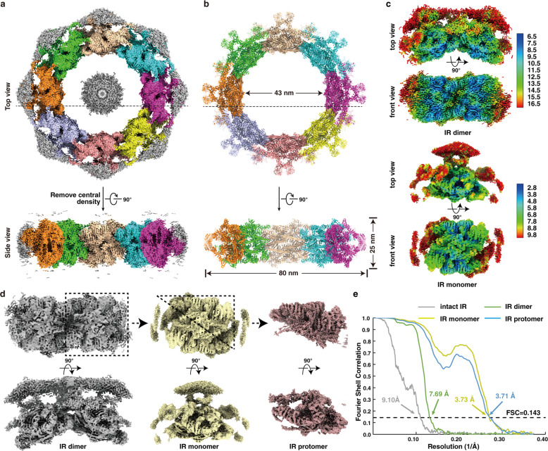
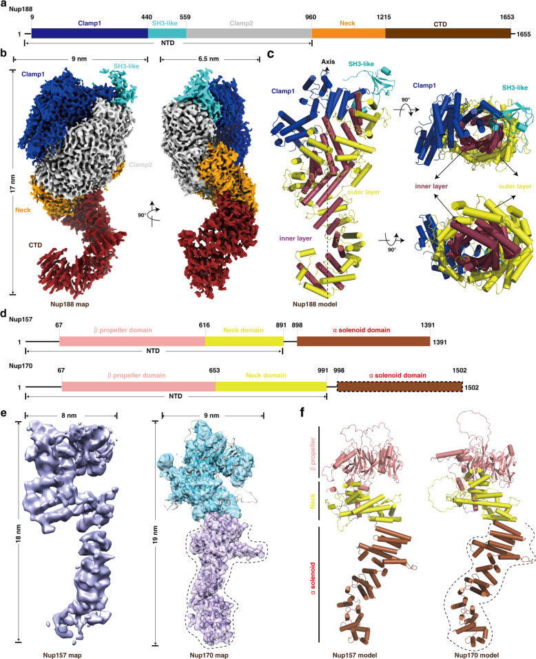
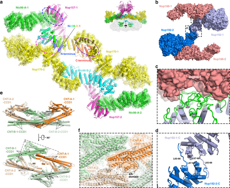
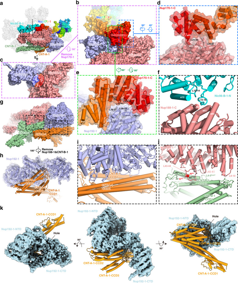
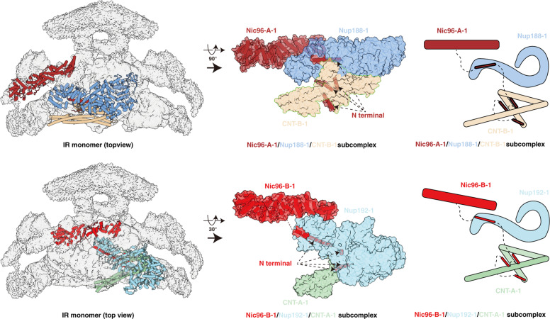
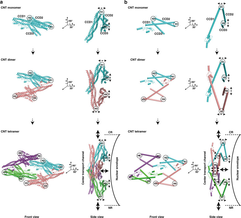
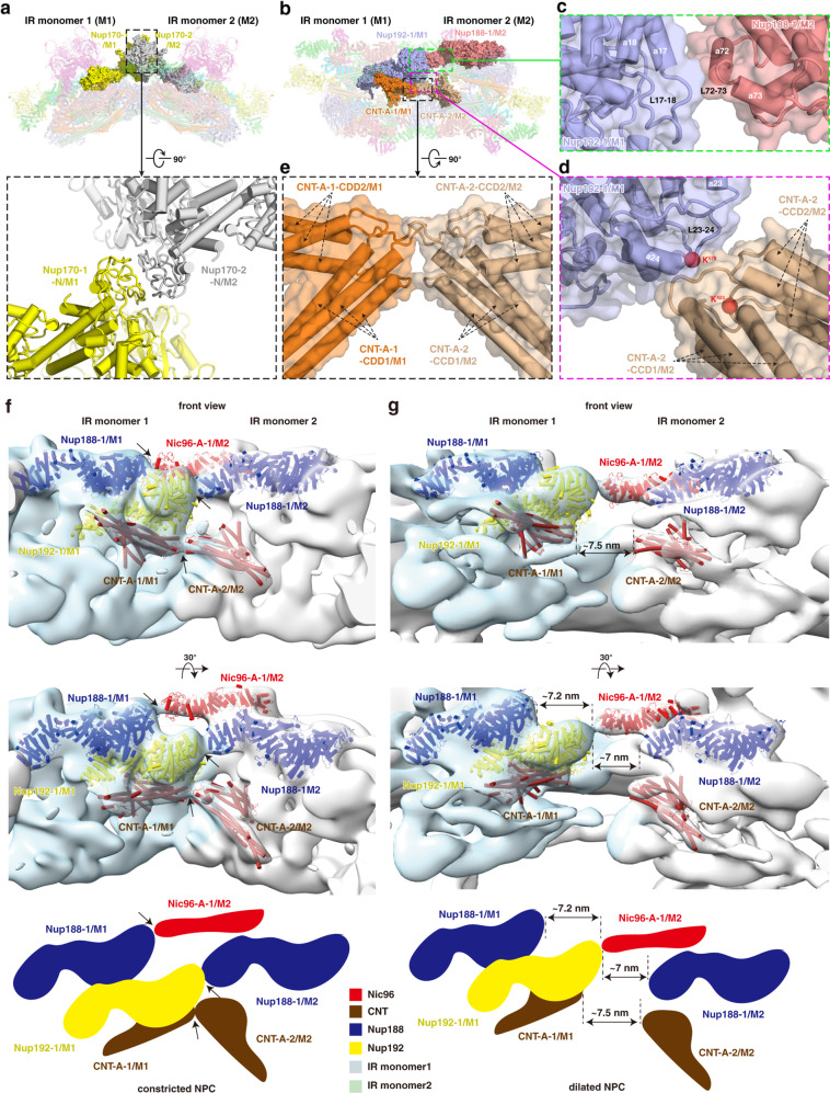
Nuclear pore complexes (NPCs) mediate bidirectional nucleocytoplasmic transport of substances in eukaryotic cells. However, the accurate molecular arrangement of NPCs remains enigmatic owing to their huge size and highly dynamic nature. Here we determined the structure of the asymmetric unit of the inner ring (IR monomer) at 3.73 Å resolution by single-particle cryo-electron microscopy, and created an atomic model of the intact IR consisting of 192 molecules of 8 nucleoporins. In each IR monomer, the Z-shaped Nup188-Nup192 complex in the middle layer is sandwiched by two approximately parallel rhomboidal structures in the inner and outer layers, while Nup188, Nup192 and Nic96 link all subunits to constitute a relatively stable IR monomer. In contrast, the intact IR is assembled by loose and instable interactions between IR monomers. These structures, together with previously reported structural information of IR, reveal two distinct interaction modes between IR monomers and extensive flexible connections in IR assembly, providing a structural basis for the stability and malleability of IR.


Conflict of interest statement

The authors declare no competing interests.

Figures

Fig. 1
Fig. 1. Cryo-EM structures of the IR of the S. cerevisiae NPC.
a, b Cryo-EM density map (a) and the atomic model (b) of the intact IR. The map and model are shown in top view (upper panel, dotted lines indicate the cross section corresponding to the side view) and side view (lower panel). Different colors represent different IR monomers. Threshold contour level is 0.22 σ. The gray blob density in the middle of the IR, most probably is a certain complex of FG-repeats, nuclear transport receptors and cargoes. c Color-coded 3D reconstructions of IR dimer (upper panel) and IR monomer (lower panel) showing local resolutions in different views. Threshold contour levels of dimer and monomer are 0.2 σ and 0.13 σ, respectively. The local resolutions were estimated with cryoSPARC and generated in ChimeraX. d Density maps of IR dimer, IR monomer and IR protomer determined in this study. e Gold standard Fourier shell correlation (FSC) curves for the maps of intact IR, IR dimer, IR monomer and IR protomer.
Fig. 2
Fig. 2. Structures of the purified full-length Nup188, Nup157 and Nup170 proteins.
a Schematic representation of the domain structures of Nup188. Domains are color coded. b Final cryo-EM map and dimensions of Nup188. The contour level is 3.95 σ. Colors of domains are the same as in a. c Model of Nup188. Colors of Clamp1 and SH3-like domains are the same as in a, and the “S” domain is divided into two layers ― inner layer and outer layer, colored in raspberry and yellow, respectively. d Schematic representation of the domain structures of Nup157 and Nup170. Domains are color coded. The α-solenoid domain (boxed by dashed line) in Nup170 indicates missing regions in our map of Nup170. e, f Final cryo-EM maps (e) and models (f) of Nup157 and Nup170, respectively. The map of Nup170 is transparent and overlapped with its model. Colors of domains and dotted line marked regions are the same as in d. Cylinders represent α-helices. NTD, N-terminal domain; CTD, C-terminal domain.
Fig. 3
Fig. 3. Intra-layer interactions in the IR monomer.
a Interactions within the outer layer. Purple, magenta and blue dashed ellipses represent interactions between Nic96-B-1 and Nup170-1, Nic96-B-1 and Nup170-2, and Nic96-A-1 and Nup157-1, respectively. b Interactions within the middle layer. Two interfaces are boxed by black dashed line and enlarged in c and d, respectively. c Details of interaction between Nup188-1 and Nup192-1. d Details of the interaction between two Nup192 proteins. e Interactions within the inner layer. The black dashed box is enlarged in f. f Details of the interaction between CNT complexes.
Fig. 4
Fig. 4. Inter-layer interactions in the IR monomer.
a Overall view of subunits involved in the inter-layer interactions, with eye symbols and arrowheads indicating view directions shown in the following panels. b Interactions of Nup170-1 with Nup188-1 and Nup192-1. Two interfaces are boxed by blue and yellowgreen dashed lines and enlarged in d and e, respectively. c The concave surface at the junction of Nup188-1 and Nup192-1. The area contacting Nup170-1 is circled. d, e Details of the interaction of the C-terminal region of Nup170-1 with Nup188-1 (d) and Nup192-1 (e). f Details of the interaction between Nic96-B-1 and Nup188-1. g Overall view of subunits involved in the middle and inner layer interactions. Two interfaces are boxed by black dashed lines and enlarged in i and j, respectively. h Loops connecting CCD2 and CCD3 of CNT-A-1 protrude into the groove located in the “S” domain from Nup192-1. i Long stalked CCD1 and CCD2 of CNT-A-1 clamp the N-terminal corner of Nup192-1 by extensive contacts via α-helices and loops. j Details of the interaction between Nup188-1 and CNT-B-1. k CNT-A-1 blocks the hole located between NTD and CTD of Nup192-1.
Fig. 5
Fig. 5. Location of N-terminal α-helices of Nic96.
The N-terminal fragment of Nic96 strings three layers of IR monomer through the interaction with Nup188, Nup192 and the CNT complex by several α-helices.
Fig. 6
Fig. 6. Conformational change of the CNT complexes.
a, b Molecular architectures and model illustrations are shown in a and b, respectively. Top and side views are shown in left and right panels, respectively. Among three CCD domains in CNT monomer, two hinge regions (H1 and H2) regulate angular variation of adjacent CCD domains to sense cargoes with different sizes. Small black arrows represent possible motions of each CCD domain around hinge region. The resultant motions of CNT tetramer are shown by larger black arrows.
Fig. 7
Fig. 7. Interactions between adjacent IR monomers and comparison of dilated NPC and constricted NPC.
a Interaction between two Nup170 molecules from adjacent IR monomers. b Overall view of subunits from middle and inner layers involved in the inter-monomer interactions. Three interfaces are boxed by green, magenta and black dashed lines and enlarged in c, d and e, respectively. c Details of the interaction between Nup192-1/M1 and the neighboring Nup188-1/M2. d Details of the interaction between Nup192-1/M1 and the neighboring CNT-A-2/M2. e Details of the interaction between CNT-A-1/M1 and the neighboring CNT-A-2/M2. f, g Distance changes between IR monomers are shown from constricted (f) to dilated (g) states. Two views are shown and schematic diagrams are drawn at the bottom. The representative subunits of IR are color coded. The interacting subunits of each interaction in the constricted NPC (black arrows) are separated by ~7 nm in the dilated NPC (dashed lines).

References

    1. Fahrenkrog B, Aebi U. The nuclear pore complex: Nucleocytoplasmic transport and beyond. Nat. Rev. Mol. Cell Biol. 2003;4:757–766. - PubMed
    1. Gorlich D, Kutay U. Transport between the cell nucleus and the cytoplasm. Annu. Rev. Cell Dev. Biol. 1999;15:607–660. - PubMed
    1. Hoelz A, Debler EW, Blobel G. The structure of the nuclear pore complex. Annu. Rev. Biochem. 2011;80:613–643. - PubMed
    1. Ptak C, Aitchison JD, Wozniak RW. The multifunctional nuclear pore complex: A platform for controlling gene expression. Curr. Opin. Cell Biol. 2014;28:46–53. - PMC - PubMed
    1. Callan HG, Randall JT, Tomlin SG. An electron microscope study of the nuclear membrane. Nature. 1949;163:280–280. - PubMed

Publication types

MeSH terms

Substances

LinkOut - more resources