

Targeting MuRF1 by small molecules in a HFpEF rat model improves myocardial diastolic function and skeletal muscle contractility
- PMID: 35301823
- PMCID: PMC9178400
- DOI: 10.1002/jcsm.12968
Targeting MuRF1 by small molecules in a HFpEF rat model improves myocardial diastolic function and skeletal muscle contractility
Abstract
Background: About half of heart failure (HF) patients, while having preserved left ventricular function, suffer from diastolic dysfunction (so-called HFpEF). No specific therapeutics are available for HFpEF in contrast to HF where reduced ejection fractions (HFrEF) can be treated pharmacologically. Myocardial titin filament stiffening, endothelial dysfunction, and skeletal muscle (SKM) myopathy are suspected to contribute to HFpEF genesis. We previously described small molecules interfering with MuRF1 target recognition thereby attenuating SKM myopathy and dysfunction in HFrEF animal models. The aim of the present study was to test the efficacy of one small molecule (MyoMed-205) in HFpEF and to describe molecular changes elicited by MyoMed-205.
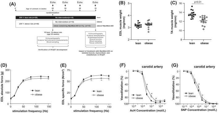
Methods: Twenty-week-old female obese ZSF1 rats received the MuRF1 inhibitor MyoMed-205 for 12 weeks; a comparison was made to age-matched untreated ZSF1-lean (healthy) and obese rats as controls. LV (left ventricle) function was assessed by echocardiography and by invasive haemodynamic measurements until week 32. At week 32, SKM and endothelial functions were measured and tissues collected for molecular analyses. Proteome-wide analysis followed by WBs and RT-PCR was applied to identify specific genes and affected molecular pathways. MuRF1 knockout mice (MuRF1-KO) SKM tissues were included to validate MuRF1-specificity.
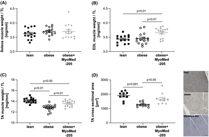
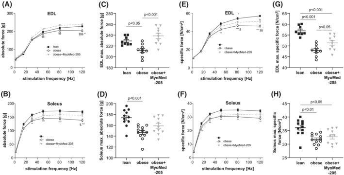
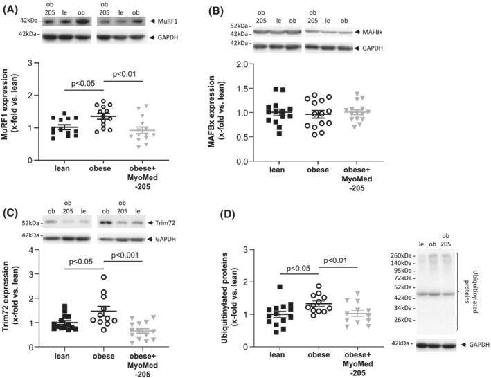
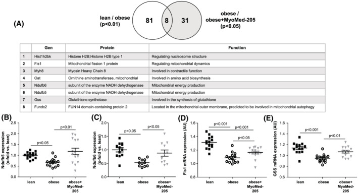
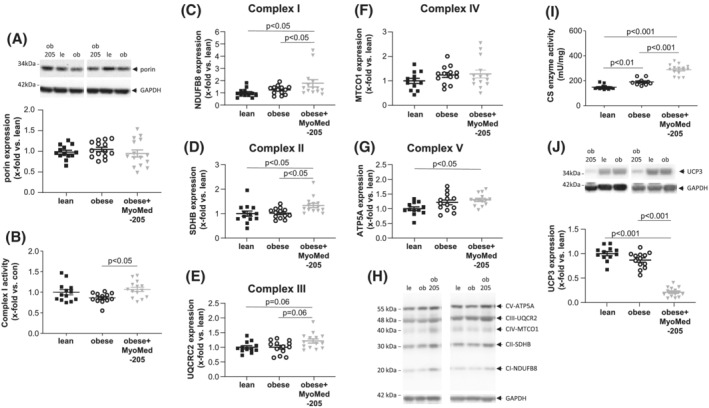
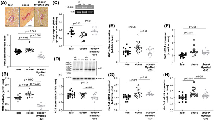
Results: By week 32, untreated obese rats had normal LV ejection fraction but augmented E/e' ratios and increased end diastolic pressure and myocardial fibrosis, all typical features of HFpEF. Furthermore, SKM myopathy (both atrophy and force loss) and endothelial dysfunction were detected. In contrast, MyoMed-205 treated rats had markedly improved diastolic function, less myocardial fibrosis, reduced SKM myopathy, and increased SKM function. SKM extracts from MyoMed-205 treated rats had reduced MuRF1 content and lowered total muscle protein ubiquitination. In addition, proteomic profiling identified eight proteins to respond specifically to MyoMed-205 treatment. Five out of these eight proteins are involved in mitochondrial metabolism, dynamics, or autophagy. Consistent with the mitochondria being a MyoMed-205 target, the synthesis of mitochondrial respiratory chain complexes I + II was increased in treated rats. MuRF1-KO SKM controls also had elevated mitochondrial complex I and II activities, also suggesting mitochondrial activity regulation by MuRF1.
Conclusions: MyoMed-205 improved myocardial diastolic function and prevented SKM atrophy/function in the ZSF1 animal model of HFpEF. Mechanistically, SKM benefited from an attenuated ubiquitin proteasome system and augmented synthesis/activity of proteins of the mitochondrial respiratory chain while the myocardium seemed to benefit from reduced titin modifications and fibrosis.
Keywords: Diastolic dysfunction; HFpEF; MuRF1; Muscle atrophy; Skeletal muscle dysfunction; ZSF1.
© 2022 The Authors. Journal of Cachexia, Sarcopenia and Muscle published by John Wiley & Sons Ltd on behalf of Society on Sarcopenia, Cachexia and Wasting Disorders.
Conflict of interest statement
Volker Adams reports a patent filing for MyoMed‐205, ID#704946 and further derivatives for its application to chronic muscle stress states (patent accession No WO2021023643A1).
Antje Schauer, Antje Augstein, Virginia Kirchhoff, Runa Draskowski, Anett Jannasch, Keita Goto, Gemma Lyall, Anita Männel, and Peggy Barthel have nothing to disclose. Norman Mangner reports personal fees from Edwards Lifesciences, Medtronic, Biotronik, Novartis, Sanofi Genzyme, AstraZeneca, Pfizer, Bayer, Abbott, Abiomed, and Boston Scientific outside the submitted work. Ephraim B. Winzer reports personal fees from Boehringer‐Ingelheim, CVRx, and Novartis outside the submitted work. Axel Linke reports grants from Novartis, personal fees from Medtronic, Abbott, Edwards Lifesciences, Boston Scientific, Astra Zeneca, Novartis, Pfizer, Abiomed, Bayer, Boehringer, and other from Picardia, Transverse Medical, and Claret Medical outside the submitted work. Siegfried Labeit reports a patent filing for MyoMed‐205, ID#704946 and further derivatives for its application to chronic muscle stress states (patent accession No WO2021023643A1).
Figures
References
-
- Bhatia RS, Tu JV, Lee DS, Austin PC, Fang J, Haouzi A, et al. Outcome of heart failure with preserved ejection fraction in a population‐based study. New Engl J Med 2006;355:260–269. - PubMed
-
- Schmederer Z, Rolim N, Bowen TS, Linke A, Wisloff U, Adams V. Endothelial function is disturbed in a hypertensive diabetic animal model of HFpEF: moderate continuous vs. high intensity interval training. Int J Cardiol 2018;273:147–154. - PubMed
-
- Akiyama E, Sugiyama S, Matsuzawa Y, Konishi M, Suzuki H, Nozaki T, et al. Incremental prognostic significance of peripheral endothelial dysfunction in patients with heart failure with normal left ventricular ejection fraction. J Am Coll Cardiol 2012;60:1778–1786. - PubMed
-
- Arena R, Myers J, Aslam SS, Varughese EB, Peberdy MA. Peak VO2 and VE/VCO2 slope in patients with heart failure: a prognostic comparison. Am Heart J 2004;147:354–360. - PubMed
Publication types
MeSH terms
Substances
Grants and funding
LinkOut - more resources
Full Text Sources
Medical
Molecular Biology Databases
Research Materials
Miscellaneous
