

Short-Term Changes in Left and Right Ventricular Cardiac Magnetic Resonance Feature Tracking Strain Following Ferric Carboxymaltose in Patients With Heart Failure: A Substudy of the Myocardial-IRON Trial
- PMID: 35301854
- PMCID: PMC9075490
- DOI: 10.1161/JAHA.121.022214
Short-Term Changes in Left and Right Ventricular Cardiac Magnetic Resonance Feature Tracking Strain Following Ferric Carboxymaltose in Patients With Heart Failure: A Substudy of the Myocardial-IRON Trial
Abstract
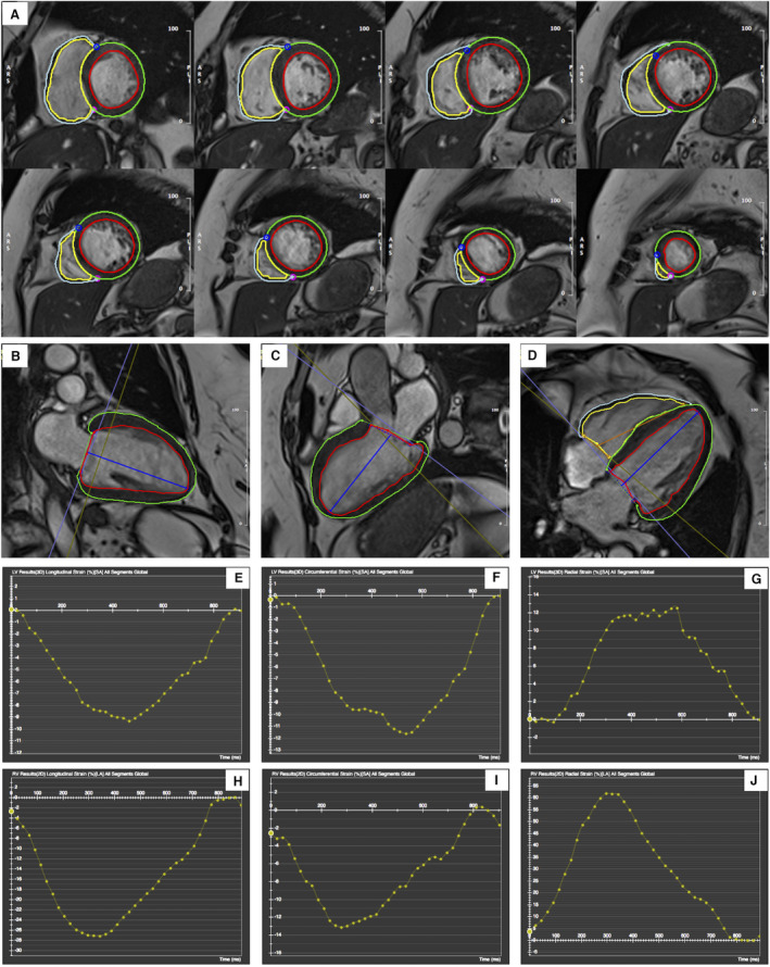
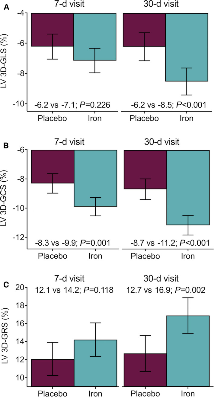
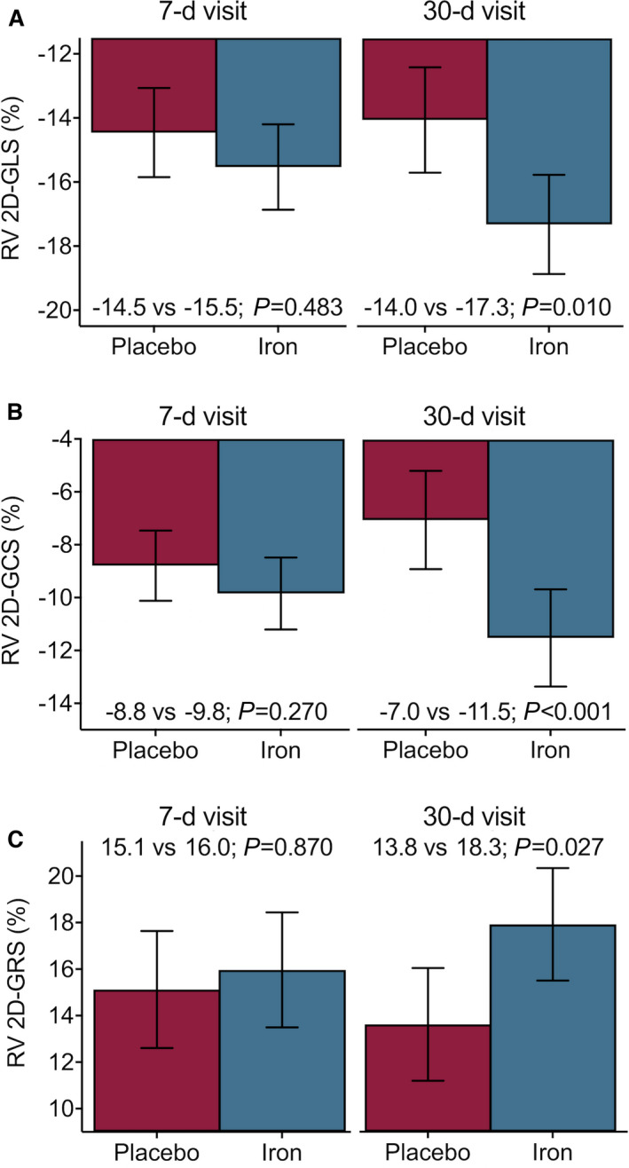
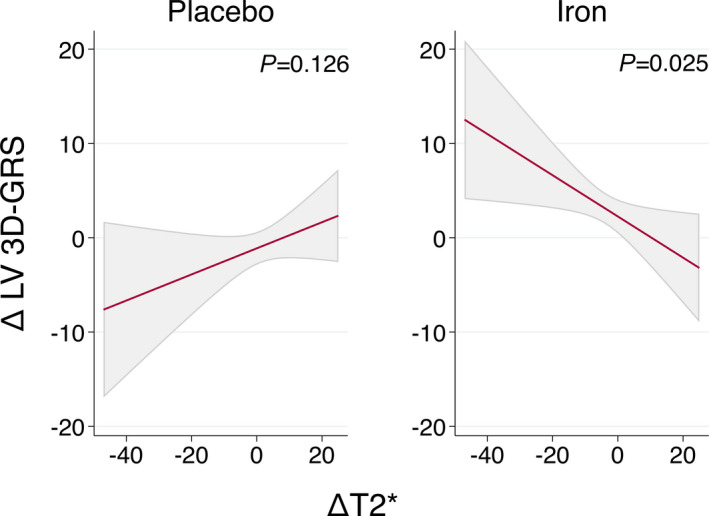
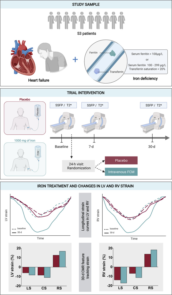
Background The mechanisms explaining the clinical benefits of ferric carboximaltose (FCM) in patients with heart failure, reduced or intermediate left ventricular ejection fraction, and iron deficiency remain not fully clarified. The Myocardial-IRON trial showed short-term cardiac magnetic resonance (CMR) changes suggesting myocardial iron repletion following administration of FCM but failed to find a significant increase in left ventricular ejection fraction in the whole sample. Conversely, the strain assessment could evaluate more specifically subtle changes in contractility. In this subanalysis, we aimed to evaluate the effect of FCM on the short-term left and right ventricular CMR feature tracking derived strain. Methods and Results This is a post hoc subanalysis of the double-blind, placebo-controlled, randomized clinical trial that enrolled 53 ambulatory patients with heart failure and left ventricular ejection fraction <50%, and iron deficiency [Myocardial-IRON trial (NCT03398681)]. Three-dimensional left and 2-dimensional right ventricular CMR tracking strain (longitudinal, circumferential, and radial) changes were evaluated before, 7 and 30 days after randomization using linear mixed-effect analysis. The median (interquartile range) age of the sample was 73 years (65-78), and 40 (75.5%) were men. At baseline, there were no significant differences in CMR feature tracking strain parameters across both treatment arms. At 7 days, the only global 3-dimensional left ventricular circumferential strain was significantly higher in the FCM treatment-arm (difference: -1.6%, P=0.001). At 30 days, and compared with placebo, global 3-dimensional left ventricular strain parameters significantly improved in those allocated to FCM treatment-arm [longitudinal (difference: -2.3%, P<0.001), circumferential (difference: -2.5%, P<0.001), and radial (difference: 4.2%, P=0.002)]. Likewise, significant improvements in global right ventricular strain parameters were found in the active arm at 30 days (longitudinal [difference: -3.3%, P=0.010], circumferential [difference: -4.5%, P<0.001], and radial [difference: 4.5%, P=0.027]). Conclusions In patients with stable heart failure, left ventricular ejection fraction <50%, and iron deficiency, treatment with FCM was associated with short-term improvements in left and right ventricular function assessed by CMR feature tracking derived strain parameters. Registration URL: https://www.clinicaltrials.gov; Unique identifier: NCT03398681.
Keywords: CMR feature tracking; ferric carboxymaltose; heart failure; iron deficiency; ventricular strain.
Figures
References
-
- Ponikowski P, Voors AA, Anker SD, Bueno H, Cleland JGF, Coats AJS, Falk V, González‐Juanatey JR, Harjola V‐P, Jankowska EA, et al. 2016 ESC guidelines for the diagnosis and treatment of acute and chronic heart failure: the Task Force for the diagnosis and treatment of acute and chronic heart failure of the European Society of Cardiology (ESC). Developed with the special contribution of the Heart Failure Association (HFA) of the ESC. Eur J Heart Fail. 2016;18:891–975. doi: 10.1002/ejhf.592 - DOI - PubMed
-
- Anker SD, Comin Colet J, Filippatos G, Willenheimer R, Dickstein K, Drexler H, Lüscher TF, Bart B, Banasiak W, Niegowska J, et al; FAIR‐HF Trial Investigators . Ferric carboxymaltose in patients with heart failure and iron deficiency. N Engl J Med. 2009;361:2436–2448. doi: 10.1056/NEJMoa0908355 - DOI - PubMed
-
- Ponikowski P, van Veldhuisen DJ, Comin‐Colet J, Ertl G, Komajda M, Mareev V, McDonagh T, Parkhomenko A, Tavazzi L, Levesque V, et al; CONFIRM‐HF Investigators . Beneficial effects of long‐term intravenous iron therapy with ferric carboxymaltose in patients with symptomatic heart failure and iron deficiency. Eur Heart J. 2015;36:657–668. doi: 10.1093/eurheartj/ehu385 - DOI - PMC - PubMed
