

pH and proton-sensitive receptors in brain ischemia
- PMID: 35301897
- PMCID: PMC9274858
- DOI: 10.1177/0271678X221089074
pH and proton-sensitive receptors in brain ischemia
Abstract
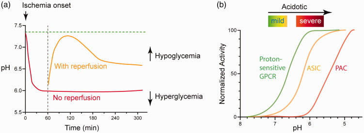
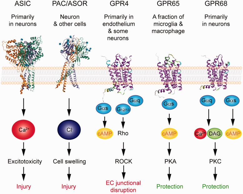
Extracellular proton concentration is at 40 nM when pH is 7.4. In disease conditions such as brain ischemia, proton concentration can reach µM range. To respond to this increase in extracellular proton concentration, the mammalian brain expresses at least three classes of proton receptors. Acid-sensing ion channels (ASICs) are the main neuronal cationic proton receptor. The proton-activated chloride channel (PAC), which is also known as (aka) acid-sensitive outwardly rectifying anion channel (ASOR; TMEM206), mediates acid-induced chloride currents. Besides proton-activated channels, GPR4, GPR65 (aka TDAG8, T-cell death-associated gene 8), and GPR68 (aka OGR1, ovarian cancer G protein-coupled receptor 1) function as proton-sensitive G protein-coupled receptors (GPCRs). Though earlier studies on these GPCRs mainly focus on peripheral cells, we and others have recently provided evidence for their functional importance in brain injury. Specifically, GPR4 shows strong expression in brain endothelium, GPR65 is present in a fraction of microglia, while GPR68 exhibits predominant expression in brain neurons. Here, to get a better view of brain acid signaling and its contribution to ischemic injury, we will review the recent findings regarding the differential contribution of proton-sensitive GPCRs to cerebrovascular function, neuroinflammation, and neuronal injury following acidosis and brain ischemia.
Keywords: Acid signaling; acidosis; brain pH; ischemia; neuroinflammation; neuronal injury.
Conflict of interest statement
Figures
References
-
- Ljunggren B, Norberg K, Siesjo BK. Influence of tissue acidosis upon restitution of brain energy metabolism following total ischemia. Brain Res 1974; 77: 173–186. - PubMed
-
- Astrup J, Symon L, Branston NM, et al. Cortical evoked potential and extracellular K+ and H+ at critical levels of brain ischemia. Stroke 1977; 8: 51–57. - PubMed
-
- Kalimo H, Rehncrona S, Soderfeldt B, et al. Brain lactic acidosis and ischemic cell damage: 2. Histopathology. J Cereb Blood Flow Metab 1981; 1: 313–327. - PubMed
-
- Chesler M. Regulation and modulation of pH in the brain. Physiol Rev 2003; 83: 1183–1221. - PubMed
Publication types
MeSH terms
Substances
Grants and funding
LinkOut - more resources
Full Text Sources
Other Literature Sources
