Review
. 2022 Apr;74(2):387-438.
doi: 10.1124/pharmrev.121.000375.

Post-Traumatic Epilepsy and Comorbidities: Advanced Models, Molecular Mechanisms, Biomarkers, and Novel Therapeutic Interventions

Affiliations
Review

Post-Traumatic Epilepsy and Comorbidities: Advanced Models, Molecular Mechanisms, Biomarkers, and Novel Therapeutic Interventions

Victoria M Golub et al. Pharmacol Rev. 2022 Apr.

Abstract

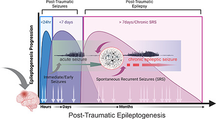
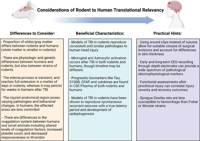
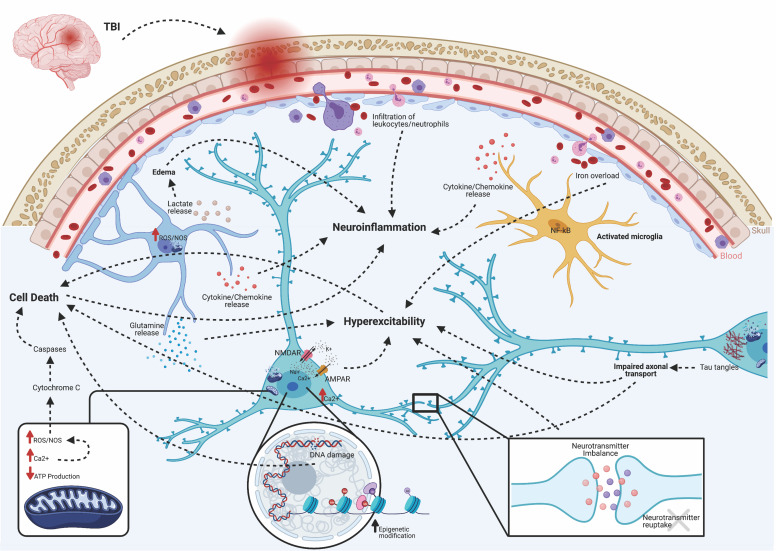
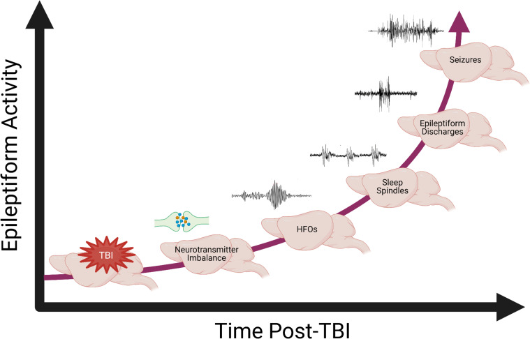
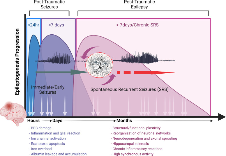
Post-traumatic epilepsy (PTE) is one of the most devastating long-term, network consequences of traumatic brain injury (TBI). There is currently no approved treatment that can prevent onset of spontaneous seizures associated with brain injury, and many cases of PTE are refractory to antiseizure medications. Post-traumatic epileptogenesis is an enduring process by which a normal brain exhibits hypersynchronous excitability after a head injury incident. Understanding the neural networks and molecular pathologies involved in epileptogenesis are key to preventing its development or modifying disease progression. In this article, we describe a critical appraisal of the current state of PTE research with an emphasis on experimental models, molecular mechanisms of post-traumatic epileptogenesis, potential biomarkers, and the burden of PTE-associated comorbidities. The goal of epilepsy research is to identify new therapeutic strategies that can prevent PTE development or interrupt the epileptogenic process and relieve associated neuropsychiatric comorbidities. Therefore, we also describe current preclinical and clinical data on the treatment of PTE sequelae. Differences in injury patterns, latency period, and biomarkers are outlined in the context of animal model validation, pathophysiology, seizure frequency, and behavior. Improving TBI recovery and preventing seizure onset are complex and challenging tasks; however, much progress has been made within this decade demonstrating disease modifying, anti-inflammatory, and neuroprotective strategies, suggesting this goal is pragmatic. Our understanding of PTE is continuously evolving, and improved preclinical models allow for accelerated testing of critically needed novel therapeutic interventions in military and civilian persons at high risk for PTE and its devastating comorbidities. SIGNIFICANCE STATEMENT: Post-traumatic epilepsy is a chronic seizure condition after brain injury. With few models and limited understanding of the underlying progression of epileptogenesis, progress is extremely slow to find a preventative treatment for PTE. This study reviews the current state of modeling, pathology, biomarkers, and potential interventions for PTE and comorbidities. There's new optimism in finding a drug therapy for preventing PTE in people at risk, such as after traumatic brain injury, concussion, and serious brain injuries, especially in military persons.


Figures

None
Graphical abstract
Fig. 1
Fig. 1
Translational relevance of rodent to human TBI. Despite significant investment in advancing technology and basic science to increase knowledge of human TBI pathology, translation from bench-to-bedside into therapeutic advances has been slower than expected. One of the factors limiting the translation of scientific knowledge from preclinical studies into the clinic is the limitation of small rodent in vivo disease models. Although these models have been developed to simulate and mimic the human condition, there are innate differences between rodents and humans, which can limit the impact of these studies. Likewise, there are many important similarities as well as practical hints that can be used to overcome these limitations. This figure discusses important considerations of rodent to human translational relevance.
Fig. 2
Fig. 2
Acute pathologies of post-traumatic epileptogenesis. Brain injury triggers several acute pathologies. Direct insult compromises the blood-brain barrier, allowing infiltration of peripherally circulating immune cells, such as leukocytes, macrophages, and neutrophils. NF-κB translocates to the nuclei of microglia, transforming them to an activated phenotype. This induces cellular proliferation and the release of inflammatory amplifiers, such as chemokines, cytokines, reactive oxygen species (ROS), and nitric oxide synthase (NOS). Macrophages participate in the cleanup of damaged cells and debris, but based on their functional activation state, may either exacerbate damage or initiate repair mechanisms. Lactate release from astrocytes contributes to water retention and edema. Excess iron from a leaky BBB can contribute to hyperexcitability. Excessive accumulation of glutamate and aspartate neurotransmitters due to spillage from damaged neurons or impaired reuptake by astrocytes activates NMDA and AMPA receptors located on postsynaptic membranes, allowing for influx of calcium ions. Together with the release of Ca2+ stores from the endoplasmic reticulum, increases in Ca2+ leads to production of ROS and activation of calpains. Damaged or dysfunctional mitochondria create a deficit of available ATP, leading to Na+/K+ pump failure, activation of Ca2+ channels, and further production of ROS/NOS. Cytochrome C released into the cytosol activates cell death pathways via caspase proteins. Epigenetic modifications, in the form of increased HDAC activity and altered DNA/histone methylation, changes transcriptionally active sites, including many genes associated with hyperexcitability and serotonin-to-melatonin conversion. Furthermore, DNA damage leads to apoptosis and cell loss. Progressive axonal damage and tau tangles lead to impaired axonal transport and results in both neurodegeneration and hyperexcitability. Together, these acute pathologies are both adaptive and maladaptive. The former contributes to functional and beneficial recovery, whereas the latter exacerbates epileptogenesis and the progression of abnormal electrographic activity.
Fig. 3
Fig. 3
Relationship of long-term neurodegeneration and spontaneous seizures. TBI induces a state of immediate inflammation and hyperexcitation in the brain, which exacerbate cell loss both ipsilateral and contralateral to the lesion. PTE was induced via a severe 2.0 mm depth CCI model of TBI. After injury, mice were tethered to 24/7-videoEEG for up to 4 months and seizures were identified by a customized MATLAB script and validated by unbiased researchers. Stereological quantification of two cell populations in the contralateral hippocampus was performed at days 1, 3, 7, 30, 60, and 120 post-TBI in subsets of these recorded mice. (A) Linear fit of remaining NeuN+ principal neurons overlayed the linear regression of average seizure output from responding mice to highlight the temporal relationship between cell loss and seizure occurrence. (B) Linear fit of remaining PV+ GABAergic interneurons overlayed the linear regression of average seizure output from responding mice to highlight the temporal relationship between cell loss and seizure occurrence.
Fig. 4
Fig. 4
Evolution of TBI-induced hyperexcitability and seizure activity. Electrographic biomarkers may predict onset of seizures, as hyperactivity in the brain progresses over time to the culmination of spontaneous recurrent seizures. TBI induces a state of heavy inflammation, disrupting both neurotransmitter and metabolic homeostasis. The emergence of these abnormal electrographic activities may reflect different stages of the epileptogenic process postinjury. Pathologic HFOs often precede seizures by weeks, followed by reduced frequency and duration of sleep spindles during the transition between stage 3 and REM sleep. Disruption of normal sleep spindles contributes to several sleep-wake disorders reported by patients with TBI. EEG spiking and discharges represent an advanced hyperactive disturbance that has been described as epileptiform abnormalities in animal brain slices and in vivo at various time-points postinjury. The final stage is the end of latency indicated by the occurrence of spontaneous seizures; however, epileptogenesis can continue to progress even after the first seizure.
Fig. 5
Fig. 5
Timeline progression of TBI-induced early seizures and late spontaneous recurrent seizures. TBI triggers acute cascades resulting in immediate or early seizure, referred to as post-traumatic seizures, and propels the process of epileptogenesis. ultimately resulting in chronic epileptic state with spontaneous recurrent seizures. The general premise about epileptogenesis can be divided into three distinct stages. The first stage occurs with an initial brain injury event. This is followed by the second latent stage that can last a varied amount of time. The third stage is the chronic period in which the patient suffers from spontaneous seizures. The time required to reach chronic stage represents a window of opportunity for testing interventions in people at high risk for epilepsy after brain injuries.

Similar articles

Cited by

References

    1. Abbasi B, Goldenholz DM (2019) Machine learning applications in epilepsy. Epilepsia 60:2037–2047. - PMC - PubMed
    1. Abdelmalik PA, Boorman DW, Tracy J, Jallo J, Rincon F (2016) Acute traumatic coagulopathy accompanying isolated traumatic brain injury is associated with worse long-term functional and cognitive outcomes. Neurocrit Care 24:361–370. - PubMed
    1. Abdullahi A, Amini-Nik S, Jeschke MG (2014) Animal models in burn research. Cell Mol Life Sci 71:3241–3255. - PMC - PubMed
    1. Adeyemo BO, Biederman J, Zafonte R, Kagan E, Spencer TJ, Uchida M, Kenworthy T, Spencer AE, Faraone SV (2014) Mild traumatic brain injury and ADHD: a systematic review of the literature and meta-analysis. J Atten Disord 18:576–584. - PubMed
    1. Agoston DV, Shutes-David A, Peskind ER (2017) Biofluid biomarkers of traumatic brain injury. Brain Inj 31:1195–1203. - PubMed

MeSH terms