. 2022 Mar 17;13(1):1436.
doi: 10.1038/s41467-022-29139-1.

LC3B is an RNA-binding protein to trigger rapid mRNA degradation during autophagy

Affiliations

LC3B is an RNA-binding protein to trigger rapid mRNA degradation during autophagy

Hyun Jung Hwang et al. Nat Commun. .

Abstract

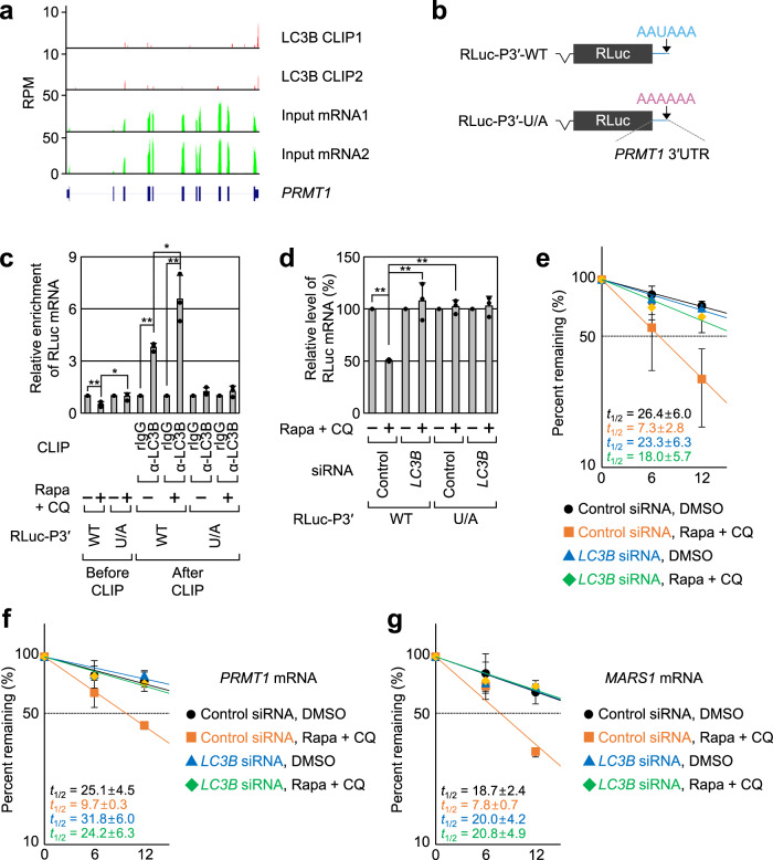
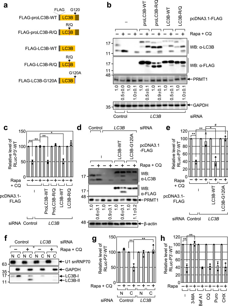
LC3/ATG8 has long been appreciated to play a central role in autophagy, by which a variety of cytoplasmic materials are delivered to lysosomes and eventually degraded. However, information on the molecular functions of LC3 in RNA biology is very limited. Here, we show that LC3B is an RNA-binding protein that directly binds to mRNAs with a preference for a consensus AAUAAA motif corresponding to a polyadenylation sequence. Autophagic activation promotes an association between LC3B and target mRNAs and triggers rapid degradation of target mRNAs in a CCR4-NOT-dependent manner before autolysosome formation. Furthermore, our transcriptome-wide analysis reveals that PRMT1 mRNA, which encodes a negative regulator of autophagy, is one of the major substrates. Rapid degradation of PRMT1 mRNA by LC3B facilitates autophagy. Collectively, we demonstrate that LC3B acts as an RNA-binding protein and an mRNA decay factor necessary for efficient autophagy.


Conflict of interest statement

The authors declare no competing interests.

Figures

Fig. 1
Fig. 1. LC3B is an RNA-binding protein with a preference for AAUAAA consensus motif.
ad CLIP-seq of endogenous LC3B on HEK293T cells treated with either DMSO or Rapa + CQ. Data obtained from two biological replicates of LC3B CLIP-seq were analyzed. The mRNAs with FPKM ≥ 1 in the DMSO-treated cells were used for the calculations. a The number of LC3B CLIP peaks in mRNAs. b Metagene profiles of LC3B peaks. Each region of the 5′UTR, CDS, or 3′UTR was binned into 50 segments. c Consensus motif for LC3B binding. The consensus RNA sequences from LC3B peaks located in the 3′UTR in the cells treated with either DMSO (left) or Rapa + CQ (right) were predicted by MEME (upper) or HOMER (lower). The E value (upper) estimates the expected number of motifs with a similarly sized set of random sequences using log likelihood ratio. The p values (lower) were calculated using cumulative binomial distributions. d Profiles of the distance between LC3B peaks located at the consensus AAUAAA motif and polyadenylation signal (PAS). p values were calculated using the two-tailed Kolmogorov–Smirnov test. e EMSA analysis using in vitro-synthesized Cy5-labeled triple repeats (×3) of either AAUAAA or AAAAAA and either purified recombinant LC3B-WT or BSA. The relative positions of the free probes and RNA-protein complexes are indicated using arrows. Representative data obtained from two independently performed biological replicates (n = 2) are shown. f Fluorescence polarization assay showing preferential interaction between purified recombinant LC3B and AAUAAA motif. Dissociation constants (Kd) are indicated at the right side of graph; n = 6; Data are presented as mean values ± SD; nd not determined. Source data are provided as a Source Data file.
Fig. 2
Fig. 2. Binding of LC3B to target mRNAs triggers rapid mRNA degradation.
ac Profiles of mRNA abundance. HEK293T cells either undepleted or depleted of endogenous LC3B were treated with either DMSO or Rapa. Total cell RNAs were purified and subjected to mRNA sequencing. The mRNAs with FPKM ≥ 1 in the cells treated with Control siRNA and DMSO were used for the calculations. a Cumulative distribution function (CDF) plots for relative changes in mRNA abundance after Rapa treatment. The mRNAs were categorized into two groups: total mRNAs either harboring (LC3B CLIP group) or lacking (non-targets group) the LC3B CLIP peak. CDF plots for the relative changes in the abundance of mRNAs upon Rapa treatment in the undepleted cells (b) or upon LC3B downregulation in Rapa-treated cells (c). The mRNAs belonging to the LC3B CLIP group were categorized into three groups: 5′UTR, CDS, and 3′UTR depending on the position of the LC3B peak. dg Profiles of mRNA half-life. HEK293T cells either undepleted or depleted of LC3B were treated with either DMSO or Rapa + CQ. The cells were harvested at three time points (0, 6, and 12 h) and total cell RNAs were subjected to mRNA sequencing experiments as described in the “Methods” section. CDF plots for the relative change in the half-life of mRNAs harboring (LC3B CLIP group) or lacking (non-targets group) LC3B peak upon Rapa + CQ treatment in the undepleted cells (d) or upon LC3B downregulation in the cells treated with Rapa + CQ (e). CDF plots for the relative change in the half-life of mRNAs belonging to the 5′UTR, CDS, or 3′UTR group upon Rapa + CQ treatment in the undepleted cells (f) or upon LC3B downregulation in the cells treated with Rapa + CQ (g). p values were calculated using the two-tailed Mann–Whitney U test. All NGS data were obtained from two independently performed biological replicates (n = 2). Source data are provided as a Source Data file.
Fig. 3
Fig. 3. PRMT1 mRNA is a de novo target of LC3B-mediated mRNA decay (LMD).
a Read densities of the LC3B CLIP-seq (LC3B CLIP1 and LC3B CLIP2) and mRNA sequencing (input mRNA1 and input mRNA2) for PRMT1 mRNA in two biological replicates. The map for PRMT1 is shown at the bottom. b Schematic representation of RLuc reporter mRNAs harboring the full-length PRMT1 3′UTR of either WT or U/A substitution. The relative positions of the LC3-binding site (AAUAAA) and its variant (AAAAAA) are indicated by arrows. c In vivo CLIP of endogenous LC3B. HEK293T cells expressing the RLuc reporter mRNA and FLuc mRNA (which served as a negative control) were either treated or not treated with Rapa + CQ. The cells were subjected to in vivo CLIP using α-LC3B antibody or nonspecific rabbit IgG (rIgG). The amounts of coimmunoprecipitated reporter mRNAs were normalized to those of the FLuc mRNAs. Then, the normalized levels obtained in IPs using rIgG in the untreated cells were arbitrarily set to 1.0. n = 3; Data are presented as mean values ± SD; p values were analyzed using two-tailed and equal-variance Student’s t test; *p < 0.05; **p < 0.01 (The exact p values are provided in Source Data file). d Effect of LC3B downregulation on the abundance of the RLuc-P3′ reporter mRNAs. HEK293T cells either undepleted or depleted of LC3B were transiently transfected with plasmids expressing RLuc and FLuc reporter mRNAs. The cells were either treated or not treated with Rapa + CQ for 12 h before cell harvest. The amounts of the RLuc mRNAs were normalized to those of FLuc mRNAs. Then, the normalized levels in the untreated cells were arbitrarily set to 100%; n = 3; Data are presented as mean values ± SD; p values were analyzed using two-tailed and equal-variance Student’s t test; **p < 0.01 (The exact p values are provided in Source Data file). e Half-life measurement of the RLuc reporter mRNA, RLuc-P3′-WT. As performed in d except that the cells were harvested at the indicated time points. The y axis represents the level of mRNA remaining (percentage) on the logarithmic scale (log2); n = 4; Data are presented as mean values ± SD. f, g Half-life of endogenous LMD substrates after treatment with Rapa + CQ or LC3B downregulation. Two endogenous LMD substrates, PRMT1 mRNA (f) and MARS1 mRNA (g) were analyzed. n = 3; Source data are provided as a Source Data file.
Fig. 4
Fig. 4. Presence of secondary or tertiary structures upstream of the AAUAAA motif promotes an association between LC3B and LMD substrates.
a Common features found in LMD substrates. Two common features, (i) a long-range looping and (ii) the secondary or tertiary structures upstream of the AAUAAA motif, are indicated by semi-transparent rectangles. b MFE values (left) and GC content (right) across the nucleotide positions upstream of the AAUAAA motif. The 3′UTR sequences of mRNAs belonging to the 3′UTR_AAUAAA or non-targets group were analyzed. p values were calculated using the two-tailed Kolmogorov–Smirnov test. c Predicted secondary structure of the 3′UTR of PRMT1-WT, M3, and M4. d In vivo CLIPs of endogenous LC3B. As performed in Fig. 3c, except that HEK293T cells were expressed with one of RLuc-P3′ reporter mRNAs (WT, M3, or M4 mRNA). n = 3; Data are presented as mean values ± SD; p values were analyzed using two-tailed and equal-variance Student’s t test; # not significant; *p < 0.05; **p < 0.01 (The exact p values are provided in Source Data file). e Relative amounts of the RLuc-P3′ reporter mRNAs upon treatment with Rapa + CQ. n = 3; Data are presented as mean values ± SD; p values were analyzed using two-tailed and equal-variance Student’s t test; *p < 0.05; **p < 0.01 (The exact p values are provided in Source Data file). Source data are provided as a Source Data file.
Fig. 5
Fig. 5. Efficient LMD involves an RNA-binding ability and PE conjugation of LC3B.
a Schematic diagram of FLAG-LC3B variants used in this study. be Complementation experiments using LC3B variants. HEK293T cells were transiently transfected with either the control siRNA or LC3B siRNA that anneals to 3′UTR of endogenous LC3B mRNA. Two days later, the cells were retransfected with an RLuc-P3′-WT reporter plasmid, a FLuc reference plasmid, and a plasmid expressing either FLAG-LC3B-WT or its variant. The cells were either treated or not treated with Rapa + CQ for 12 h before cell harvest. b, d Western blotting showing specific downregulation of endogenous LC3B and proper expression of FLAG-LC3B or its variant at a level comparable to that of endogenous LC3B. c, e Effect of LC3B-WT or its variant on LMD. The amounts of RLuc mRNAs were normalized to those of FLuc mRNAs. Then, the normalized levels in the untreated cells were arbitrarily set to 100%. n = 3; # not significant; *p < 0.05; **p < 0.01. f, g Measurement of LMD efficiency in the nucleus and cytoplasm. f Western blotting showing specific separation between the nuclear (N) and cytoplasmic (C) fractions. g Efficiency of LMD of RLuc-P3′-WT reporter mRNAs. n = 3; **p < 0.01. h Effect of treatment with the indicated chemical inhibitor on LMD of RLuc-P3′-WT reporter mRNAs. n = 3; **p < 0.01. In c, e, g, h, data are presented as mean values ± SD from three biological replicates. p values were analyzed using two-tailed and equal-variance Student’s t test; The exact p values are provided in Source Data file. Source data are provided as a Source Data file.
Fig. 6
Fig. 6. CCR4-NOT complex associates with LC3B, promoting efficient LMD.
a IPs of endogenous LC3B. The extracts of HEK293T cells either treated or not treated with Rapa + CQ were subjected to IPs using α-LC3B antibody or nonspecific rIgG. The intensities of each western blot image were quantitated. The intensities of coimmunoprecipitated proteins were normalized to those of immunoprecipitated LC3B. The relative levels obtained in untreated cells were arbitrarily set to 1.0. n = 3. b, c IPs of endogenous LC3B upon downregulation of both ATG5 and ATG7. As performed in a, except that the cells were either undepleted or depleted of ATG5 and ATG7. b Western blotting proving specific downregulation of endogenous ATG5 and ATG7. c Western blotting of cellular proteins before or after IPs of LC3B. d The proximity ligation assay (PLA) between endogenous LC3B and either CNOT1 or CNOT7. PLA experiments using the indicated antibodies were performed on HeLa cells treated with either DMSO or Rapa + CQ. The PLA images are shown in Supplementary Fig. 10. The number of PLA spots per cell was quantified and is presented in this panel. Box-whiskers show maximum, third quartile to first quartile, median and minimum; n = 767 cells examined over three independent experiments. e, f Effect of CNOT1 or CNOT7 downregulation on LMD. e Western blotting showing specific downregulation of endogenous CNOT1 and CNOT7. f LMD efficiency of RLuc-P3′-WT reporter mRNA, endogenous PRMT1 mRNA, and MARS1 mRNA. n = 3; Data are presented as mean values ± SD; p values were analyzed using two-tailed and equal-variance Student’s t test; *p < 0.05; **p < 0.01 (The exact p values are provided in Source Data file). Source data are provided as a Source Data file.
Fig. 7
Fig. 7. Association between LC3B and the CCR4-NOT complex is initiated outside of autophagic puncta.
a Intracellular distributions of PLA spots involving endogenous LC3B and CNOT1. Intracellular distributions of PLA spots showing the specific interaction between endogenous LC3B and CNOT1 were determined using PLA experiments. In addition, the intracellular distributions of FLAG-LC3B and endogenous p62 (both of which were used for markers for autophagosome), FLAG-DCP1A (a marker for processing bodies), or endogenous G3BP1 (a marker for stress granules) were determined by immunostaining with α-FLAG antibody, α-p62 antibody, α-G3BP1 antibody, and α-G3BP1 antibody, respectively. Nuclei were stained with 4′,6-diamidino-2-phenylindole (DAPI; blue). An enlarged view of the white boxed area is provided in the lower right corner of each image. n = 3; Scale bar = 10 μm. b IPs of ATG5, ATG12, and ATG16L1. HEK293T cells were transiently transfected with a plasmid expressing FLAG, FLAG-ATG5, FLAG-ATG12, or FLAG16L1. Two days later, the cells were treated with Rapa + CQ for 12 h before cell harvest. Total cell extracts were subjected to IPs with the α-FLAG antibody; n = 2; Source data are provided as a Source Data file.
Fig. 8
Fig. 8. Rapid degradation of the PRMT1 mRNA via LMD promotes autophagy.
a Schematic diagram of siRNA-resistant (R) FLAG-PRMT1R reporters, WT, M3, or M4 mRNA. These reporter mRNAs are the same as RLuc-P3′-WT, M3, and M4 mRNA described in Fig. 4c, except that the FLAG-PRMT1R reporter mRNAs encode the full-length PRMT1 protein instead of RLuc. bd Complementation experiment using the FLAG-PRMT1R reporter mRNAs. HeLa cells stably expressing GFP-LC3B were transiently transfected with either PRMT1 siRNA or nonspecific Control siRNA. Two days later, the cells were retransfected with a plasmid expressing either FLAG or one of the FLAG-PRMT1R reporter mRNAs. The cells were treated with Rapa + CQ for 12 h before immunostaining with the α-GFP antibody. n = 3. b Relative level of PRMT1 mRNAs. n = 3. Data are presented as mean values ± SD; # not significant; **p < 0.01 (The exact p values are provided in Source Data file). c Immunostaining of GFP-LC3B. Scale bar = 10 μm. d Quantitation of GFP-LC3B puncta per cell. Box-whiskers show maximum, third quartile to first quartile, median and minimum. A one-way ANOVA test was conducted to calculate the p values; n = 865 cells examined over three independent experiments; # not significant; **p < 0.01. e Proposed model illustrating the role of LMD in autophagy. Source data are provided as a Source Data file.

References

    1. Pohl C, Dikic I. Cellular quality control by the ubiquitin-proteasome system and autophagy. Science. 2019;366:818–822. - PubMed
    1. Morishita H, Mizushima N. Diverse cellular roles of autophagy. Annu. Rev. Cell Dev. Biol. 2019;35:453–475. - PubMed
    1. Dikic I. Proteasomal and autophagic degradation systems. Annu. Rev. Biochem. 2017;86:193–224. - PubMed
    1. Levine B, Kroemer G. Biological functions of autophagy genes: a disease perspective. Cell. 2019;176:11–42. - PMC - PubMed
    1. Li X, He S, Ma B. Autophagy and autophagy-related proteins in cancer. Mol. Cancer. 2020;19:12. - PMC - PubMed

Publication types

MeSH terms

Substances