Do Radiographic Results of Transforaminal Lumbar Interbody Fusion Vary with Cage Position in Patients with Degenerative Lumbar Diseases?
- PMID: 35302296
- PMCID: PMC9002072
- DOI: 10.1111/os.13224
Do Radiographic Results of Transforaminal Lumbar Interbody Fusion Vary with Cage Position in Patients with Degenerative Lumbar Diseases?
Abstract
Objective: To investigate whether the radiographic results are affected by cage position in single-level transforaminal lumbar interbody fusion (TLIF).
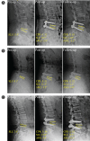
Method: Between January 2016 and June 2018, 130 patients (62 males and 68 females, average age: 55.28 ± 10.11 years) who underwent single-level TLIF were analyzed retrospectively. Standing lateral radiographs of the lumbar spine were collected and evaluated preoperatively, postoperatively, and at the time of last follow-up. Cage position in the fused segment was recorded using a central point ratio (CPR), which indicated the cage position. CPR is calculated by dividing the distance between the cage center point and the posterior extent of the superior endplate of the inferior vertebra by the length of the superior endplate of the inferior vertebra. Based on cage positions, the patients were divided into three groups: Anterior Group (n = 38); Middle Group (n = 68); and Posterior Group (n = 24). Segmental lumbar lordosis (SLL), foraminal height (FH), posterior disc height (PDH), and anterior disc height (ADH) were evaluated. A subanalysis was also performed on cage height within each group.
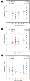
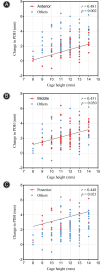
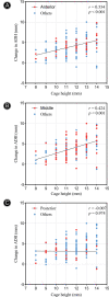
Results: The average follow-up time of the patients was 35.20 ± 4.43 months. The mean values of CPR in Anterior Group, Middle Group, and Posterior Group were 0.64, 0.51, and 0.37, respectively. The FH, PDH, and ADH were significantly increased after TLIF in all groups (P < 0.05). There were significant differences in increase of SLL in Anterior Group (4.4°) and Middle Group (3.0°), but not in Posterior Group (0.3°). Furthermore, in the comparison of the three groups, the increase of SLL, FH, and PDH was statistically different (P < 0.05), while not for ADH (P > 0.05). The significant correlations in surgery were: CPR and ΔSLL (r = 0.584, P < 0.001), CPR and ΔFH (r = -0.411, P < 0.001), and CPR and ΔPDH (r = -0.457, P < 0.001). However, ADH had a positive correlation with cage height when the cage was located in anterior and middle of the endplate. Moreover, cage height had a positive correlation with SLL when the cage was located anteriorly and had a negative correlation with SLL when the cage was located posteriorly. FH and PDH both had a positive correlation with cage height in any cage position.
Conclusion: The cage located in different positions has different effects on radiographic results in single-level TLIF. A thicker cage located anteriorly will gain maximum SLL and avoid the reduction of FH and PDH.
Keywords: Cage position; Disc height; Foraminal height; Segmental lumbar lordosis; Transforaminal lumbar interbody fusion.
© 2022 The Authors. Orthopaedic Surgery published by Tianjin Hospital and John Wiley & Sons Australia, Ltd.
Figures


References
-
- Rosenberg WS, Mummaneni PV. Transforaminal lumbar interbody fusion: technique, complications, and early results. Neurosurgery. 2001;48:569–75. - PubMed
-
- Talia AJ, Wong ML, Lau HC, Kaye AH. Comparison of the different surgical approaches for lumbar interbody fusion. J Clin Neurosci. 2015;22:243–51. - PubMed
-
- Humphreys SC, Hodges SD, Patwardhan AG, Eck JC, Murphy RB, Covington LA. Comparison of posterior and transforaminal approaches to lumbar interbody fusion. Spine (Phila PA 1976). 2001;26:567–71. - PubMed
-
- Salehi SA, Tawk R, Ganju A, LaMarca F, Liu JC, Ondra SL. Transforaminal lumbar interbody fusion: surgical technique and results in 24 patients. Neurosurgery. 2004;54:368–74. - PubMed
-
- Liang Y, Shi W, Jiang C, et al. Clinical outcomes and sagittal alignment of single‐level unilateral instrumented transforaminal lumbar interbody fusion with a 4 to 5‐year follow‐up. Eur Spine J. 2015;24:2560–6. - PubMed
MeSH terms
LinkOut - more resources
Full Text Sources
Miscellaneous

