. 2022 May;47(5):1862-1872.
doi: 10.1007/s00261-022-03490-9. Epub 2022 Mar 18.

Conspicuity and muscle-invasiveness assessment for bladder cancer using VI-RADS: a multi-reader, contrast-free MRI study to determine optimal b-values for diffusion-weighted imaging

Affiliations

Conspicuity and muscle-invasiveness assessment for bladder cancer using VI-RADS: a multi-reader, contrast-free MRI study to determine optimal b-values for diffusion-weighted imaging

Andrea Delli Pizzi et al. Abdom Radiol (NY). 2022 May.

Abstract

Objective: To (1) compare bladder cancer (BC) muscle invasiveness among three b-values using a contrast-free approach based on Vesical Imaging-Reporting and Data System (VI-RADS), to (2) determine if muscle-invasiveness assessment is affected by the reader experience, and to (3) compare BC conspicuity among three b-values, qualitatively and quantitatively.

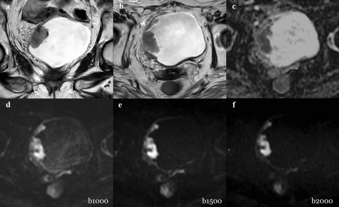
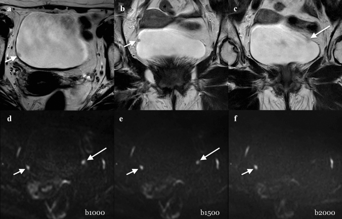
Methods: Thirty-eight patients who underwent a bladder MRI on a 3.0-T scanner were enrolled. The gold standard was histopathology report following transurethral resection of BC. Three sets of images, including T2w and different b-values for DWI, set 1 (b = 1000 s/mm2), set 2 (b = 1500 s/mm2), and set 3 (b = 2000 s/mm2), were reviewed by three differently experienced readers. Descriptive statistics and Intraclass Correlation Coefficient (ICC) were calculated. Comparisons among readers and DWI sets were performed with the Wilcoxon test. Receiver operating characteristic (ROC) analysis was performed. Areas under the curves (AUCs) and pairwise comparison were calculated.

Results: AUCs of muscle-invasiveness assessment ranged from 0.896 to 0.984 (reader 1), 0.952-0.968 (reader 2), and 0.952-0.984 (reader 3) without significant differences among different sets and readers (p > 0.05). The mean conspicuity qualitative scores were higher in Set 1 (2.21-2.33), followed by Set 2 (2-2.16) and Set 3 (1.82-2.14). The quantitative conspicuity assessment showed that mean normalized intensity of tumor was significantly higher in Set 2 (4.217-4.737) than in Set 1 (3.923-4.492) and Set 3 (3.833-3.992) (p < 0.05).

Conclusion: Muscle invasiveness can be assessed with high accuracy using a contrast-free protocol with T2W and DWI, regardless of reader's experience. b = 1500 s/mm2 showed the best tumor delineation, while b = 1000 s/mm2 allowed for better tumor-wall interface assessment.

Keywords: Bladder cancer; Diffusion-weighted imaging; MRI; VI-RADS.


Conflict of interest statement

Domenico Mastrodicasa had no activities related to the present article. Activities not related to the present article: shareholder of Segmed, Inc. and consultant for Segmed, Inc. All the other authors of this manuscript declare no relationships with any companies whose products or services may be related to the subject matter of the article. Andrea Delli Pizzi is a member of the Scientific Editorial Board of Insight Into Imaging.

Figures

Fig. 1
Fig. 1
Three-point scale used to assess tumor conspicuity on DWI: a hyperintense lesion, but only slightly demarcated from the background and/or poor tumor–wall interface, b hyperintense lesion, well demarcated from the background with good tumor–wall interface, and c hyperintense lesion with excellent background suppression and optimal tumor–wall interface. BC = bladder cancer; arrow = bladder wall; BL = bladder lumen
Fig. 2
Fig. 2
Quantitative analysis. Three circular 5-mm2 regions of interest (ROIs) of the tumor were placed on DWI images. Another circular 5-mm2 ROI was also placed in the bladder content to normalize data. Tumor conspicuity was defined as the ratio between the mean tumor intensity and the bladder content intensity
Fig. 3
Fig. 3
Example of muscle-invasive BC classified correctly. A 79-year-old woman with hematuria and a bladder mass reported after flexible cystoscopy underwent bladder MRI before primary TURB. Coronal (a) and axial (b) T2W imaging showed a thickened right-lateral wall. All gradient strengths (b1000, b1500, and b2000) and ADC map (c) confirmed the restricted diffusion of the tumor extending to the muscular layer. The tumor conspicuity on b1000 (d) was higher than b1500 (e) and b2000 (f) due to better tumor–wall interface visualization. The T stage after TURB was HG-T2 (TURB). DWI = diffusion-weighted imaging; HG = high grade; MRI = magnetic resonance imaging; T2W = T2 weighted; TURB = transurethral resection of the bladder; VI-RADS = Vesical Imaging-Reporting and Data System
Fig. 4
Fig. 4
Example of a not muscle-invasive BC classified incorrectly. A 73-year-old man with hematuria and two polyps, documented after flexible cystoscopy, underwent MRI before primary TURB. Axial (a) and coronal (b, c) T2W imaging (T2) showed a small (4 mm) polypoid lesion on the right wall of the bladder (short arrow in a and b). The lesion was well detected by the three readers regardless the image set and was scored as VI-RADS 1 (short arrow in d, e, and f). Another slightly visible small (4 mm) non-muscular invasive lesion (VI-RADS 1) on the left wall of the bladder was suspected on T2 images (long arrow in c). However, it was definitely detected by the three readers only when reading b1000 and b1500 images (long arrow in d and e), but not on b2000 images. T stage after TURB was LG-T1 (TURB). After four weeks, Re-TURB was performed and it confirmed the absence of residual tumor. DWI = diffusion-weighted imaging; HG = high grade; MRI = magnetic resonance imaging; T2W = T2 weighted; TURB = transurethral resection of the bladder; VI-RADS = Vesical Imaging-Reporting and Data System

References

    1. Bray F, Ferlay J, Soerjomataram I, Siegel RL, Torre LA, Jemal A. Global cancer statistics 2018: GLOBOCAN estimates of incidence and mortality worldwide for 36 cancers in 185 countries. CA Cancer J Clin. 2018;68:394–424. doi: 10.3322/caac.21492. - DOI - PubMed
    1. Richters A, Aben KKH, Kiemeney L. The global burden of urinary bladder cancer: an update. World J Urol. 2019. - PMC - PubMed
    1. Palumbo C, Mistretta FA, Knipper S, Pecoraro A, Tian Z, Shariat SF, et al. How cancer-specific mortality changes over time after radical cystectomy: Conditional survival of patients with nonmetastatic urothelial carcinoma of the urinary bladder. Urol Oncol. 2019;37:893–899. doi: 10.1016/j.urolonc.2019.05.020. - DOI - PubMed
    1. Zhang N, Wang X, Wang C, Chen S, Wu J, Zhang G, et al. Diagnostic Accuracy of Multi-Parametric Magnetic Resonance Imaging for Tumor Staging of Bladder Cancer: Meta-Analysis. Front Oncol. 2019;9:981. doi: 10.3389/fonc.2019.00981. - DOI - PMC - PubMed
    1. Panebianco V, Narumi Y, Altun E, Bochner BH, Efstathiou JA, Hafeez S, et al. Multiparametric Magnetic Resonance Imaging for Bladder Cancer: Development of VI-RADS (Vesical Imaging-Reporting And Data System) Eur Urol. 2018;74:294–306. doi: 10.1016/j.eururo.2018.04.029. - DOI - PMC - PubMed

MeSH terms