. 2022 May 9;32(9):1895-1908.e5.
doi: 10.1016/j.cub.2022.02.066. Epub 2022 Mar 17.

Astrocytic GABA transporter controls sleep by modulating GABAergic signaling in Drosophila circadian neurons

Affiliations

Astrocytic GABA transporter controls sleep by modulating GABAergic signaling in Drosophila circadian neurons

Ratna Chaturvedi et al. Curr Biol. .

Abstract

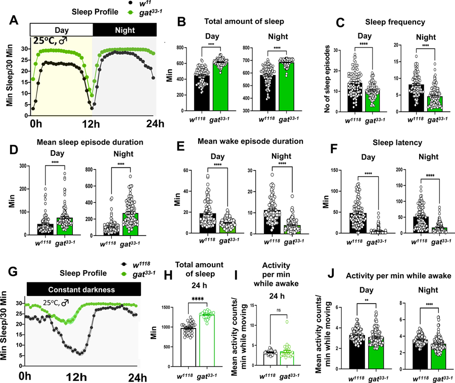
A precise balance between sleep and wakefulness is essential to sustain a good quality of life and optimal brain function. GABA is known to play a key and conserved role in sleep control, and GABAergic tone should, therefore, be tightly controlled in sleep circuits. Here, we examined the role of the astrocytic GABA transporter (GAT) in sleep regulation using Drosophila melanogaster. We found that a hypomorphic gat mutation (gat33-1) increased sleep amount, decreased sleep latency, and increased sleep consolidation at night. Interestingly, sleep defects were suppressed when gat33-1 was combined with a mutation disrupting wide-awake (wake), a gene that regulates the cell-surface levels of the GABAA receptor resistance to dieldrin (RDL) in the wake-promoting large ventral lateral neurons (l-LNvs). Moreover, RNAi knockdown of rdl and its modulators dnlg4 and wake in these circadian neurons also suppressed gat33-1 sleep phenotypes. Brain immunohistochemistry showed that GAT-expressing astrocytes were located near RDL-positive l-LNv cell bodies and dendritic processes. We concluded that astrocytic GAT decreases GABAergic tone and RDL activation in arousal-promoting LNvs, thus determining proper sleep amount and quality in Drosophila.

Keywords: GABA; GAT; astrocytes; large ventral lateral neurons; sleep.


Conflict of interest statement

Declaration of interests The authors declare no competing interests.

Figures

Figure 1.
Figure 1.. gat33-1 is a hypomorphic mutant of GABA transporter (GAT):
(A) Schematic of the gat gene locus. TALEN targeting site in the first exon is indicated. (B) Nucleotide sequence of the targeted region of gat. The grey boxes indicate the target sites for TALEN-mediated mutagenesis in the 1st exon, the start codon is indicated in green. gat33-1 mutants harbor a 7 nucleotides deletion that removes a Bcc1 endonuclease restriction site and leads to a frameshift and early termination of the GAT protein. (C) Protein sequence of the GAT N-terminus. Amino acids encoded by exon 1 and 2 are indicated in orange and blue respectively. The beginning of the first transmembrane domain (TM) is indicated by a light blue box. The frameshift in gat33-1 should lead to an early termination after 20 amino acids (red), however downstream methionines (Green boxes) could act as alternative start sites. (D) Schematic overview of the putative GAT membrane topology. The sites of the two putative alternative start sites in gat33-1 mutants are indicated (Blue arrows) and they would lead to 22aa or 64aa truncations of the cytoplasmic N-terminus. (E) Western blot analysis of GAT protein of wild type and gat331 mutant fly heads using C- and N-terminal anti-GAT antibodies. The C-terminal anti-GAT antibody recognizes a strongly reduced and truncated GAT specific band (white arrow) in gat33-1 mutant extracts while the N-terminal anti-GAT antibody only recognized a GAT specific band in wild type control flies (black arrow). See also Figure S1.
Figure 2.
Figure 2.. GABA transporter (GAT) is required for normal sleep-wake behavior
(A) Sleep profiles of gat33-1 (green, n=110) and control w1118 (black, n=93) male flies in 12/12 h LD cycle showing enhanced amount of sleep in the gat hypomorphic mutant during both day and night. (B-E) Total sleep amounts (min) (B), number of sleep episodes (C), mean sleep episode’s durations (min) (D), and mean wake episode’s durations (min) (E) during daytime and during night showing increase in sleep consolidation in gat33-1. (F) Latency to sleep (min) during the day and nighttime indicates accelerated sleep initiation in gat33-1. (G) Sleep profile of gat33-1 (green, n=32) and control w1118 (black, n=32) male flies in constant darkness (DD). Results show that enhancement of gat33-1 sleep is not light dependent. (H) Total amount of sleep during DD. (I) Mean activity per min while moving during DD. (J) Mean activity per min while moving during LD cycle. Error bar represents SEM. For normally distributed data, a two-tailed Student’s t-test was used. In case of non-gaussian distribution, a Mann–Whitney U-test was performed, and statistical significance is shown as ***P < 0.001; **P < 0.01; and ‘ns’ as not significant. See also figure S2–3 and Video S1–2.
Figure 3.
Figure 3.. gat33-1 sleep defects are rescued by expressing gat in astrocytes
(A) Sleep profile of male flies with genotype gat33-1 (green, n=90), control w1118 (black, n=45), UAS-gat;; gat33-1 (olive green, n=64), alrm-Gal4;; gat33-1 (sea-green, n=44), alrm-Gal4/UAS-genomic-gat;; gat33-1 (red, n=96), and 1b12;; gat33-1 (crimson red, n=37) in 12/12 h LD cycle. The results show that GAT functions in astrocytes. (B-D) Total sleep amounts (min) (B), mean sleep episode’s durations (min) (C), and number of sleep episodes (beam breaks) (D) during day and night. (E) Latency to sleep (min) during day and nighttime. (F) Mean activity per min while moving during day and night. Error bar represents SEM. For normally distributed data, one-way ANOVA followed by a Tukey post-hoc test was performed. For non-gaussian distribution, a Kruskal-Wallis test with Dunn’s post-hoc test was performed. Different alphabetical letters (a, b, c, and d) illustrate statistically significant differences between groups. The same statistical tests were used in Figures 5–7, as well as Supplemental Figures S4–S7. See also Figure S4.
Figure 4.
Figure 4.. RDL and PDF expressing wake promoting clock neurons (l-LNvs) are in close proximity with GAT expressing astrocytes.
(A) Confocal analysis of w1118 wild type fly brains for localization of RDL (blue) in PDF (red) positive l-LNvs. Astrocytes are expressing membrane bound GFP (green) (alarm-Gal4/UAS-CD8-GFP). (a-a3) Labeling in large ventral lateral neurons (l-LNvs). (b-b3) Labeling in medulla region of optic lobe. l-LNvs neurons also extend its projections in the medulla. (c) 3D view of image-stacks demonstrate projections of astrocytes (green) are extending towards /close to the cell body of l-LNvs and co-localized with RDL, and PDF. (B) Magnified view of l-LNvs labeled with RDL (blue) (a), astrocytes (green) (b), PDF (red) in l-LNvs (c). Merged image (d) was used for orthogonal projection (d’ and d”) where yellow arrows point to astrocytes situated very closely to RDL, and PDF expressing in l-LNvs. (C) Magnified view of the medulla region in the optic lobe labeled for RDL (blue) (a), astrocytes (green) (b), and PDF (red) (c). Merged image (d) was used for orthogonal projections (d’ and d”) where yellow arrows point to astrocytes processes approaching RDL and PDF positive l-LNvs projections innervating into medulla. (D) Confocal analysis of w1118 brains for localization of GAT (blue), PDF positive circadian neurons (red), and astrocytes expressing membrane bound GFP (green) (alrm-Gal4/UAS-CD8-GFP). (a-a3) Labeling in large ventral lateral neurons (l-LNvs). (b-b3) Labeling in the medulla region of the optic lobe. (E) Magnified view of l-LNvs region labeled with GAT (blue) (a), astrocytes (green) (b), PDF (red) in l-LNvs (c). Merged image (d) was used for orthogonal projection (d’ and d”) where yellow arrows point to GAT present in astrocytes and closely placed to PDF expressing in l-LNvs. (F) Magnified view of the medulla region of the optic lobe labeled for GAT (blue) (a), astrocytes (green) (b), and PDF (red) (c). Merged image (d) was used for orthogonal projections (d’ and d”) where yellow arrows point to GAT in astrocytes processes approaching to PDF positive l-LNvs. Scale bars are represented in respective image panels. See also Video S3–7.
Figure 5.
Figure 5.. A wide-awake (wake) mutation suppresses gat33-1 sleep phenotypes
(A) Sleep profile of male flies with genotype gat33-1 (green, n=34), control w1118 (black, n=38), wide-awake mutant waked2 (orange, n=38), and double-mutant with waked2; gat331 (pink, n=31) in 12/12 h LD cycle. The results show that excessive sleeping in gat33-1 mutants is suppressed in the double-mutant flies (B) Quantification of total sleep amounts (min) during day and during night. (C) Quantification of mean sleep-episode’s durations lengths (min). (D) Number of sleep episodes indicates fragmented sleep in waked2 and double-mutant flies. (E) Higher latency to sleep (min) shows difficulty in initiation of sleep in waked2 and double-mutant flies compared to gat33-1 and w1118 controls. (F) Mean activity per min while moving during day and night did not show any significant difference between waked2 flies, double-mutants, and controls. Error bar represents SEM. Statistics as in figure 3. See also Figure S5 and S7.
Figure 6.
Figure 6.. GABAA receptor RDL depletion in PDF neurons suppresses the gat33-1 sleep phenotypes.
(A) Sleep profile of male flies with genotype PD2/+;; gat33-1 (brown, n=21), UAS-rdlRNAi/+;; gat33-1 (navy blue, n=25), PD2/UAS-rdlRNAi;; gat33-1 (red, n=30), and PD2 /UAS-rdlRNAi (lavender, n=33) in 12/12 h LD cycle. PD2 is short for pdf-GAL4 combined with UAS-dcr2 (see material and methods). The results show that excessive sleeping in gat33-1 mutants is suppressed by rdl RNAi knockdown in PDF neurons (B) Quantification of total sleep amount (min) during day and night. (C) Quantification of mean sleep episode’s durations (min). (D) Number of sleep episodes indicate fragmented sleep in rdl RNAi knockdown flies with or without gat33-1. (E) Latency to sleep (min) in rdl RNAi knockdown flies with and without gat33-1 (F) Mean activity per min while moving during day and night. Error bar represents SEM. Statistics as in Figure 3. See also Figure S6
Figure 7.
Figure 7.. DNLG4 depletion in PDF neurons suppresses the gat33-1 sleep phenotypes.
(A) Sleep profile of male flies with genotype PD2/+;; gat33-1 (brown, n=23), UAS-dnlg4RNAi/+;;; gat33-1 (teal, n=24), UAS-dnlg4RNAi; PD2/+;; gat33-1 (purple, n=23), and UAS-dnlg4RNAi; PD2/+ (blue, n=24) in 12/12 h LD cycle. The results show that excessive sleeping in gat33-1 mutants is suppressed during nighttime by dnlg4 RNAi knockdown in PDF neurons (B) Quantification of total sleep amounts (min) during day and night. Results show reduction in sleep during night in dnlg4 knockdown flies with and without gat33-1. (C) Quantification of mean sleep-episode’s durations (min). (D) Number of sleep episodes. (E) Higher latency to sleep (min) shows difficulty in initiation of sleep in dnlg4 RNAi knockdown flies with and without gat33-1. (F) Mean activity per min while moving during day and night. Error bar represents SEM. Statistics as in Figure 3. See also Figure S6.

Similar articles

Cited by

References

    1. Miyazaki S, Liu CY, and Hayashi Y (2017). Sleep in vertebrate and invertebrate animals, and insights into the function and evolution of sleep. Neurosci. Res. 118, 3–12. doi:10.1016/j.neures.2017.04.017. - DOI - PubMed
    1. Keene AC, and Duboue ER (2018). The origins and evolution of sleep. J. Exp. Biol. 221(Pt 11), jeb159533. doi:10.1242/jeb.159533. - DOI - PMC - PubMed
    1. Anafi RC, Kayser MS, and Raizen DM (2019). Exploring phylogeny to find the function of sleep. Nat. Rev. Neurosci. 20, 109–116. doi:10.1038/s41583-018-0098-9. - DOI - PubMed
    1. Sehgal A, and Mignot E (2011). Genetics of sleep and sleep disorders. Cell 146, 194–207. doi:10.1016/j.cell.2011.07.004. - DOI - PMC - PubMed
    1. Jagannath A, Taylor L, Wakaf Z, Vasudevan SR, and Foster RG (2017). The genetics of circadian rhythms, sleep and health. Hum. Mol. Genet. 26, R128–R138. doi:10.1093/hmg/ddx240. - DOI - PMC - PubMed

Publication types

Substances

LinkOut - more resources