. 2022 Mar 18;18(3):e1010338.
doi: 10.1371/journal.ppat.1010338. eCollection 2022 Mar.

Mechanism of Borrelia immune evasion by FhbA-related proteins

Affiliations

Mechanism of Borrelia immune evasion by FhbA-related proteins

Konstantin Kogan et al. PLoS Pathog. .

Abstract

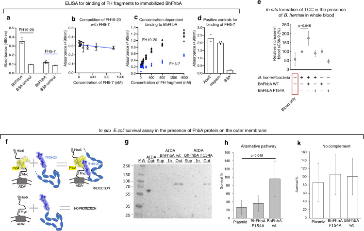
Immune evasion facilitates survival of Borrelia, leading to infections like relapsing fever and Lyme disease. Important mechanism for complement evasion is acquisition of the main host complement inhibitor, factor H (FH). By determining the 2.2 Å crystal structure of Factor H binding protein A (FhbA) from Borrelia hermsii in complex with FH domains 19-20, combined with extensive mutagenesis, we identified the structural mechanism by which B. hermsii utilizes FhbA in immune evasion. Moreover, structure-guided sequence database analysis identified a new family of FhbA-related immune evasion molecules from Lyme disease and relapsing fever Borrelia. Conserved FH-binding mechanism within the FhbA-family was verified by analysis of a novel FH-binding protein from B. duttonii. By sequence analysis, we were able to group FH-binding proteins of Borrelia into four distinct phyletic types and identified novel putative FH-binding proteins. The conserved FH-binding mechanism of the FhbA-related proteins could aid in developing new approaches to inhibit virulence and complement resistance in Borrelia.


Conflict of interest statement

The authors have declared that no competing interests exist.

Figures

Fig 1
Fig 1. 2.2 Å crystal structure of BhFhbA in complex with FH19-20.
(a) A cartoon representation of the BhFhbA:FH19-20 complex. BhFhbA forms a bundle of nine α-helices resembling an arc-like or L-shaped structure. α-helices 1–5 (in green) wrap around the core formed by α-helices 6–9 (in teal). The connecting loop of α-helices 7 and 8 create a hinge-like structure close to C-terminus of BhFhbA. Together the L-shaped cavity (α-helices 3, 8 and 9) and the hinge-loop coordinate the binding of FH20 (in blue). (b) Close-up of the binding site with key sidechains represented as sticks. FH20 Trp1183 and Val1200 (in blue, indicated by *) coordinate binding by a group of hydrophobic residues in BhFhbA. Trp1183 of FH20 is inserted to the hydrophobic pocket formed by BhFhbA Phe154, Met137, I171 and Tyr170. Further, FH20 Val1200 is within van der Waals distance of BhFhbA Tyr170 and Tyr199. 2Fo-Fc density, contoured at 1 σ, is shown around the residues.
Fig 2
Fig 2. Binding of FH20 to BhFhbA is mediated by a hydrophobic binding pocket.
(a) The sequence of BhFhbA from Borrelia hermsii YOR (UniprotID: W5SB08) with mutated residues marked in red. (b) Positions of the mutations mapped on the structure of the BhFhbA:FH19-20 complex. BhFhbA α-helices 1–5 are shown in green, 6–9 in teal (cartoon), and FH19-20 is shown as an electrostatic surface model. (c) Binding of FH19-20 to wild type BhFhbA and (d) Phe154Ala mutant BhFhbA detected by MST. Kd-values ± SEM (μM) calculated from three technical replicates (normalized fluorescence values) and individual data points are shown.
Fig 3
Fig 3. BhFbA affects complement activation and survival.
(a) Binding of FH19-20 and FH5-7 to immobilized BhFhbA detected by ELISA. Individual data points are shown with bars indicating mean values. Values shown in panels a-e are absorbances measured by ELISA. (b) Effect of FH5-7 on binding of FH19-20 to immobilized BhFhbA detected by ELISA. n = 5 or more. Error bars represent S.D. (c) Concentration-dependent binding of FH19-20 and FH5-7 to BhFhbA. Individual data points from several ELISA assays are shown. (d) Controls for FH5-7 fragment confirmed by ELISA. Binding to ApoE [65] protein and heparin were used as positive controls, and BSA as negative control. Individual data points from assays are shown, and bars indicate mean values. (e) Complement activation measured by formation of soluble terminal complement complexes (TCC) in whole blood. Proteins added to reactions are shown below the graph. Data are presented as relative TCC-amount (%) compared to the sample where bacteria alone were incubated in blood (n = 4, error bars represent S.D.) Difference between bacteria alone to bacteria incubated with wild type FhbA (* p<0.05) calculated by paired t-test (Mann-Whitney U-test for independent samples). (f) Schematic representation of the survival assay using the AIDA-1 transport system44, where binding of membrane expressed FhbA to serum FH protects E. coli against complement attack. (g) Western blot showing the presence of His-tagged BhFhbA on the outer surface of E. coli. Outer membrane sample of E. coli expressing an empty AIDA1 system shows a band present at about 63 kDa, consistent with the expected molecular weight (lane 2). BhFhbA wt and Fhe154Ala mutant proteins are present only in outer membrane (out) at the expected molecular weight of 83 kDa (lanes 5 and 8). The anti-His signal was not detected in the supernatant fraction (sup) or in the inner membrane (in). (h) Serum survival of E. coli clones expressing BhFhbA, BhFhbA mutant F154A and empty vector (AIDA) as a control. Result was calculated as a percentage of bacteria that survived after 15 min incubation in serum as compared to the number of bacteria at time point zero (n = 4, error bars represent S.D.) Difference between BhFhbA clone to control significant (* p<0.05) calculated by one-way ANOVA supplemented with Dunnet’s test for unequal variances. (i) Survival of E. coli clones expressing BhFhbA, BhFhbA mutant F154A and empty vector as a control in the presence of inactivated (with 10 mM EDTA) serum without complement. Result was calculated as a percentage of colonies surviving after 15 min incubation in media as compared to the number of bacteria at time point zero (n = 3, error bars represent S.D.).
Fig 4
Fig 4. A new family of immune evasion proteins in borreliae revealed by bioinformatics searches.
(a) Multiple sequence alignment of the FhbA-related proteins. The signal sequence is marked in magenta and secondary structure elements derived from the crystal structure of BhFhbA:FH19-20 are shown in blue above the sequence. The conserved hinge region is marked with a red rectangle, and an arrowhead points at the key Phe residue important for tight binding to FH20. The sequence alignment consists of protein sequences, predicted protein sequences and translated genomic regions that match the BhFhbA used as the search sequence (see Materials and Methods for details). Asterisk ‘*’ represents a stop codon and question mark ‘?’ stands for an incomplete codon, where a frameshift appears to have occurred. In generating the protein alignment, the frameshift was ignored, and the translation frame was preserved to allow further protein alignment after that problematic codon. Sequence parts with grey background are translated genomic sequences, which most probably are not present in derived proteins due to stop codons or frameshifts. Accession numbers and references for the sequences used in the alignment: B. hermsii YOR (W5SB08), B. coriaceae Co53 (W5T1N6), B. duttonii CR2A (W6TXL9), B. parkeri (D5GU46), B. turicatae (B0L8C8), B. miyamotoi (A0A075BUA1), B. recurrentis (C1L349), B. persica No12 Bp4780 (contig: NZ_AYOT01000066.1, nucleotides: 4020 -> 4611), B. hispanica CRI Bhis_2727 (contig: NZ_AYOU01000105.1, nucleotides: 3668 -> 4235), B. crocidurae str. Achema (contig: NC_017778.1, nucleotides: 46553 -> 45974), B. valaisiana VS116 (C0R979), Bo. afzelii (WP_011703930.1), B. bissettiae DN127 (contig: NC_015916.1, nucleotides: 3284 -> 2685). (b) Phylogenetic tree of the FhbA-family proteins constructed from the sequences shown in (a). The numbers represent the substitutions per position and the length of the lines is equivalent to these numbers. The three colours emphasize that the sequences cluster into three distinct groups.
Fig 5
Fig 5. FhbA-related protein from B. duttonii (BdFhbA) utilizes the same binding mechanism to FH20 as BhFhbA.
(a) Sequence alignment of FhbA proteins from B. duttonii and B. hermsii shows a difference in the region between helices 3 and 4 (dashed red rectangle). (b) Structure-based homology model of BdFhbA shows almost identical overall structure to BhFhbA except shortening of helices 3 and 4. (c) Elution profiles of the proteins alone and in combination with each other on the size exclusion column. FH19-20 protein elution profile in magenta, wild type and Phe130A variant BdFhbA in light blue, and a combination of the two proteins in black curves. In each chromatography run, 100 μl of sample containing 20 nmole of each of the tested protein(s) was injected. (d) Binding of FH19-20 to BdFhbA and BdFhbA/Phe130A variant as detected by MST. Kd-values with SEM (μM) calculated from three technical replicates (normalized fluorescence values) are shown for the wild type BdFhbA. For the BdFhbA/Phe130A variant, the MST software could not reliably determine a value for Kd. (e) Cofactor activity of FH in factor I mediated cleavage of C3b. FH from serum or after purification was bound to BdFhbA coated on microwell plate. After washing, C3b and/or Factor I were added. C3b and its cleavage fragments (α’46 and α’43) were detected using anti-C3d antibody. (f) Binding of FH19-20 and FH5-7 to surface coated BdFhbA. Individual data points from ELISA are shown, and the bars represent mean values.
Fig 6
Fig 6. A working model for how BhFhbA recruits FH to mediate immune evasion.
Membrane-bound BhFhbA (bright green) recruits FH (cartoon representation) of the host through binding site on domain 20 of FH (in red). When bound to BhFhbA, FH domains 1–4 are free to bind to C3b (yellow) and inhibit complement. When bound to microbial protein via domain 20, FH can also bind C3d fragment (in pink) via domain 19. Full-length FH model was manually constructed in Pymol by combining existing structures of different subcomplexes. Model for FH1-4 bound to C3b was from PDB2wii [15]. Structures of FH19-20 (in red) bound to C3d were obtained from PDB entries 5nbq [56] and 2xqw [66].

Similar articles

Cited by

References

    1. Barbosa A.S., I L. Complement Immune Evasion by Spirochetes in Spirochete Biology: The Post Genomic Era. A B., editor: Springer, Cham; 2017.
    1. Adeolu M, Gupta RS. A phylogenomic and molecular marker based proposal for the division of the genus Borrelia into two genera: the emended genus Borrelia containing only the members of the relapsing fever Borrelia, and the genus Borreliella gen. nov. containing the members of the Lyme disease Borrelia (Borrelia burgdorferi sensu lato complex). Antonie Van Leeuwenhoek. 2014;105(6):1049–72. doi: 10.1007/s10482-014-0164-x - DOI - PubMed
    1. Margos G, Gofton A, Wibberg D, Dangel A, Marosevic D, Loh SM, et al.. The genus Borrelia reloaded. PLoS One. 2018;13(12):e0208432. doi: 10.1371/journal.pone.0208432 - DOI - PMC - PubMed
    1. Steere AC. Lyme disease. N Engl J Med. 2001;345(2):115–25. doi: 10.1056/NEJM200107123450207 - DOI - PubMed
    1. Rizzoli A, Hauffe H, Carpi G, Vourc HG, Neteler M, Rosa R. Lyme borreliosis in Europe. Euro Surveill. 2011;16(27). - PubMed

Publication types

Substances