. 2022 Mar 18;24(1):21.
doi: 10.1186/s13058-022-01513-3.

Quantitative glycoproteomics analysis identifies novel FUT8 targets and signaling networks critical for breast cancer cell invasiveness

Affiliations

Quantitative glycoproteomics analysis identifies novel FUT8 targets and signaling networks critical for breast cancer cell invasiveness

Cheng-Fen Tu et al. Breast Cancer Res. .

Abstract

Background: We recently showed that fucosyltransferase 8 (FUT8)-mediated core fucosylation of transforming growth factor-β receptor enhances its signaling and promotes breast cancer invasion and metastasis. However, the complete FUT8 target glycoproteins and their downstream signaling networks critical for breast cancer progression remain largely unknown.

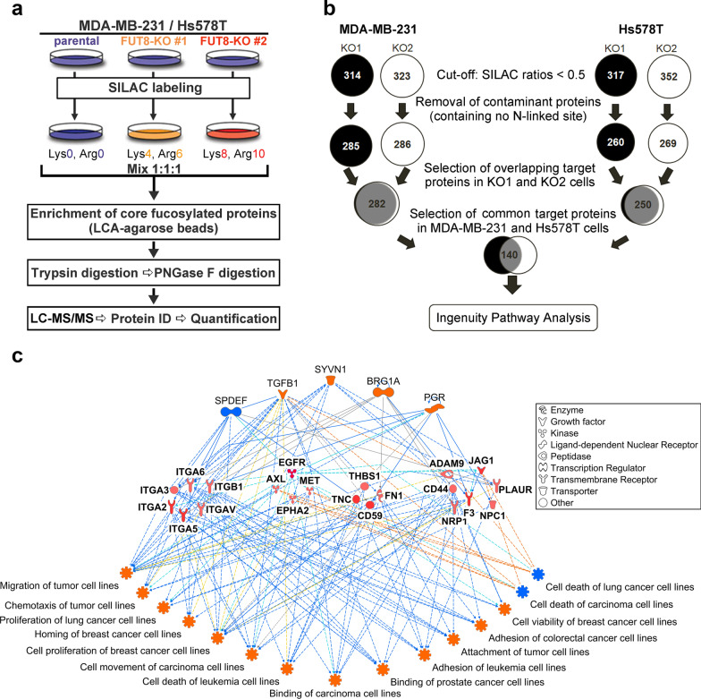
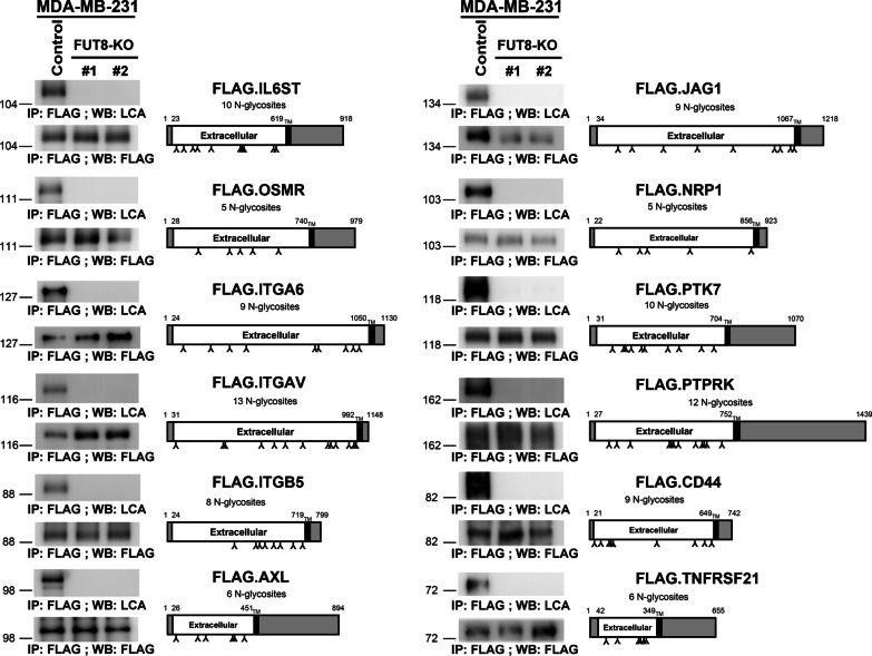
Method: We performed quantitative glycoproteomics with two highly invasive breast cancer cell lines to unravel a comprehensive list of core-fucosylated glycoproteins by comparison to parental wild-type and FUT8-knockout counterpart cells. In addition, ingenuity pathway analysis (IPA) was performed to highlight the most enriched biological functions and signaling pathways mediated by FUT8 targets. Novel FUT8 target glycoproteins with biological interest were functionally studied and validated by using LCA (Lens culinaris agglutinin) blotting and LC-MS/MS (liquid chromatography-tandem mass spectrometry) analysis.

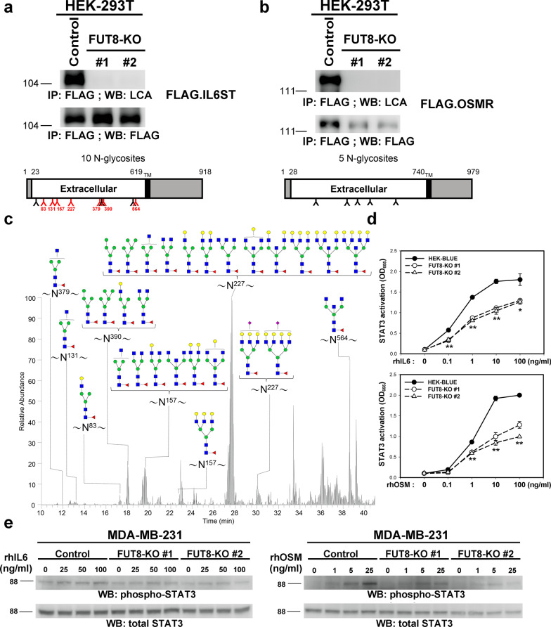
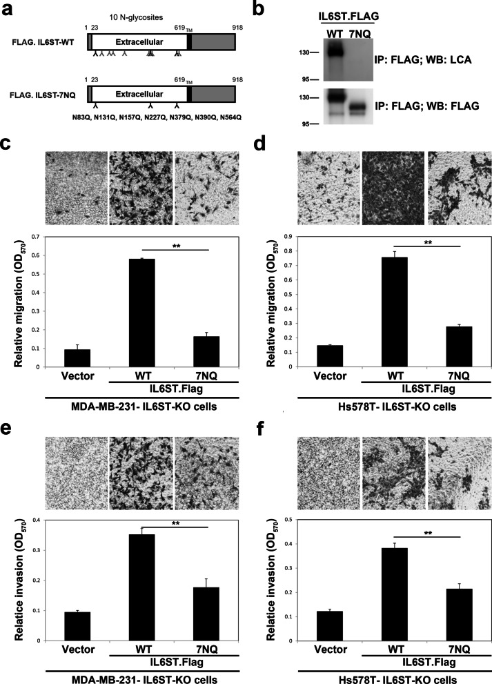
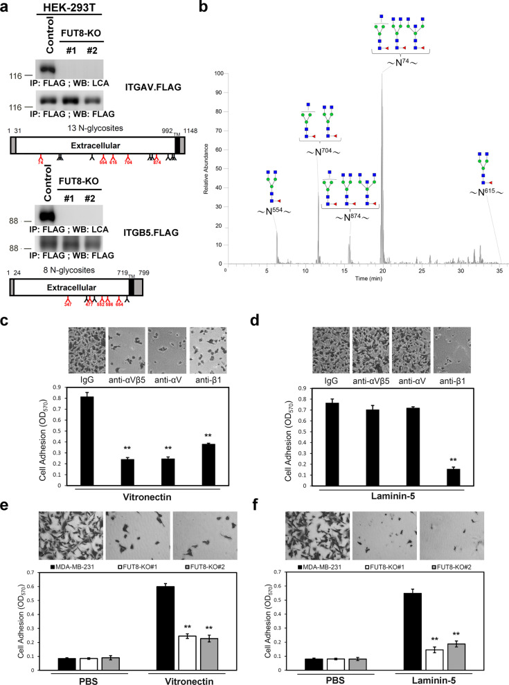
Results: Loss-of-function studies demonstrated that FUT8 knockout suppressed the invasiveness of highly aggressive breast carcinoma cells. Quantitative glycoproteomics identified 140 common target glycoproteins. Ingenuity pathway analysis (IPA) of these target proteins gave a global and novel perspective on signaling networks essential for breast cancer cell migration and invasion. In addition, we showed that core fucosylation of integrin αvβ5 or IL6ST might be crucial for breast cancer cell adhesion to vitronectin or enhanced cellular signaling to interleukin 6 and oncostatin M, two cytokines implicated in the breast cancer epithelial-mesenchymal transition and metastasis.

Conclusions: Our report reveals a comprehensive list of core-fucosylated target proteins and provides novel insights into signaling networks crucial for breast cancer progression. These findings will assist in deciphering the complex molecular mechanisms and developing diagnostic or therapeutic approaches targeting these signaling pathways in breast cancer metastasis.

Keywords: Breast cancer; Core fucosylation; FUT8; Glycoproteomics; Metastasis.


Conflict of interest statement

The authors declare that they have no competing interests.

Figures

Fig. 1
Fig. 1
Knockout of FUT8 suppresses the invasive ability of two highly metastatic breast carcinoma cell lines. a and b Establishment of FUT8-knockout (KO) cells by CRISPR-Cas9-mediated genome editing. Protein domain organization of FTU8 is depicted on the top of each panel. TM, transmembrane domain. Two independent CRISPR-Cas9 clones targeting exon 3 or 6 of FUT8 were established (KO#1 or #2) in two invasive breast cancer cells, MDA-MB-231 (a) and Hs578T (b). Insertion (+) or deletion (Δ) mutations of each clone were verified by sequencing and are shown in parentheses. Inactivation of FUT8 gene and consequent loss of core fucosylation were validated by western blot analysis (lower-left panel) and core-specific Lens culinaris agglutinin (LCA) binding assay (lower-right panel). kDa, kiloDalton. Cell migration (c, d) and invasiveness (e, f) of control and FUT8-KO MDA-MB-231 (c, e) and Hs578T cells (d, f) were measured by Transwell assay with (invasion assay) and without Matrigel coating (migration assay). Data are mean ± SD. **p < 0.01, FUT8-KO versus parental cells. OD optical density
Fig. 2
Fig. 2
Quantitative glycoproteomics analysis. a Workflow of SILAC-based quantitative glycoproteomics in two highly invasive breast cancer cell lines (MDA-MB-231 and Hs578T). b Identification of core fucosylated glycoproteins. Using 0.5 as the threshold ratio, 282 and 250 candidate core-fucosylated glycoproteins were identified in MDA-MB-231 and Hs578T cells, respectively: 140 were common to both cell lines. c Functional network analysis of identified core fucosylated glycoproteins. Functional networks based on IPA predicted the activation state and subsequent effect on cellular functionality. Upstream regulators are in the top tier, and functions are in the bottom tier. FUT8 target proteins are in the middle tier. Orange lines indicate activation and blue lines suppression
Fig. 3
Fig. 3
Prognostic role of identified FUT8 targets. Possible prognostic values of 140 FUT8 targets related to expression in 17 different human cancer types were assessed by using The Human Protein Atlas database. Kaplan–Meier plots are presented in the Human Protein Atlas, and genes with significantly high expression (p < 0.001) associated with patient survival were defined as prognostic genes. a In all, 109 of 140 FUT8 targets showed clinical prognostic values in a number of cancer types in The Human Protein Atlas. b Prognostic value of ANO6 expression in breast cancer patients (The Human Protein Atlas at https://www.proteinatlas.org). c Prognostic value of SCARB2 expression in breast cancer patients (The Human Protein Atlas at https://www.proteinatlas.org)
Fig. 4
Fig. 4
Validation of the SILAC results of identified glycoproteins. Core fucosylation of the selected proteins was eliminated by FUT8 knockout (KO). Recombinant proteins in the control or two FUT8-KO MDA-MB-231 cell lines were probed with biotinylated LCA, then detected with streptavidin-conjugated horseradish peroxidase. Protein domain organization of these selected proteins was depicted according to the Uniprot database (https://www.uniprot.org); potential N-linked glycosylation sites are marked. TM transmembrane domain
Fig. 5
Fig. 5
FUT8 modulates core fucosylation of IL6ST protein and its downstream signaling. a and b IL6ST and OSM receptor (OSMR) proteins core fucosylated by FUT8 in HEK-293 T cells. Recombinant IL6ST or OSMR proteins in control or two FUT8-KO 293 T cell lines were probed with biotinylated LCA, then detected with streptavidin-conjugated horseradish peroxidase. Protein domain organization of these selected proteins is illustrated according to Uniprot data (http://uniprot.org). Amino acid number and potential N-linked glycosylation sites are marked on the upper and lower side, respectively. Core-fucosylated sites confirmed by LC–MS/MS analyses are in red. TM, transmembrane domain. c Core-fucosylation sites of IL6ST protein. Shows LC–MS/MS summed extracted ion chromatogram (XIC) of identified core-fucosylated glycopeptides for IL6ST. The number on asparagine (N) indicates the position in the protein sequence. d FUT8 KO impaired IL-6 and OSM signaling in STAT3 reporter HEK cells. Parental or FUT8-KO HEK-Blue™ IL-6 cells (InvivoGen) were treated with recombinant IL-6 (upper) or OSM (lower). Levels of STAT3-inducible secreted embryonic alkaline phosphatase (SEAP) indicating STAT3 activity were monitored by using QUANTI-Blue. Data are mean ± SD. **p < 0.01, *p < 0.05, FUT8-KO #1 or FUT8-KO#2 versus parental HEK-Blue™ IL-6 cells. e FUT8 KO impaired IL-6 and OSM signaling in MDA-MB-231 cells. Control and FUT8-KO MDA-MB-231 cells were treated with the indicated concentrations of recombinant IL-6 (left panel) or OSM (right panel) for 15 min. Cell lysates underwent western blot analysis with antibody for phosphorylated STAT3 or total STAT3
Fig. 6
Fig. 6
Establishment of IL6ST-knockout cells by CRISPR-Cas9-mediated genome editing. Two independent CRISPR-Cas9 clones targeting exon 3 and 8 of IL6ST were established (KO#1 or #2) in two invasive breast cancer cells, MDA-MB-231 (a) or Hs578T (b), respectively. Depletion of IL6ST gene were validated by western blot. kDa, kiloDalton. Cell migration (c, d) and invasiveness (e, f) of control and IL6ST-KO MDA-MB-231 (c, e) and Hs578T cells (d, f) by Transwell assay with (invasion assay) and without Matrigel coating (migration assay). Data are mean ± SD. **p < 0.01, IL6ST-KO versus parental cells. OD optical density
Fig. 7
Fig. 7
Core fucosylation sites of IL6ST and its biological function. a Schematic diagram of human IL6ST wild type (WT) and its core fucosylation site mutant IL6ST-7NQ. TM, transmembrane domain. b Western blot and LCA blot analysis of immunoprecipitated FLAG.IL6ST-WT and FLAG.IL6ST-7NQ. cf Effect of IL6ST core fucosylation site mutant on the migratory and invasive ability of breast cancer cells. Cell migration (c, d) and invasiveness (e, f) of IL6ST-KO MDA-MB-231 (c, e) and Hs578T cells (d, f) re-expressing FLAG.IL6ST-WT or FLAG.IL6ST-7NQ were measured by Transwell assay with (invasion assay) and without Matrigel coating (migration assay). Data are mean ± SD. **p < 0.01. OD optical density
Fig. 8
Fig. 8
FUT8 modulates core fucosylation and adhesive capability of αvβ5 integrins. a Integrins αv and β5 were core fucosylated by FUT8 in HEK-293 T cells. Recombinant integrin αv (upper panel) and integrin β5 (lower panel) in the control or two FUT8-KO 293 T cell lines were probed with biotinylated LCA, then detected with streptavidin-conjugated horseradish peroxidase. Protein domain organization of these selected proteins are depicted according to Uniprot data. Amino acid number and potential N-linked glycosites are marked on the upper and lower side, respectively. Core-fucosylated sites verified by LC–MS/MS analysis are in red. TM, transmembrane domain. b Core fucosylation of integrin αv. LC–MS/MS summed extracted ion chromatogram (XIC) of identified core-fucosylated glycopeptides for integrin αv. The number on asparagine (N) indicates the position in the protein sequence. c and d Functional blocking antibodies abolished cell adhesion to vitronectin and laminin-5 in MDA-MB-231 cells. Plates were pre-coated with 5 μg/ml vitronectin (c) or laminin-5 (d). MDA-MB-231 cells were pre-incubated with anti-integrin antibodies (10 μg/ml) before cells were placed on coated plates for 10 min at 37 °C. Adherent cells were quantified after fixation and stained with crystal violet. Data are mean ± SD. **p < 0.01 compare with control IgG. e and f FUT8 KO reduced cell adhesion to vitronectin and laminin-5 in MDA-MB-231 cells. Parental or FUT8-KO MDA-MB-231 cells were allowed to attach to vitronectn-coated (e) or laminin-5-coated (f) plates. Adherent cells were quantified after fixation and stained with crystal violet. Data are mean ± SD. **p < 0.01, FUT8-KO versus parental cells

References

    1. Bray F, Ferlay J, Soerjomataram I, Siegel RL, Torre LA, Jemal A. Global cancer statistics 2018: GLOBOCAN estimates of incidence and mortality worldwide for 36 cancers in 185 countries. CA Cancer J Clin. 2018;68:394–424. doi: 10.3322/caac.21492. - DOI - PubMed
    1. Weigelt B, Peterse JL, van Veer LJ. Breast cancer metastasis: markers and models. Nat Rev Cancer. 2005;5:591–602. - PubMed
    1. Ohtsubo K, Marth JD. Glycosylation in cellular mechanisms of health and disease. Cell. 2006;126:855–867. - PubMed
    1. Mehta A, Herrera H, Block T. Glycosylation and liver cancer. Adv Cancer Res. 2015;126:257–279. - PMC - PubMed
    1. Miyoshi E, Moriwaki K, Nakagawa T. Biological function of fucosylation in cancer biology. J Biochem. 2008;143:725–729. - PubMed

Publication types