. 2022 Mar 18;12(1):33.
doi: 10.1186/s13578-022-00769-8.

BRD4 inhibitor GNE987 exerts anti-cancer effects by targeting super-enhancers in neuroblastoma

Affiliations

BRD4 inhibitor GNE987 exerts anti-cancer effects by targeting super-enhancers in neuroblastoma

Yan-Ling Chen et al. Cell Biosci. .

Abstract

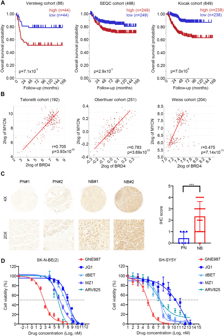
Background: Neuroblastoma (NB) is a common extracranial malignancy with high mortality in children. Recently, super-enhancers (SEs) have been reported to play a critical role in the tumorigenesis and development of NB via regulating a wide range of oncogenes Thus, the synthesis and identification of chemical inhibitors specifically targeting SEs are of great urgency for the clinical therapy of NB. This study aimed to characterize the activity of the SEs inhibitor GNE987, which targets BRD4, in NB.

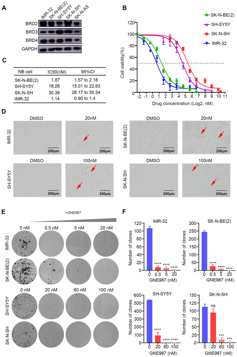
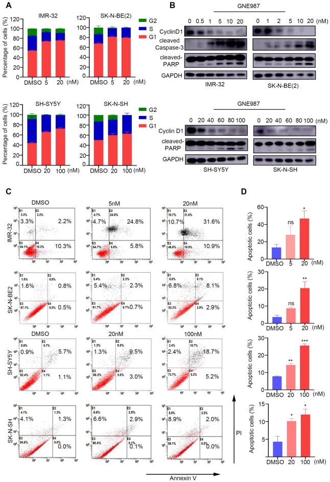
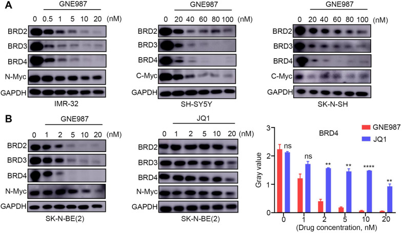
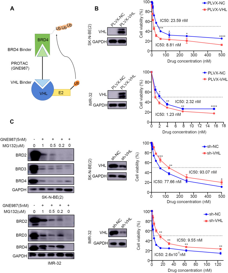
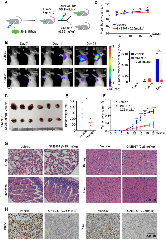
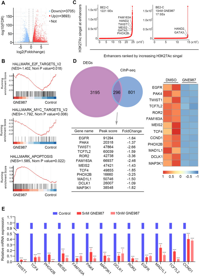
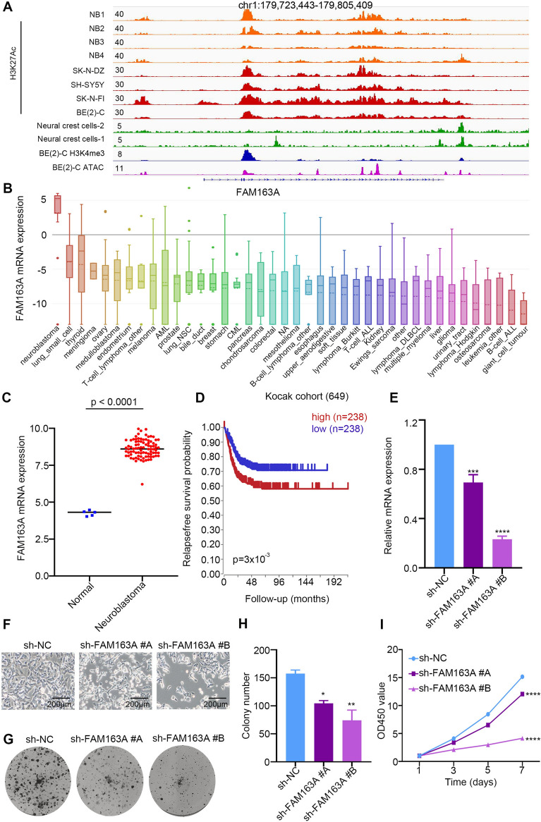
Results: In this study, we found that nanomolar concentrations of GNE987 markedly diminished NB cell proliferation and survival via degrading BRD4. Meanwhile, GNE987 significantly induced NB cell apoptosis and cell cycle arrest. Consistent with in vitro results, GNE987 administration (0.25 mg/kg) markedly decreased the tumor size in the xenograft model, with less toxicity, and induced similar BRD4 protein degradation to that observed in vitro. Mechanically, GNE987 led to significant downregulation of hallmark genes associated with MYC and the global disruption of the SEs landscape in NB cells. Moreover, a novel candidate oncogenic transcript, FAM163A, was identified through analysis of the RNA-seq and ChIP-seq data. FAM163A is abnormally transcribed by SEs, playing an important role in NB occurrence and development.

Conclusion: GNE987 destroyed the abnormal transcriptional regulation of oncogenes in NB by downregulating BRD4, which could be a potential therapeutic candidate for NB.

Keywords: BRD4; Broad H3K27ac domain; Neuroblastoma; PROTAC; Super-enhancer.


Conflict of interest statement

The authors declare that they have no conflict of interest.

Figures

Fig. 1
Fig. 1
High BRD4 expression in NB was associated with poor prognosis. A Kaplan–Meier analysis of the correlation of BRD4 high or low expression with the OS of the patients with NB [generated from R2 Genomics Analysis and Visualization Platform (http://r2.amc.nl)]. Median survival time was used as a cutoff point for defining high or low expression; p < 0.001. B Correlation between BRD4 and MYCN mRNA expression in NB [generated from R2 Genomics Analysis and Visualization Platform (http://r2.amc.nl)]; p < 0.001. C Representative images of IHC staining of the BRD4 protein on a tissue microarray constructed from 27 NB tissues and seven matched normal tissues. Histological scores were determined according to the intensity of BRD4 staining. PN, peripheral neuron; NB, neuroblastoma. ***p < 0.001. D Dose–response curves of five BRD4 inhibitors (GNE987, JQ1, dBET, MZ1, ARV825) at the indicated concentrations in SK-N-BE (2) or SH-SY5Y cells. Data are represented as the mean ± SD
Fig. 2
Fig. 2
GNE987 inhibited NB cell viability and proliferation. A BET protein levels in NB cells were assessed using Western blotting. B Survival rate curves of the SK-N-BE (2), IMR-32, SK-N-SH, and SH-SY5Y cells after treatment with vehicle (DMSO) or increasing concentrations of GNE987 for 24 h. Data are shown by percent cell viability relative to that of the DMSO-treated cells. C The IC50 value of GNE987 in different NB cell lines. D Microscopic images of the SK-N-BE (2), IMR-32, SK-N-SH and SH-SY5Y cells treated with vehicle (DMSO) and the specified concentrations of GNE987 for 24 h (red arrows indicate dead cells after GNE987 treatment). E, F The clonal formation ability of the NB cells treated with the vehicle or a series of concentrations of GNE987 as assessed using a clone formation assay. ***p < 0.001, ****p < 0.0001; ns, not significant
Fig. 3
Fig. 3
GNE987 inhibited the NB cell cycle and induced apoptosis. A NB cells were treated with GNE987 for 24 h and the cell cycle distribution of NB cells was analysed using flow cytometry. B Western blotting analysis showing the expression of cyclin D2, cleaved caspase-3, and cleaved PARP in the NB cells treated with vehicle (DMSO) or increasing concentrations of GNE987 for 24 h. C, D Annexin-V/PI staining and flow cytometry were performed to detect apoptosis in the NB cells treated with vehicle (DMSO) or the specified concentrations of GNE987. *p < 0.05, **p < 0.01, ***p < 0.001
Fig. 4
Fig. 4
GNE987 induced the degradation of a BET protein and N-myc or C-myc. A Western blotting analysis of BET proteins and N-myc or C-myc levels in the NB cells treated with vehicle (DMSO) or. increasing doses of GNE987 for 24 h. B Protein levels of BET and N-myc in the SK-N-BE (2) cells treated with vehicle (DMSO), GNE987, or JQ1. The BRD4 protein level was normalized against that of GAPDH. *p < 0.05, **p < 0.01, ***p < 0.001, ****p < 0.0001; ns, not significant
Fig. 5
Fig. 5
GNE987 is a BET-targeting PROTAC based on VHL, for which the BET protein degradation is proteasome-dependent. A Bifunctional PROTAC molecules bind to the targeting-protein (BRD4, dark green) with one end (a motif, light green) while the other end (a motif, light blue) binds to an E3-ubiquitin ligase (dark blue) to form a ternary complex. The recruited E3 ligase then mediates the transfer of ubiquitin from an E2 enzyme to the targeted protein (direction of arrow). B Analysis of VHL expression in the IMR-32 and SK-N-BE (2) cells treated with negative control sh-NC (or PLVX-NC) and sh-VHL (or PLVX-VHL) examined by Western blotting. Then the sensitivity to GNE987 of the NB cells transfected with sh-VHL (or PLVX-VHL) and NB cells transfected with sh-NC (or PLVX-NC) was compared. *p < 0.05, **p < 0.01, ***p < 0.001, ****p < 0.0001. C Western blotting analysis of the BET protein in SK-N-BE (2) and IMR-32 cells treated with the 5 nM GNE987 and series concentrations of MG132, and their combination for 24 h
Fig. 6
Fig. 6
In vivo GNE987 treatment caused tumour necrosis in NB xenograft models. A Schematic of the experimental design. Mice bearing luciferase-labelled NB xenograft tumours were treated with intraperitoneal injections of 0.25 mg/kg GNE987 every other day once their tumour luminescence flux reached 3–5 × 105 photons/second via bioluminescent imaging (BLI). B Representative sequential BLI images of the tumour bearing mice in the control group or the GNE987 treatment group taken at 7, 14, and 21 days after subcutaneous implantation of NB cells. Scale is in photons/second. n = 6. *p < 0.05. C Photograph of excised tumours from the mice in the GNE987 treatment group and the control group at d 21. Scale bar, 1 cm. D Weight changes in the tumour bearing mice in the GNE987 treatment group and the control group. E The average tumor weight of the excised tumours at d 21. F Tumour growth curves of tumour-bearing mice in the GNE987 treatment group and the control group in response to different treatments. ***p < 0.001, ****p < 0.0001. G H&E staining of organs in the mice in the GNE987 treatment group. Scale bar, 200 μm. H Representative images of IHC staining with the indicated antibodies in tumours harvested from the mice in the GNE987 treatment group and the control group. Scale bar, 200 μm
Fig. 7
Fig. 7
Comprehensive analysis of RNA-seq and ChIP-seq identified novel candidate oncogenic transcripts. Volcano plot of gene expression differences between the GNE987-treated and DMSO-treated SK-N-BE (2) cells. The blue and red dots highlight all the statistically significant transcripts with downregulated/upregulated expression (log2FoldChange < − 1.0 or > 1, adjusted p < 0.05). B Enrichment analysis results of differentially expressed genes by using GSEA Pathway Database to investigate the function of GNE987 in gene regulation. C Enhancers were ranked by increasing H3K27ac signal in the GNE 987 treated and nontreated SK-N-BE (2) cells. The number of SEs is shown for each group. D Venn diagram showing the 297 overlapping genes between the RNA-seq decreased genes and SE-related genes calculated from ChIP-seq results. The relative expression levels of the representative overlapping genes are depicted in the heatmap, of which the peak score and fold change are shown in the table. E RT-qPCR analysis of genes related to SE from SK-N-BE (2) treated with DMSO, 5 nM or 10 nM for 24 h. ****p < 0.0001
Fig. 8
Fig. 8
FAM163A is a potential oncogene of NB. A IGV plots showing ChIP-seq profiles of FAM163A in NB gene locus. The ChIP-seq gene tracks represent the H3K27ac signal in normal neural crest cells (green) (GSE90683), NB cell lines (red) (GSE90683) and clinical samples from NB (orange) (GSE90805). B FAM163A mRNA expression level in a broad range of tumours (generated from CCLE: https://portals.broadinstitute.org/ccle). C Relative mRNA expression of FAM163A in normal nerve cells and NB cells based on data available from the GEO database. D Overall survival (OS) in patients with NB with high or low levels of FAM163A plotted using Kaplan–Meier method (generated from R2 Genomics Analysis and Visualization Platform: r2.amc.nl). p < 0.001. E SK-N-BE (2) cells were transfected with sh-NC, sh-FAM163A #A, or sh-FAM163A #B and positive cells were screened using puromycin for 6 days. The mRNA expression of FAM163A was assessed using qRT-PCR. F Microscopic images of the SK-N-BE (2) cells and SK-N-BE (2) cells transfected with sh-NC, sh-FAM163A #A, or sh-FAM163A #B. G–H Representative images and statistical results of a clone formation assay showing the colony formation ability of the SK-N-BE (2) cells stably transfected with sh-NC, sh-FAM163A #A, or sh-FAM163A #B. *p < 0.05, **p < 0.01. I Proliferation rate of the SK-N-BE (2) cells transfected with sh-NC, sh-FAM163A #A, or sh-FAM163A #B. ****p < 0.0001

Similar articles

Cited by

References

    1. Nakagawara A, Li Y, Izumi H, Muramori K, Inada H, Nishi M. Neuroblastoma. Jpn J Clin Oncol. 2018;48(3):214–241. - PubMed
    1. Whittle SB, Smith V, Doherty E, Zhao S, McCarty S, Zage PE. Overview and recent advances in the treatment of neuroblastoma. Expert Rev Anticancer Ther. 2017;17(4):369–386. - PubMed
    1. Wienke J, Dierselhuis MP, Tytgat GAM, Kunkele A, Nierkens S, Molenaar JJ. The immune landscape of neuroblastoma: challenges and opportunities for novel therapeutic strategies in pediatric oncology. Eur J Cancer. 2021;144:123–150. - PubMed
    1. Moreno L, Guo D, Irwin MS, Berthold F, Hogarty M, Kamijo T, Morgenstern D, Pasqualini C, Ash S, Potschger U, et al. A nomogram of clinical and biologic factors to predict survival in children newly diagnosed with high-risk neuroblastoma: an international neuroblastoma risk group project. Pediatr Blood Cancer. 2021;68(3):e28794. - PubMed
    1. Lin CY, Loven J, Rahl PB, Paranal RM, Burge CB, Bradner JE, Lee TI, Young RA. Transcriptional amplification in tumor cells with elevated c-Myc. Cell. 2012;151(1):56–67. - PMC - PubMed