. 2022 Sep;150(3):640-648.
doi: 10.1016/j.jaci.2022.02.027. Epub 2022 Mar 15.

Signaling sphingolipids are biomarkers for atopic dermatitis prone to disseminated viral infections

Affiliations

Signaling sphingolipids are biomarkers for atopic dermatitis prone to disseminated viral infections

Evgeny Berdyshev et al. J Allergy Clin Immunol. 2022 Sep.

Abstract

Background: Life-threatening viral diseases such as eczema herpeticum (EH) and eczema vaccinatum (EV) occur in <5% of individuals with atopic dermatitis (AD). The diagnosis of AD, however, excludes all individuals with AD from smallpox vaccination.

Objectives: We sought to identify circulatory and skin lipid biomarkers associated with EH and EV.

Methods: Stratum corneum and plasma samples from 15 subjects with AD and a history of EH, 13 age- and gender-matched subjects with AD and without EH history, and 13 healthy nonatopic (NA) controls were analyzed by liquid chromatography tandem mass spectrometry for sphingolipid content. Sphingosine-1-phosphate (S1P) and ceramide levels were validated in plasma samples from the Atopic Dermatitis Vaccinia Network/Atopic Dermatitis Research Network repository (12 NA, 12 AD, 23 EH) and plasma from 7 subjects with EV and 7 matched subjects with AD. S1P lyase was downregulated in human primary keratinocytes to evaluate its effect on herpes simplex virus 1 (HSV-1) replication in vitro.

Results: The stratum corneum of patients with EH demonstrated significantly higher levels of free sphingoid bases than those in patients who were NA, indicating enhanced sphingolipid turnover in keratinocytes (P < .05). Plasma from 2 independent cohorts of patients with EH had a significantly increased S1P/ceramide ratio in subjects with EH versus those with AD and or who were NA (P < .01). The S1P level in plasma from subjects with EV was twice the level in plasma from subjects with AD (mean = 1,533 vs 732 pmol/mL; P < .001). Downregulation of S1P lyase expression with silencing RNA led to an increased S1P level and doubled HSV-1 titer in keratinocytes.

Conclusions: Our data point to long-term abnormalities in the S1P signaling system as a biomarker for previous disseminated viral diseases and a potential treatment target in recurring infections.

Keywords: Eczema vaccinatum; S1P lyase; S1P/ceramide ratio; ceramide; eczema herpeticum; human primary keratinocytes; plasma; sphingosine-1-phosphate; stratum corneum.


Conflict of interest statement

Conflict of Interest: The authors have declared that no conflict of interest exists.

Figures

Fig 1.
Fig 1.. Non-lesional skin of EH subjects demonstrate disturbed sphingolipid profile.
The levels of short-chain (N-16:0-) (A), long-chain (N-26:0-) NS-CER (B), and EOS-CER/NS-CER ratio (C) were the most abnormal in skin samples of EH subjects as compared to AD subjects and NA controls. (D) The stratum corneum of EH but not AD subjects demonstrate substantially increased total sphingomyelin content, with all sphingomyelin molecular species being unselectively increased (E). All lipid molecular species were quantified by targeted LC-ESI-MS/MS and normalized by sample total protein content. Some data were expressed as relative percentage within each lipid subclass. For the total list of analyzed molecular species, see Supplemetary Tables E4 and E5. Here and further, where applicable, data are presented as box and whiskers plots with individual values shown, with box indicating 25–75% interqualrtile range and whiskers showing minimum and maximum values. * p<0.05, ** p<0.01, *** p<0.001. This and further data are compared using one-way Anova with a correction for multiple comparisons (Tukey) unless otherwise specified. EH non-lesional skin – n=13, AD non-lesional skin – n=13, NA skin – n=15.
Fig 2.
Fig 2.. Non-lesional skin samples of EH subjects have increased levels of free sphingoid bases.
Non-lesional stratum corneum even-chain (C18-) (A) and odd-chain (C17-) (B) sphingosines are upregulated in EH skin compared to AD skin. Each sphingoid base was quantified by targeted LC-ESI-MS/MS and normalized by sample total protein content. EH non-lesional skin – n=13, AD non-lesional skin – n=13, NA skin – n=15.
Fig 3.
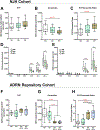
Fig 3.. Atopic subjects with a history of EH are distinct from AD subjects without history of EH by circulatory levels of ceramides and S1P.
Plasma lipids from two cohorts of subjects (A-E – NJH cohort; F-H – ADRN repository cohort) were extracted and analyzed by the LC-ESI-MS/MS for the content of S1P and ceramides. Both cohorts demonstrate that EH patients have a slight increase in circulatory levels of S1P (A,F) with a concomitant significant decrease in ceramide content (B,G), that leads to a substantial shift in S1P / ceramide ratio (C,H). Both NS-ceramides (ceramides, D) and NDS-ceramides (dihydroceramides, E) are affected in plasma samples of EH subjects. Note that plasma samples from EH subjects with prior history of one to two EH episodes (Gr1–2) or three and more episodes (Gr3+) are identical in sphingolipid abnormalities (F-H). NJH cohort: NA – n=13; AD - n=12; EH - n=15. ADRN repository cohort: NA – n=12; AD – n=12; EH Gr1–2 – n=11; EH Gr3+ - n=12. Analyzed species of NS-ceramides in the ADRN repository cohort are the same as presented in Figure 3D and E.
Fig 4.
Fig 4.. AD severity does not affect plasma S1P / ceramide ratio.
EASI scores are substantially different in NJH (moderate to severe) and ADVN repository (mild to moderate) cohorts. However, AD severity has no effect on plasma S1P to total NS-ceramide ratio in these subjects. Analyzed species of NS-ceramides in the NJH and ADVN cohorts are the same as presented in Figure 3D and E.
Fig 5.
Fig 5.. S1P-to-ceramide ratio in plasma of a patient who clinically transitioned from AD to EH category.
Plasma was collected from the subject who enrolled in ADRN studies in 2006 as AD patient, then was diagnosed with EH in 2008 and later enrolled in ADRN studies as EH subject who had three or more episodes of EH. Lipids were extracted and analyzed for the S1P and ceramide content by the ESI-LC-MS/MS. Years indicate when plasma was collected. Shadowed boxes provide 25–75% interquartile range of ratio values determined for the NJH cohort as reported in Fig 4C. Analyzed species of NS-ceramides are the same as presented in Figure 3D and E,
Fig 6.
Fig 6.. AD patients with a history of EV have upregulated levels of plasma S1P and ceramides.
(A) S1P and DHS1P levels in plasma samples from AD and EV subjects. (B) Ceramide (NS-CER) and dihydroceramide (NDS-CER) levels in plasma samples from AD and EV subjects. Sphingolipid levels were measured by LC-ESI-MS/MS. n=7 for each group. Statistical difference was determined using a two-tailed Student’s t-test. Analyzed species of NS- and NDS-ceramides are the same as presented in Figure 3D and E.
Fig 7.
Fig 7.. S1P promotes herpes simplex virus 1 replication in keratinocytes in vitro.
Primary human keratinocytes were silenced with control non-targeting siRNA, SGPL1, SPHK1 or SPHK2 siRNA, and induced to differentiate for five days in the presence of 1.3mM CaCl2. Cells for lipid analysis were taken at this point. Then, keratinocytes were infected with herpes simplex virus 1 (0.1 MOI) and cell lysates were prepared 24 hours post-infection. Inhibition of S1P lyase (SGPL1) expression drastically upregulates intracellular S1P and DHS1P levels (A) and the number of viral particles in cells lysates as measured by RT-PCR (B) or plaque assay (C). Data from one of the two independent experiments are shown, each done in triplicate. *** - p < 0.0001 versus corresponding control, one-way ANOVA.

Comment in

References

    1. Bieber T, D’Erme AM, Akdis CA, Traidl-Hoffmann C, Lauener R, Schappi G, et al. Clinical phenotypes and endophenotypes of atopic dermatitis: where are we, and where should we go? J Allergy Clin Immunol 2017;139:S58–S64. - PubMed
    1. Langan SM, Irvine AD, Weidinger S. Atopic dermatitis. Lancet 2020;396:345–60. - PubMed
    1. Beck LA, Boguniewicz M, Hata T, Schneider LC, Hanifin J, Gallo R, et al. Phenotype of atopic dermatitis subjects with a history of eczema herpeticum. J Allergy Clin Immunol 2009;124:260–9,269.e1–7. - PMC - PubMed
    1. Wang V, Boguniewicz J, Boguniewicz M, Ong PY. The infectious complications of atopic dermatitis. Ann Allergy Asthma Immunol 2021;126:3–12. - PMC - PubMed
    1. Reed JL, Scott DE, Bray M. Eczema vaccinatum. Clin Infect Dis 2012;54:832–40. - PubMed

Publication types