Review
. 2022 May:184:114233.
doi: 10.1016/j.addr.2022.114233. Epub 2022 Mar 15.

Immunoengineering strategies to enhance vascularization and tissue regeneration

Affiliations
Review

Immunoengineering strategies to enhance vascularization and tissue regeneration

Jana Zarubova et al. Adv Drug Deliv Rev. 2022 May.

Abstract

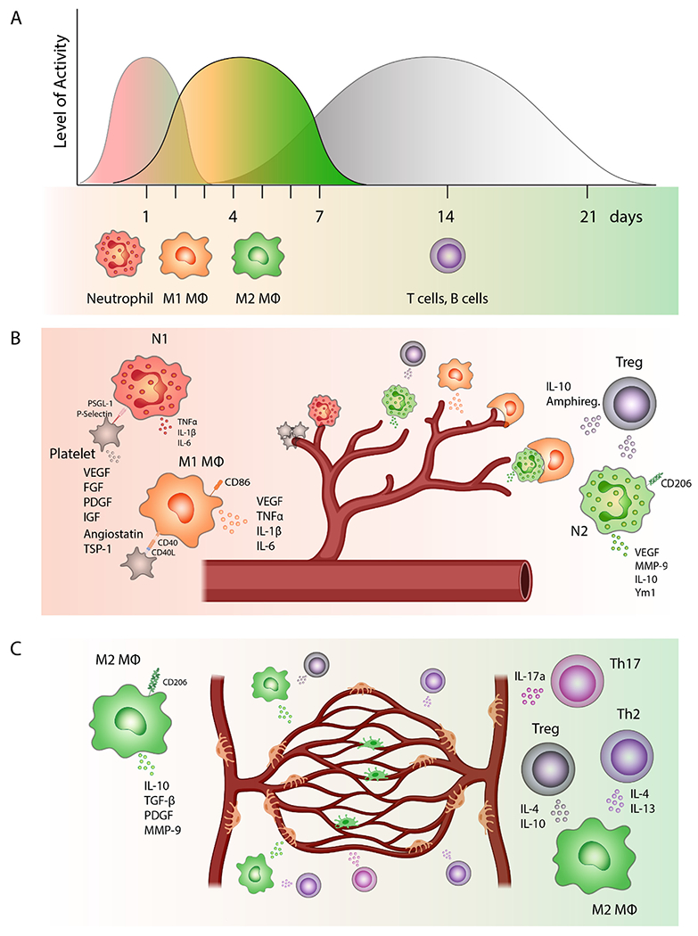
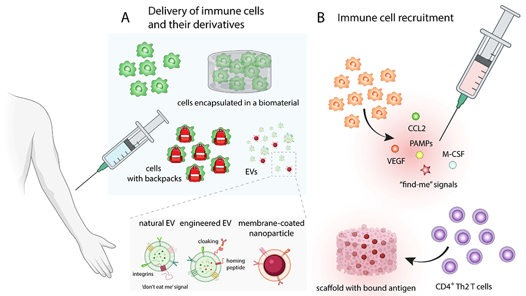
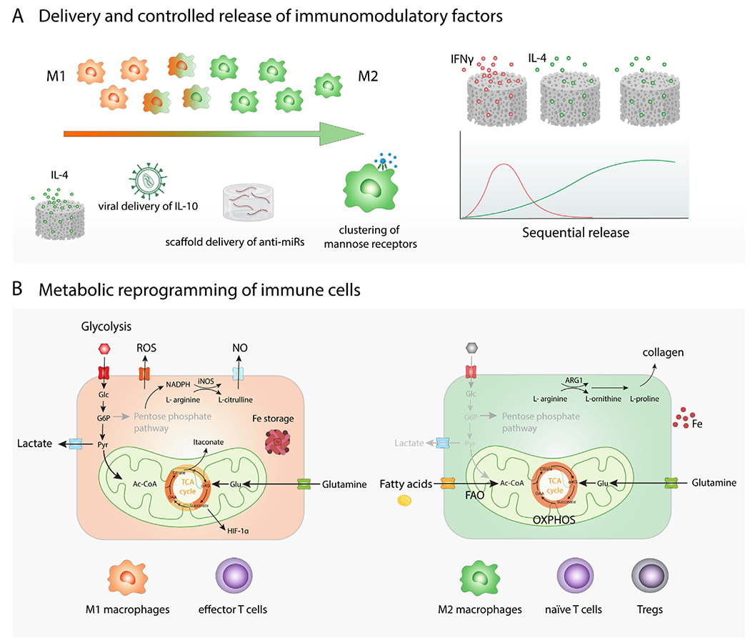
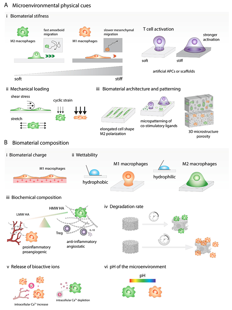
Immune cells have emerged as powerful regulators of regenerative as well as pathological processes. The vast majority of regenerative immunoengineering efforts have focused on macrophages; however, growing evidence suggests that other cells of both the innate and adaptive immune system are as important for successful revascularization and tissue repair. Moreover, spatiotemporal regulation of immune cells and their signaling have a significant impact on the regeneration speed and the extent of functional recovery. In this review, we summarize the contribution of different types of immune cells to the healing process and discuss ways to manipulate and control immune cells in favor of vascularization and tissue regeneration. In addition to cell delivery and cell-free therapies using extracellular vesicles, we discuss in situ strategies and engineering approaches to attract specific types of immune cells and modulate their phenotypes. This field is making advances to uncover the extraordinary potential of immune cells and their secretome in the regulation of vascularization and tissue remodeling. Understanding the principles of immunoregulation will help us design advanced immunoengineering platforms to harness their power for tissue regeneration.

Keywords: Biomaterial; Cell delivery; Extracellular vesicles; Immune cell metabolism; Immunomodulation; Macrophages; Neutrophils; Patterning; Stiffness; T cells.


Conflict of interest statement

Declaration of Competing Interest The authors declare that they have no known competing financial interests or personal relationships that could have appeared to influence the work reported in this paper.

Figures

Fig. 1.
Fig. 1.. Engagement and polarization of immune cells in different phases of re-vascularization and tissue regeneration.
(A) Physiological timeline of sequential recruitment of different types of immune cells to the damaged tissue and an average duration of their involvement in revascularization and tissue repair. Neutrophils are among the first immune cells to be recruited to the wound bed followed by macrophages, which are first polarized into pro-inflammatory M1-like phenotypes. Gradually over the course of the healing process, the balance is shifted to M2-like anti-inflammatory macrophages. Finally, after approximately 3–5 days, the activated effector T cells and B cells travel to the site of injury to modulate tissue regeneration. (B) The beginning of the healing process. P-selectin on the surface of activated platelets helps to recruit free-floating neutrophils to the affected site. Neutrophils are first polarized into pro-inflammatory N1 phenotype and secrete cytokines like TNFα, IL-1β or IL-6. Activated platelets also express CD40 ligand (CD40L) on their surface that interacts with the receptor on the surface of macrophages. This interaction can stimulate VEGF secretion by macrophages, which promotes angiogenesis. M1-like macrophages are important at the beginning of the angiogenic process when they are in a close contact with tip cells of sprouting capillaries. Neutrophils, which are polarized in the presence of TGF-β into an anti-inflammatory N2-like phenotype, also secrete VEGF and metalloproteinases, which release growth factors bound to ECM. Apoptotic N2-like neutrophils are phagocytosed by macrophages, which induce their polarization into M2-like anti-inflammatory state. Regulatory T cells (Tregs) represent a subset of CD4 + T cells that are involved in the regulation of the healing process by suppressing proinflammatory immune responses. Tregs also secrete amphiregulin, which can promote angiogenesis by inducing VEGF synthesis in other cell types. (C) Blood vessel stabilization and tissue remodeling. M2-like anti-inflammatory macrophages, which express high levels of CD206, dominate in later stages of the healing process where they secrete cytokines like IL-10 or growth factors such as TGF-β or PDGF. M2-like macrophages also promote anastomoses, stabilize growing blood vessels, and consequently are involved in vascular network remodeling. CD4 + T helper 2 (Th2) cells, Tregs, and Th17 cells can promote regeneration and angiogenesis either directly, by secreting angiogenic factors that enhance EC proliferation and migration, or indirectly by secreting cytokines like IL-4 which can induce macrophage polarization into M2-like phenotype.
Fig. 2.
Fig. 2.. Delivery or recruitment of immune cells to enhance angiogenesis and regeneration.
(A) Delivery of ex vivo expanded immune cells, mainly M2-like pro-regenerative macrophages or immune cells equipped with backpacks bound to their surface, which gradually release compounds that continuously guide the polarization of immune cells. In order to improve immune cell retention at the site of the injury and increase cell viability, cells can be encapsulated various types of implantable or injectable biomaterials. Moreover, extracellular vesicles (EVs) secreted by cells can replicate the therapeutic effect of delivered cells. EVs can be furthermore modified by various techniques such as cloaking, loaded with drugs, or conjugated with homing peptides to further increase their pro-healing effects and homing specificity. (B) Macrophage recruitment can be promoted by cytokines like macrophage colony-stimulating factor (M-CSF), growth factors such as VEGF, chemokines like CCL2, or “find-me” signals, which are normally secreted by apoptotic cells. Biomaterials can provide not only the sustained and prolonged presentation of therapeutics but they can also enhance their stability and localization. Biomaterial scaffold with bound antigen can increase the local concentration of antigen-specific CD4 + Th2 T cells and promote angiogenesis and regeneration.
Fig. 3.
Fig. 3.. Modulation of immune cell phenotype and behavior in situ
(A) Delivery and controlled release of immunomodulatory factors. To promote phenotypic switch towards M2-like macrophages, various immune-instructive biomaterials releasing growth factors or cytokines like IL-4 can be used. Sequential release of multiple immunomodulatory agents from a biomaterial, such as initial fast release of M1-promoting IFN-γ followed by sustained release of IL-4, was developed to better control dynamic phenotypic switch of macrophages. Genetic material delivery using different types of viruses or biomaterials has been employed to provide instructive signaling for immune cells. Nanoparticle-triggered clustering of mannose receptors on the surface of macrophages promote their polarization into M2-like phenotype. (B) Metabolic reprograming of immune cells. Glycolysis-based energy metabolism is generally connected with inflammatory reactions while metabolism based on oxidative phosphorylation is associated with anti-inflammatory processes.
Fig. 4.
Fig. 4.. In situ modulation of immune cell function by changing biomaterial properties and mechanical stimulation
(A) Microenvironmental physical cues. (i) Stiff biomaterials prime macrophages towards M1-like phenotype, while softer ones direct macrophages into anti-inflammatory state. Macrophages cultured on the softer substrates display fast amoeboid migration, while macrophages on stiff gels adopt slower mesenchymal migration. T cell activation by interactions with antigen presenting cell (APC) can be replicated by biomaterials functionalized with ligands that bind to receptors on the T cell surface and biomaterial stiffness is one of the parameters that might significantly affect the level of T cell activation. Generally, stronger T cell activation is observed on stiffer materials. (ii) Immune cell phenotype can be also affected by mechanical loading such as tensile, compressive and shear forces. Moderate cyclic strain can promote M2-like phenotype while higher cyclic strain triggered induction of M1-like macrophages. (iii) Biomaterial architecture and patterning can modulate cell shape and cytoskeletal organization, which impact the immune cell behavior. Macrophage elongated morphology induces M2-like polarization and reduces secretion of proinflammatory cytokines by these cells. Micropatterning can also affect T cell activation, for example, focal presentation of antibodies against CD3 and CD28 increases T cell activation. Microstructure of 3D scaffolds such as pore size and geometry affect immune cell infiltration and polarization. (B) Biomaterial composition. (i) Positively charged materials are more likely to trigger proinflammatory immune cell responses. (ii) Hydrophobic biomaterials tend to induce proinflammatory M1-like macrophage activation while hydrophilic or neutral surfaces create more anti-inflammatory microenvironment. (iii) Low molecular weight hyaluronic acid (LMW HA) fragments promote angiogenesis, high molecular weight HA have angiostatic properties, suppress M1-like polarization and promote Treg formation. (iv) Slowly degrading biomaterials induce prolonged proinflammatory reactions while scaffolds that degrade faster result in constructive tissue remodeling. (v) Release of bioactive ions: calcium signaling plays important role in the proinflammatory activation of macrophages and intracellular Ca2+ oscillations also occur in migrating ECs during capillary sprouting, on the contrary, calcium ion depletion leads to M2-like polarization. (vi) Biomaterials can change the pH of the microenvironment, which can contribute to the immune cell phenotypic changes, alkaline microenvironment can promote M1-like macrophage polarization while acidic pH polarizes macrophages into M2-like phenotype.

References

    1. Faber JE, Chilian WM, Deindl E, van Royen N, Simons M, A brief etymology of the collateral circulation, Arterioscler. Thromb. Vasc. Biol 34 (2014) 1854–1859. - PMC - PubMed
    1. Carmeliet P, Jain RK, Molecular mechanisms and clinical applications of angiogenesis, Nature 473 (2011) 298–307. - PMC - PubMed
    1. Stratman AN, Schwindt AE, Malotte KM, Davis GE, Endothelial-derived PDGF-BB and HB-EGF coordinately regulate pericyte recruitment during vasculogenic tube assembly and stabilization, Blood 116 (2010) 4720–4730. - PMC - PubMed
    1. Stratman AN, Saunders WB, Sacharidou A, Koh W, Fisher KE, Zawieja DC, Davis MJ, Davis GE, Endothelial cell lumen and vascular guidance tunnel formation requires MT1-MMP-dependent proteolysis in 3-dimensional collagen matrices, Blood 114 (2009) 237–247. - PMC - PubMed
    1. Davis GE, Stratman AN, Sacharidou A, Koh W, Molecular basis for endothelial lumen formation and tubulogenesis during vasculogenesis and angiogenic sprouting, Int Rev Cell Mol Biol 288 (2011) 101–165. - PMC - PubMed

Publication types

Substances