. 2022 Mar 18;13(1):1483.
doi: 10.1038/s41467-022-29146-2.

Genome-wide maps of nucleolus interactions reveal distinct layers of repressive chromatin domains

Affiliations

Genome-wide maps of nucleolus interactions reveal distinct layers of repressive chromatin domains

Cristiana Bersaglieri et al. Nat Commun. .

Abstract

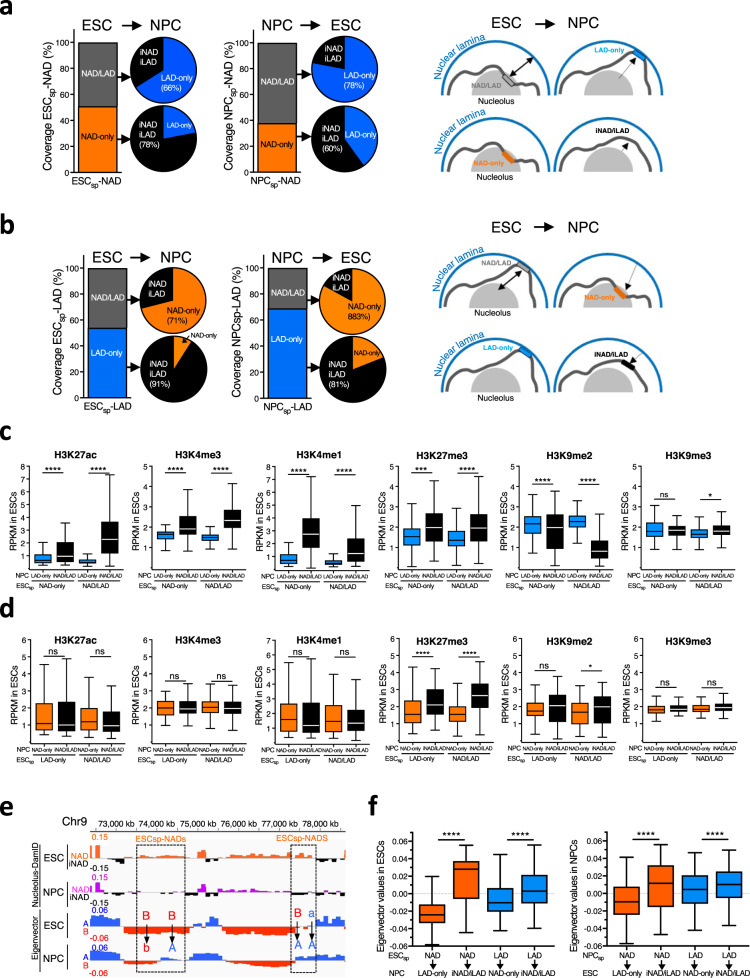
Eukaryotic chromosomes are folded into hierarchical domains, forming functional compartments. Nuclear periphery and nucleolus are two nuclear landmarks contributing to repressive chromosome architecture. However, while the role of nuclear lamina (NL) in genome organization has been well documented, the function of the nucleolus remains under-investigated due to the lack of methods for the identification of nucleolar associated domains (NADs). Here we have established DamID- and HiC-based methodologies to generate accurate genome-wide maps of NADs in embryonic stem cells (ESCs) and neural progenitor cells (NPCs), revealing layers of genome compartmentalization with distinct, repressive chromatin states based on the interaction with the nucleolus, NL, or both. NADs show higher H3K9me2 and lower H3K27me3 content than regions exclusively interacting with NL. Upon ESC differentiation into NPCs, chromosomes around the nucleolus acquire a more compact, rigid architecture with neural genes moving away from nucleoli and becoming unlocked for later activation. Further, histone modifications and the interaction strength within A and B compartments of NADs and LADs in ESCs set the choice to associate with NL or nucleoli upon dissociation from their respective compartments during differentiation. The methodologies here developed will make possible to include the nucleolar contribution in nuclear space and genome function in diverse biological systems.


Conflict of interest statement

The authors declare no competing interests.

Figures

Fig. 1
Fig. 1. Establishment of Nucleolar-DamID.
a Scheme representing the strategy for the establishment of Nucleolar-DamID. b Live cell imaging of NIH3T3 cells 24 h post-transfection with plasmids expressing H2B-GFP and H2B-GFP-NoLS under the minimal CMV promoter. Phase contrast images serve to visualize the nucleoli. c Representative immunofluorescence images of NIH3T3 cells transfected with plasmids expressing H2B-GFP and H2B-NoLS-GFP. Nucleoli can be visualized with the nucleolar protein NPM1. Scale bars represent 5 μm. d Chromatin-bound (Chrom.) and soluble (Sol.) fractions of equivalent number of NIH3T3 cells transfected with H2B-GFP and H2B-GFP-NoLS were analyzed by western blot using anti-GFP antibodies. Coxa4 and histones are shown as loading and fractionation control. e Representative immunofluorescence images of NIH3T3 cells transfected with plasmids expressing H2B-GFP, TTF1-GFP, and H2B-NoLS-GFP and treated for 24 h with or without ActD (50 ng/ml). ActD was added to cells 24 h post-transfection. The nucleolar protein Fibrillarin serve to visualize nucleoli in untreated cells and nucleolar caps in ActD-treated cells. Large nucleoli can be visualized by the low DAPI intensity. Scale bars represent 5 μm. f Scheme representing the constructs used for double inducible expression of Dam-fused H2B and nucleolar H2B (H2B-NoLS) proteins. g qRT-PCR showing similar expression levels of H2B-Dam and H2B-NoLS-Dam in ESCs. Mean values from data from three biologically independent experiments. Error bars represent s.d. h m6A levels at rRNA genes and Tuba1a in ESCs with and without 15 h treatment with 100 ng Doxycycline (Dox) and 1 μM Shield1. m6A levels were measured by digestion of genomic DNA with DpnII, which is blocked by m6A, followed by quantitative amplification with primers encompassing the Dam GATC element. Normalization was achieved through measurements with primers encompassing sequences lacking GATC. Mean values from data of four biologically independent experiments. Error bars represent s.d. Statistical significance (P-values) for the experiments was calculated using the paired two-tailed t test (*** <0.001). Source data are provided as a Source Data file.
Fig. 2
Fig. 2. Nucleolar-DamID identifies NADs.
a Chromosomal view of NADs, LADs, A and B compartments, and early and late replicating DNA in ESCs. NADs are measured as log2 ratio of m6A levels between H2B-Dam-NoLS and H2B-Dam. iNAD: domains not contacting nucleoli. iLAD: domains not contacting NL. b Bars represent NAD coverage values at mouse chromosomes. Red lines showed NAD coverage values in the centromere-proximal end of each chromosome. Dotted line shows NAD whole genome coverage. c Nucleolar H2B-Dam chromosomal interaction maps. d rDNA-contacts obtained from HiC maps. Data represent HiC scores of unique contacts for each chromosome. Statistical significance (P-values) was calculated using the unpaired two-tailed t test (**** <0.0001). Lines are mean values. e Representative images showing normalized count score of rDNA-contacts obtained from HiC-rDNA on chromosomes 9 and 18. f Distribution of rDNA-contacts. Values represent the proportion of the number of unique contacts for each chromosome quintile. Statistical significance (P-values) was calculated using the unpaired two-tailed t test (**** <0.0001). g NADs identified by Nucleolar-DamID are enriched in rDNA-contacts. Data represent the proportion of unique HiC-rDNA contacts at NADs and regions non contacting the nucleolus (iNAD). h Hi-C normalized count score of identified unique rDNA-contacts at NADs and iNADs. Statistical significance (P-values) was calculated using the unpaired two-tailed t test (**** <0.0001). Box plots depict the minimum and maximum values. The horizontal line within the boxes represents the mean value. i, j Upper panels represent NAD and LAD profiles of chromosome 1 (i) and 5 (j) and the DNA-FISH probes (orange bar) hybridizing to regions identified by the Nucleolar-DamID as NAD-only. Lower panels. Example images from immunofluorescences for nucleolin (red) combined with the corresponding DNA-FISH probe (green) and DAPI (blue). Scale bar is 5μm. k Distance of the indicated DNA-FISH probes from nucleoli (micron). Statistical significance (P-values) was calculated using the unpaired two-tailed t test (**** <0.0001). l Quantification of the number of cells displaying at least one DNA-FISH probe signal contacting the nucleolus. Data are from the measurements of 70–120 cells for each condition. Source data are provided as a Source Data file.
Fig. 3
Fig. 3. Distinct layers of repressive chromatin states distinguish genomic domains according to their interaction with the nucleolus, nuclear lamina, or both.
a Venn diagram showing the proportion of NAD-only and NAD/LAD regions in NADs identified by Nucleolar-DamID. b Venn diagram showing the proportion of LAD-only and NAD/LAD regions in LADs of ESCs. c Lamin B1-DamID scores plotted over NAD-only and NAD/LAD boundaries in ESCs. d Gene density of total genome, NAD subclasses, and LAD-only. e Amounts (%) of NAD-only, LAD-only and NAD/LAD in A and B compartments. f Amounts (%) of early and late replicating DNA of NAD-only, LAD-only, and NAD/LAD sequences. g Expression values (RPKM) of genes within A compartment (A Comp.), NAD-only, LAD-only, and NAD/LAD. Statistical significance (P-values) was calculated using the unpaired two-tailed t test (***<0.001). Box plots depict the minimum and maximum values. The horizontal line within the boxes represents the mean value. h Levels of active histone marks (H3K4me3, H3K27ac, H3K4me1) and repressive histone marks (H3K27me3, H3K9me2, H3K9me3) at genomic regions located at the A compartment (A Comp.) and NAD-only, LAD-only, and NAD/LAD regions. Dataset used in this analysis is listed in Supplementary Data 12. Values are shown as average RPKM. Statistical significance (P-values) was calculated using the unpaired two-tailed t test (*<0.05, ***<0.001, ****<0.0001, ns: non-significant). Box plots depict the minimum and maximum values. The horizontal line within the boxes represents the mean value. i. Occupancy (average RPKM) of histone modifications, Ezh2, Ring1b, CTCF and early and late-DNA replication plotted over the boundaries of NAD-only (orange lane), NAD/LAD (blue line), and NAD/LAD (gray line), respectively. Source data are provided as a Source Data file.
Fig. 4
Fig. 4. The chromatin state of rRNA genes regulates H3K9me2 levels at sequences adjacent to the nucleolus.
a qRT-PCR showing 45 S pre-rRNA levels in ESCs transfected with pRNA or RNA-control. Values were normalized to β-actin mRNA and to ESCs transfected with RNA-control. Data are from two independent experiments. b H3K9me2 and H3K9me3 ChIP in ESCs transfected with pRNA or RNA-control. Data were measured by qPCR and normalized to input and ESC + RNA-control. Data are from two independent experiments. c Addition of pRNA in ESCs caused an increase in H3K9me2 at several genomic regions. Scatter plot showing H3K9me2 and H3K9me3 levels (reads/20 kb bin) between ESC + pRNA and ESC + RNA-control. d Chromosomal interaction map showing the distribution of regions with increased H3K9me2 levels in ESC + pRNA compared to ESC + RNA-control. e Heterochromatinization of rRNA genes promotes H3K9me2 expansion at regions neighboring NADs. H3K9me2 fold changes in ESC + pRNA vs. ESC + RNA-control plotted over the boundaries of NAD-only, LAD-only, and NAD/LAD. f Representative images showing the increase of H3K9me2 at regions neighboring NADs. Source data are provided as a Source Data file.
Fig. 5
Fig. 5. ESC and NPC differ in their chromosome organization around the nucleolus.
a Nucleolar-DamID. Chromosomal view of NADs in ESCs and NPCs. NADs are measured as log2 ratio of m6A levels between H2B-Dam-NoLS and H2B-Dam. iNAD: regions not contacting the nucleolus. iLAD: regions not contacting the NL. Chromosomes 4 and 11 are shown. b Representative images showing HiC-score of rDNA contacts at chromosome 18, which contains rRNA genes and chromosome 7. c, d Genomic contacts with the nucleolus are more frequent in NPCs than in ESCs. HiC-score of rDNA contacts (c) and Nucleolar-DamID values of NADs (d) in ESCs and NPCs. Statistical significance (P-values) was calculated using the unpaired two-tailed t test (****<0.0001). Box plots depict the minimum and maximum values. The horizontal line within the boxes represents the mean value. e ESCs have more rDNA contacts than NPC. Number of unique rDNA contacts at chromosomes in ESCs and NPCs. f rDNA contacts are enriched in the centromeric-proximal regions of chromosomes of NPCs relative to ESCs. To allow a better comparison, data of ESCs of Fig. 2f were plotted together with data of NPCs. Values represent the proportion of rDNA-contacts for each chromosome quintile. Statistical significance (P-values) was calculated using the unpaired two-tailed t test (****<0.0001). g Representative images of ESCsp- and NPCsp-rDNA contacts and their eigenvector values for A and B compartment. B to A and A to B represent switch of compartments. B to b and b to B indicate a decrease or increase of eigenvector values between ESCs and NPCs. h ESCsp- and NPCsp-rDNA contacts are in the repressive B compartment. Amounts (%) of ESCsp- and NPCsp-rDNA contacts in A and B compartments. i Values represent the number of ESCsp- and NPCsp-rDNA contacts and their corresponding location in A and B compartments of ESCs and NPCs. j Box plots showing eigenvector values of ESCsp- and NPCsp-rDNA contacts in the active A and repressive B compartments. Statistical significance (P-values) was calculated using the paired two-tailed t test (****<0.0001). Box plots depict the minimum and maximum values. The horizontal line within the boxes represents the mean value. Source data are provided as a Source Data file.
Fig. 6
Fig. 6. Chromatin features of cell type specific NADs.
a Coverage of ESCsp- and NPCsp-NAD types in ESCs and NPCs. Venn diagrams represent the proportion of ESCsp- and NPCsp-NAD types as iNAD/iLAD and LAD-only in ESCs and NPCs. Right panel. Model showing the location of ESCsp-NAD-only and -NAD/LAD in ESCs and in NPCs. Dotted arrows represent the relocation of ESCsp-NAD that lost the interaction with the nucleolus in NPC. b Coverage of ESCsp- and NPCsp-LAD types in ESCs and NPCs. Venn diagrams represent the proportion of ESCsp- and NPCsp-LAD types as iNAD/iLAD and NAD-only in NPCs and ESCs. Right panel. Model showing the location of ESCsp-LAD-only and -NAD/LAD in ESCs and in NPCs. Dotted arrows represent the relocation of a ESCsp-LAD that lost the interaction with NL in NPC. c Levels of histone modifications at ESCsp-NAD-only and -NAD/LAD in ESCs according to their location in NPCs. Values are average RPKM. Statistical significance (P-values) was calculated using the unpaired two-tailed t test (*<0.05, ***<0.001, ****<0.0001, ns: non-significant). Box plots depict the minimum and maximum values. The horizontal line within the boxes represents the mean value. d Levels of histone modifications at ESCsp-LAD-only and -NAD/LAD in ESCs according to their location in NPCs. Values are average RPKM. Statistical significance (P-values) was calculated using the unpaired two-tailed t test (*<0.05, ****<0.0001, ns: non-significant). Box plots depict the minimum and maximum values. The horizontal line within the boxes represents the mean value. e Representative image of ESCsp-NAD and their eigenvector values for A and B compartment in ESCs and NPCs. Arrows highlight changes in eigenvector values between ESCs and NPCs. B to A represents regions switching from B (ESCs) to A (NPCs) compartment. B to b and a to A indicate higher eigenvector values in ESCs compared to NPCs. f Box plots showing eigenvectors values of ESCsp-NADs, ESCsp-LADs, NPCsp-NADs, and NPCsp-LADs in ESCs and NPCs, respectively, that became LAD-only, NAD-only, and iNAD/iLAD in NPCs and ESCs. Statistical significance (P-values) was calculated using the unpaired two-tailed t test (****<0.0001). Box plots depict the minimum and maximum values. The horizontal line within the boxes represents the mean value. Source data are provided as a Source Data file.
Fig. 7
Fig. 7. NAD detachment from the nucleolus unlock genes for activation in later stages of differentiation.
a Scatter plot showing expression levels between ESC and NPCs. Expression of genes located at ESCsp- and NPCsp-rDNA contacts are highlighted in orange and magenta, respectively, whereas total genes are represented in black. Dotted lines indicate RPKM value as 1. b Top 10 gene ontology terms of genes located at ESCsp-rDNA contacts. c Scatter plot showing gene expression levels between ESC and NPCs. Expression of genes located at ESCsp-NAD-only and ESCsp-NAD/LAD that become LAD-only or iNAD/iLAD in NPCs. Genes located at ESCsp-NAD are highlighted in orange, whereas total genes are represented in black. Dotted lines indicate RPKM value as 1. d Number of genes located at ESCsp-NAD and NPCsp-NAD. The proportion of low or not expressing (<1 RPKM) and expressing genes in ESCs and NPCs is indicated. e Expression levels of genes located at ESCsp-NAD-only that become iNAD/iLAD in NPCs. Values are from genes that were expressed (> RPKM 1) in ESCs or NPCs. Statistical significance (P-values) was calculated using the paired two-tailed t test (****<0.0001). Source data are provided as a Source Data file.

References

    1. Belmont AS. Mitotic chromosome scaffold structure: new approaches to an old controversy. Proc. Natl Acad. Sci. USA. 2002;99:15855–15857. - PMC - PubMed
    1. Misteli T. The inner life of the genome. Sci. Am. 2011;304:66–73. - PMC - PubMed
    1. Nicodemi M, Pombo A. Models of chromosome structure. Curr. Opin. Cell Biol. 2014;28:90–95. - PubMed
    1. Dekker J, Misteli T. Long-Range Chromatin Interactions. Cold Spring Harb. Perspect. Biol. 2015;7:a019356. - PMC - PubMed
    1. Kempfer, R. & Pombo, A. Methods for mapping 3D chromosome architecture. Nat. Rev. Genet.21, 207–226 (2019). - PubMed

Publication types