Review
. 2022 Mar 31;40(3):227-238.
doi: 10.1093/stmcls/sxac003.

Cell Death and the p53 Enigma During Mammalian Embryonic Development

Affiliations
Review

Cell Death and the p53 Enigma During Mammalian Embryonic Development

Sonam Raj et al. Stem Cells. .

Abstract

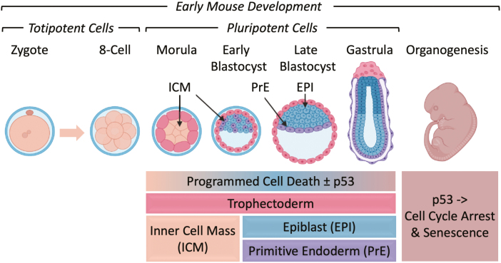
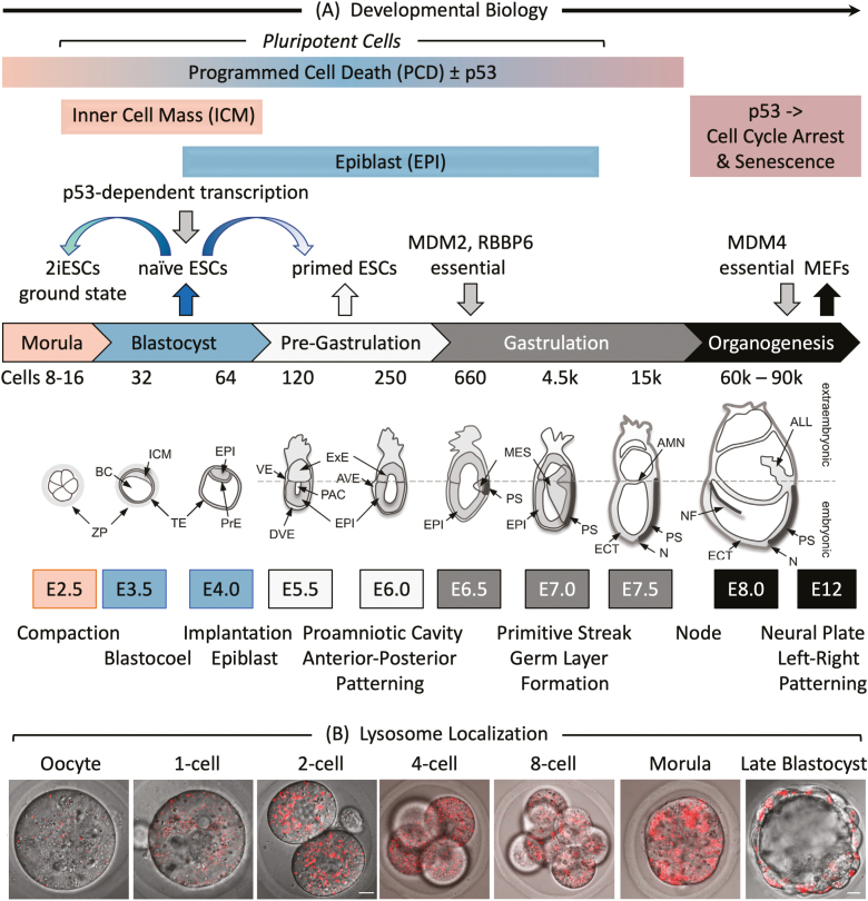
Twelve forms of programmed cell death (PCD) have been described in mammalian cells, but which of them occurs during embryonic development and the role played by the p53 transcription factor and tumor suppressor remains enigmatic. Although p53 is not required for mouse embryonic development, some studies conclude that PCD in pluripotent embryonic stem cells from mice (mESCs) or humans (hESCs) is p53-dependent whereas others conclude that it is not. Given the importance of pluripotent stem cells as models of embryonic development and their applications in regenerative medicine, resolving this enigma is essential. This review reconciles contradictory results based on the facts that p53 cannot induce lethality in mice until gastrulation and that experimental conditions could account for differences in results with ESCs. Consequently, activation of the G2-checkpoint in mouse ESCs is p53-independent and generally, if not always, results in noncanonical apoptosis. Once initiated, PCD occurs at equivalent rates and to equivalent extents regardless of the presence or absence of p53. However, depending on experimental conditions, p53 can accelerate initiation of PCD in ESCs and late-stage blastocysts. In contrast, DNA damage following differentiation of ESCs in vitro or formation of embryonic fibroblasts in vivo induces p53-dependent cell cycle arrest and senescence.

Keywords: apoptosis; cell cycle; differentiation; embryo; p53; pluripotent; programmed cell death; stem cells.


Figures

None
Neither cell cycle arrest nor programmed cell death requires p53 prior to gastrulation, at which stage DNA damage induces p53-dependent cell cycle arrest and senescence.
Figure 1.
Figure 1.
Early mouse embryonic development. (A) The number of cells, days post-coitum (E2.5-E12), and morphogenetic events are indicated. ALL, allantois; AMN, amnion; AVE, anterior visceral endoderm; BC, blastocyst cavity; DVE, distal visceral endoderm; ECT, ectoderm; EPI, epiblast; ExE, extraembryonic ectoderm; ICM, inner cell mass; MES, mesoderm; N, node; NF, neural fold; PAC, proamniotic cavity; PrE, primitive endoderm; PS, primitive streak; TE, trophectoderm; VE, visceral endoderm; ZP, zona pellucida. Adapted from Ref. . Preimplantation development begins with totipotent blastomeres (1-8 cell stage) encapsulated by the zona pellucida. Totipotent cells can give rise to both placental and embryonic cells. When the blastomeres develop cell-to-cell adhesion (compaction), the outer blastomeres differentiate into the trophectoderm while the remaining blastomeres form the inner cell mass. The epithelial trophoblast cells (trophectoderm) are multipotent; they differentiate only into cells required for implantation and placentation. The inner cell mass (recognized upon formation of a blastocoel cavity) differentiates into the epiblast and the primitive endoderm. Postimplantation development begins when the primitive endoderm differentiates into multipotent visceral and parietal endoderm. Mesoderm and ectoderm are derived from the epiblast during gastrulation. Gastrulation begins at the primitive streak, from which mesoderm and endoderm progenitor’s ingress and begin to differentiate. Mouse embryonic fibroblasts (MEFs) are derived from E12-E14 embryos. Ablation of the Mdm2, Rbbp6, or Mdm4 gene is lethal in embryos at the indicated times. Mouse embryonic stem cells (mESCs) are derived from the epiblast in blastocysts. mESCs cultured in the presence of serum and LIF interleukin-6 are considered “naïve” pluripotent cells, because they can give rise to all the cells of the embryo, but not to the trophectoderm. Naïve mESCs cultured in defined medium (no serum) containing 2 metabolic inhibitors are considered totipotent “ground-state” ESCs (2iESCs), because they give rise to both extraembryonic and embryonic cells. Naïve mESCs cultured in the presence of activin and fibroblast growth factor generate pluripotent “primed” ESCs, because they give rise to the same cells as “naïve mESCs,” but they cannot generate chimeric animals. Human embryonic stem cells (hESCs) and mouse epiblast stem cells (EpiSCs) are derived from the epiblast of post-implantation blastocysts. (B) Images of LysoTracker Red stained oocytes and preimplantation embryos revealed that the number of lysosomes increased after fertilization. Scale bar is 10 µm.
Figure 2.
Figure 2.
Cell cycle arrest and apoptosis in naïve ESCs are not dependent on p53. (A) Doxorubicin/Adriamycin (Dox) induced DNA damage (γH2AX expression), DNA damage response (PARP to c-PARP cleavage) and apoptosis (CASP3 to c-CASP3 cleavage) in ESCs derived from p53+/+ and p53−/− mouse blastocysts (BD-ESCs, “chronic phenotype”). ESCs were cultured with or without 500 nM Dox. At the times indicated, attached and unattached cells were combined, and total cellular proteins analyzed by immunoblotting. (B) PCD was detected by translocation of AIFM (red) from cytoplasm to nuclei (blue) in BD-ESCs cultured with 500 nM Dox for 16 h. Scale bar is 15 µm. (C) A transient accumulation of cells with 4N DNA content is characteristic of a DNA damage-induced G2-arrest. The G2-checkpoint was activated within 24 h and apoptosis within 72 h by 50 nM Dox in both conditional knockout p53−/− ESCs and their p53+/+ parent (cKO-ESCs, “acute phenotype”). Attached and unattached cells were combined, and their DNA content quantified by fluorescence-activated cell sorting. Cells with <2N DNA content (apoptotic cells) and cells with 4N DNA content (G2/M phase cells) are indicated. Equivalent results were obtained with BD-ESCs. (D) Cells with <2N DNA content were quantified as a function of time cultured with Dox and normalized to 0% at zero hours. Error bars indicate ±SEM. Panels A and B are from Fig. 3B and C in Ref. and panels C and D are from Figs. 2 and S2 in Ref. .
Figure 3.
Figure 3.
p53 activity and MEF PCD response at the beginning of mouse development. (A) p53 activity assayed in embryos isolated from mice homozygous for reporter genes expressing enhanced green fluorescence protein driven by either the Cdkn1a/p21 or the Bbc3/Puma gene’s p53 response element. At embryonic day E3.5, fluorescence was detected in the inner cell mass (ICM), and trophectoderm (TE) of blastocysts. The large blastocoel cavity identifies these examples as late-stage blastocysts containing early epiblast (Fig. 1). At embryonic day E6.5, fluorescence was detected in the epiblast but not in the extraembryonic tissue of gastrula. (B) MEFs cultured for 24 h with doxorubicin and then stained for “apoptosis-inducing factor” AIFM. Scale bar is 15 μm. Translocation of AIFM from mitochondria to nucleus occurred in both p53+/+ and p53−/− cells, thereby confirming non-canonical apoptosis in MEFs treated with 500 nM doxorubicin, but not in MEFs with 50 nM doxorubicin.

References

    1. Galluzzi L, Bravo-San Pedro JM, Vitale I, et al. . Essential versus accessory aspects of cell death: recommendations of the NCCD 2015. Cell Death Differ. 2015;22(1):58-73. https://doi.org/10.1038/cdd.2014.137 - DOI - PMC - PubMed
    1. Wyllie AH, Kerr JF, Currie AR.. Cell death: the significance of apoptosis. Int Rev Cytol. 1980;68:251-306. https://doi.org/10.1016/s0074-7696(08)62312-8 - DOI - PubMed
    1. Hardy K, Handyside AH, Winston RM.. The human blastocyst: cell number, death and allocation during late preimplantation development in vitro. Development. 1989;107(3):597-604. - PubMed
    1. Fuchs Y, Steller H.. Programmed cell death in animal development and disease. Cell. 2011;147(4):742-758. https://doi.org/10.1016/j.cell.2011.10.033 - DOI - PMC - PubMed
    1. Munoz-Espin D, Canamero M, Maraver A, et al. . Programmed cell senescence during mammalian embryonic development. Cell. 2013;155(5):1104-1118. https://doi.org/10.1016/j.cell.2013.10.019 - DOI - PubMed

Substances