. 2023 Nov;84(6):362-372.
doi: 10.1007/s00056-022-00385-8. Epub 2022 Mar 18.

Significance of bone morphology and quality on the primary stability of orthodontic mini-implants: in vitro comparison between human bone substitute and artificial bone

Affiliations

Significance of bone morphology and quality on the primary stability of orthodontic mini-implants: in vitro comparison between human bone substitute and artificial bone

Sachin Chhatwani et al. J Orofac Orthop. 2023 Nov.

Abstract

Aim: This study evaluated artificial bone models against a human bone substitute to assess the primary stability of orthodontic mini-implants (OMIs) at varying implant sites with different morphologies and qualities.

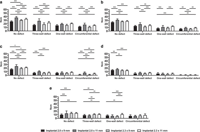
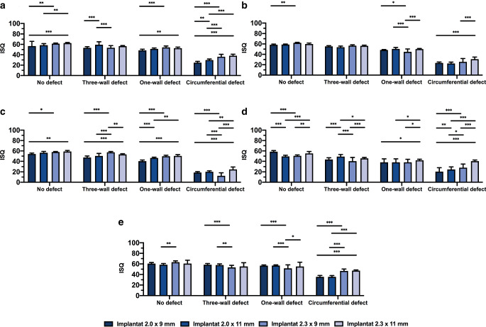
Materials and methods: A total of 1200 OMI placements of four types were inserted into four artificial bone models of different density (D1, D2, D3, D4) and into a human bone substitute (HB). The implants varied in diameter (2.0 and 2.3 mm) and length (9 and 11 mm). Each specimen had four implant sites: no defect, one-wall defect, three-wall defect, and circular defect. The implant stability quotient (ISQ) values were measured using resonance frequency analysis (RFA) and insertion placement torque values (IPT) were assessed for primary stability. Correlation analysis was performed to evaluate the different models.

Results: The highest IPT value was registered for the 2.0 mm × 11 mm implant inserted into D1 with no defect (37.53 ± 3.02 Ncm). The lowest ISQ value was measured for the 2.3 mm × 9 mm OMI inserted into D3 with a circular defect (12.33 ± 5.88) and the highest for the 2.3 mm × 9 mm implant inserted into HB with no defect (63.23 ± 2.57). A strong correlation (r = 0.64) for IPT values and a very strong correlation (r = 0.8) for ISQ values was found between D2 and HB.

Conclusion: Bone defects and bone quality affected the primary stability of implants in terms of ISQ and IPT values. Results for bone model D2 correlated very well with the HB substitution material.

Zusammenfassung: ZIEL: In dieser Studie wurden synthetische Knochenmodelle mit einem humanen Knochenersatzmaterial verglichen, um die Primärstabilität kieferorthopädischer Mini-Implantate (OMIs) an verschiedenen Implantatpositionen mit unterschiedlichen Morphologien und Qualitäten zu beurteilen.

Materialien und methoden: Insgesamt wurden 1200 OMIs von vier verschiedenen Typen in synthetische Knochenmodelle von unterschiedlicher Dichte (D1, D2, D3, D4) und in ein humanes Knochenersatzmaterial (HB) inseriert. Die Implantate variierten im Durchmesser (2,0 und 2,3 mm) und in der Länge (9 und 11 mm). Jedes Exemplar hatte 4 Implantatstellen: kein Defekt, einwandiger Defekt, dreiwandiger Defekt und zirkulärer Defekt. Der Implantatstabilitätsquotient (ISQ), gemessen mit Hilfe der Resonanzfrequenzanalyse (RFA), und die Werte des Insertionsdrehmoments (IPT) wurden für die Primärstabilität bewertet. Zur Evaluation der verschiedenen Modelle wurde eine Korrelationsanalyse durchgeführt.

Ergebnisse: Der höchste IPT-Wert wurde für das 2,0 × 11 mm Implantat registriert, für die Insertion in D1 ohne Defekt (37,53 ± 3,02 Ncm). Der niedrigste ISQ-Wert wurde für das 2,3 × 9 mm OMI gemessen, bei Einbringen in D3 mit einem zirkulärem Defekt (12,33 ± 5,88), und der höchste für das 2,3 × 9 mm Implantat, das in HB ohne Defekt eingesetzt wurde (63,23 ± 2,57). Es wurde eine hohe Korrelation (r = 0,64) für IPT-Werte und eine sehr hohe Korrelation (r = 0,8) für ISQ-Werte zwischen D2 und HB festgestellt.

Schlussfolgerung: Knochendefekte und Knochenqualität beeinflussten die Primärstabilität von Implantaten in Bezug auf ISQ- und IPT-Werte. Die Ergebnisse für das Knochenmodell D2 korrelierten sehr stark mit dem HB-Ersatzmaterial.

Keywords: Bone defects; Bone quality; Insertion placement torque; Resonance frequency analysis; Skeletal anchorage.


Conflict of interest statement

S. Chhatwani, O. Kouji-Diehl, K. Kniha, A. Modabber, F. Hölzle, J. Szalma, G. Danesh and S.C. Möhlhenrich declare that they have no competing interests.

Figures

Fig. 1
Fig. 1
Orthodontic mini-implants (OMI) placed in artificial bone (a no defect, c three-wall defect, e one-wall defect, g circular defect) and in human bone allograft (b no defect, d three-wall defect, f one-wall defect, h circular defect) Kieferorthopädische Mini-Implantate (OMI) in synthetischem Knochen (a kein Defekt, c dreiwandiger Defekt, e einwandiger Defekt, g zirkulärer Defekt) und in menschlichem Knochenallotransplantat (b kein Defekt, d dreiwandiger Defekt, f einwandiger Defekt, h zirkulärer Defekt)
Fig. 2
Fig. 2
Bar chart with mean values (standard deviation) for insertion placement torque in Ncm depending on bone quality, defect size, implant diameter and implant length. Artificial bone models of different density (a D1, b D2, c D3, d D4). Human bone substitute (e). ***p < 0.001, **p < 0.005, *p < 0.05 Balkendiagramm mit Mittelwerten (Standardabweichung) für das Eindrehmoment in Ncm in Abhängigkeit von Knochenqualität, Defektgröße, Implantatdurchmesser und Implantatlänge. Synthetische Knochenmodelle mit unterschiedlicher Dichte (a D1, b D2, c D3, d D4). Humanes Knochenersatzmaterial (e). ***p < 0,001, **p < 0,005, *p < 0,05
Fig. 3
Fig. 3
Significance of p-values for post hoc Tukey multiple comparison tests for pairwise comparison of bone defect sizes. D1–D4 Artificial bone models of different density, HB human bone, ISQ implant stability quotient, Ncm for insertion placement torque Signifikanz der p-Werte für Post-hoc-Tukey-Mehrfachvergleichstests zum paarweisen Vergleich der Knochendefektgrößen. D1–D4 Synthetische Knochenmodelle unterschiedlicher Dichte, HB humaner Knochen, ISQ Implantatstabilitätsquotienten, Ncm für das Eindrehmoment
Fig. 4
Fig. 4
Significance of p-values for post hoc Tukey multiple comparison tests for pairwise comparison of bone qualities. D1–D4 Artificial bone models of different density, HB human bone substitute, ISQ implant stability quotient, Ncm for insertion placement torque Signifikanz der p-Werte für Post-hoc-Tukey-Mehrfachvergleichstests zum paarweisen Vergleich der Knochenqualität. D1–D4 Synthetische Knochenmodelle unterschiedlicher Dichte, HB humaner Knochen, ISQ Implantatstabilitätsquotienten, Ncm für das Eindrehmoment
Fig. 5
Fig. 5
Bar chart with mean values (standard deviation) of implant stability quotient (ISQ) depending on bone quality, defect size, implant diameter and implant length. Artificial bone models of different density (a D1, b D2, c D3, d D4). Human bone substitute (e). ***p < 0.001, **p < 0.005, *p < 0.05 Balkendiagramm mit Mittelwerten (Standardabweichung) des Implantatstabilitätsquotienten (ISQ) in Abhängigkeit von Knochenqualität, Defektgröße, Implantatdurchmesser und Implantatlänge. Synthetische Knochenmodelle mit unterschiedlicher Dichte (a D1, b D2, c D3, d D4). Humanes Knochenersatzmaterial (e). ***p < 0,001, **p < 0,005, *p < 0,05
Fig. 6
Fig. 6
Correlation of insertion placement torque in Ncm (a) and implant stability quotient (ISQb) for human bone and artificial bone models Korrelation des Insertionsdrehmoments in Ncm (a) und des Implantatstabilitätsquotienten (ISQb) für humanen Knochen und synthetische Knochenmodelle

Similar articles

Cited by

References

    1. Augthun M, Conrads G. Microbial findings of deep peri-implant bone defects. Int J Oral Maxillofac Implant. 1997;12(1):1–12. - PubMed
    1. Baumgaertel S. Temporary skeletal anchorage devices: the case for miniscrews. Am J Orthod Dentofac Orthop. 2014;145:560. doi: 10.1016/j.ajodo.2014.03.009. - DOI - PubMed
    1. Bayarchimeg D, Namgoong H, Kim BK, et al. Evaluation of the correlation between insertion torque and primary stability of dental implants using a block bone test. J Periodontal Implant Sci. 2013;43:30. doi: 10.5051/jpis.2013.43.1.30. - DOI - PMC - PubMed
    1. Becker W, Sennerby L, Bedrossian E, et al. Implant stability measurements for implants placed at the time of extraction: a cohort, prospective clinical trial. J Periodontol. 2005;76:391–397. doi: 10.1902/jop.2005.76.3.391. - DOI - PubMed
    1. Chen ST, Buser D. Clinical and esthetic outcomes of implants placed in postextraction sites. Int J Oral Maxillofac Implant. 2009;24:186–217. - PubMed

LinkOut - more resources