. 2022 Jul;49(8):2716-2722.
doi: 10.1007/s00259-022-05764-2. Epub 2022 Mar 19.

Spatiotemporal dynamics of brain function during the natural course in a dental pulp injury model

Affiliations

Spatiotemporal dynamics of brain function during the natural course in a dental pulp injury model

Feiyan Yu et al. Eur J Nucl Med Mol Imaging. 2022 Jul.

Abstract

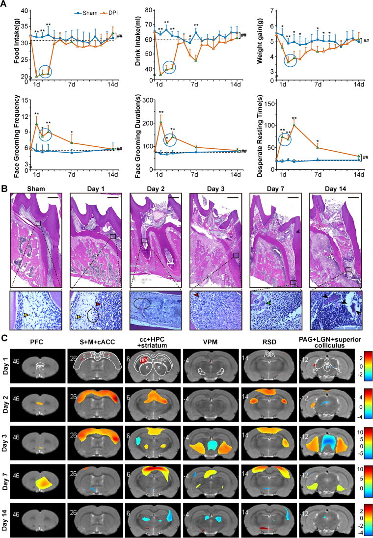
Purpose: Toothache, a common disorder afflicting most people, shows distinct features at different clinical stages. This study aimed to depict metabolic changes in brain and investigate the potential mechanism involved in the aberrant affective behaviors during the natural process of toothache.

Methods: We investigated the spatiotemporal patterns of brain function during the natural course of toothache in a rat model of dental pulp injury (DPI) by using positron emission tomography (PET).

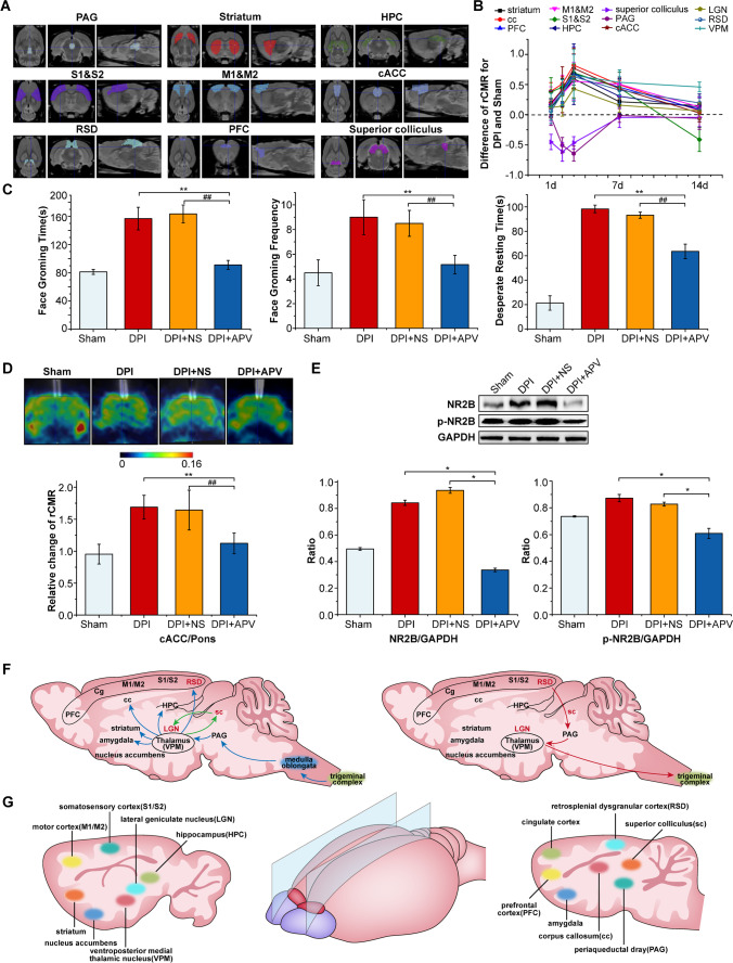
Results: Glucose metabolism peaked on the 3rd day and gradually decreased in several brain regions after DPI, which was in line with the behavioral and histological results. PET imaging showed that visual pathway was involved in the regulation of toothache. Meanwhile, the process of emotional regulation underlying toothache was mediated by N-methyl-D-aspartic receptor subunit 2B (NR2B) in the caudal anterior cingulate cortex (cACC).

Conclusion: Our results revealed the spatiotemporal neurofunctional patterns during toothache process and preliminarily elucidated the role of NR2B in cACC in the regulation of toothache-related affective behaviors.

Keywords: Caudal anterior cingulate cortex (cACC); Dental pulp injury (DPI); Positron emission tomography (PET); Toothache.


Conflict of interest statement

The authors declare no competing interests.

Figures

Fig. 1
Fig. 1
Establishment of DPI verified by behavioral assessment, Histology and PET molecular imaging. A Behavioral changes of food intake, drink intake, average weight gain, face grooming frequency, face grooming time, and freezing time in DPI. B HE staining images showed the development changes of pulpitis during the natural course of DPI (bar = 250 μm). The yellow arrows represent normal pulp cells, the red arrows indicate neutrophils, the green arrows indicate osteoclasts, the black circles represent the border of normal pulp and inflammatory tissue, and the black arrows indicate necrotic tooth tissue (400 ×). C Brain regions that showed significant glucose metabolism changes on days 1, 2, 3, 7, and 14 after DPI operation in rats (data are shown as mean ± SD, n = 6 in each group; p < 0.01, d day)
Fig. 2
Fig. 2
Nerve conduction pathway related to toothache in DPI. A The transverse (left), coronal (middle), and sagittal (right) of different brain regions of the rat using different colors, mainly including PAG, striatum, HPC, S1& S2, M1&M2, cACC, RSD, PFC, superior colliculus et al. B The rCMR of each ROI of the 12 brain regions after DPI by PMOD. The p values of relative change of rCMR in each group were shown in Table S2 (two independent samples t-test; n = 6 in each group, d day). CE Face grooming time and frequency, and desperate resting time, glucose metabolism after injecting APV in cACC region, and the expression of NR2B and p-NR2B proteins were analyzed in DPI + APV group compared to DPI and DPI + NS groups (data are shown as mean ± SD, **, ##: p < 0.01, one-way ANOVA test. DPI dental pulp injury; NS normal saline). F Schematic of major ascending (bottom-up) pathways from the medulla oblongata to the brain that are activated by noxious stimuli related to toothache (left) and descending (top-down) pathways that modulate transmission of ascending nociceptive signals (right). G Schematic of the location of brain regions that showed significant glucose metabolism changes in sagittal view after DPI

Similar articles

Cited by

References

    1. Clark GT. Persistent orodental pain, atypical odontalgia, and phantom tooth pain: when are they neuropathic disorders? J Calif Dent Assoc. 2006;34(8):599–609. - PubMed
    1. Danyluk H, Ishaque A, Ta D, Yang YH, Wheatley BM, Kalra S, et al. MRI texture analysis reveals brain abnormalities in medically refractory trigeminal neuralgia. Front Neurol. 2021;12:626504. doi: 10.3389/fneur.2021.626504. - DOI - PMC - PubMed
    1. Tohyama S, Hung PS, Cheng JC, Zhang JY, Halawani A, Mikulis DJ, et al. Trigeminal neuralgia associated with a solitary pontine lesion: clinical and neuroimaging definition of a new syndrome. Pain. 2020;161(5):916–925. doi: 10.1097/j.pain.0000000000001777. - DOI - PMC - PubMed
    1. Danyluk H, Andrews J, Kesarwani R, Seres P, Broad R, Wheatley BM, et al. The thalamus in trigeminal neuralgia: structural and metabolic abnormalities, and influence on surgical response. BMC Neurol. 2021;21(1):290. doi: 10.1186/s12883-021-02323-4. - DOI - PMC - PubMed
    1. Tian M, He X, Jin C, He X, Wu S, Zhou R, et al. Transpathology: molecular imaging-based pathology. Eur J Nucl Med Mol Imaging. 2021;48(8):2338–2350. doi: 10.1007/s00259-021-05234-1. - DOI - PMC - PubMed

LinkOut - more resources