. 2022 May:199:114993.
doi: 10.1016/j.bcp.2022.114993. Epub 2022 Mar 15.

(+)-Catharanthine potentiates the GABAA receptor by binding to a transmembrane site at the β(+)/α(-) interface near the TM2-TM3 loop

Affiliations

(+)-Catharanthine potentiates the GABAA receptor by binding to a transmembrane site at the β(+)/α(-) interface near the TM2-TM3 loop

Hugo R Arias et al. Biochem Pharmacol. 2022 May.

Abstract

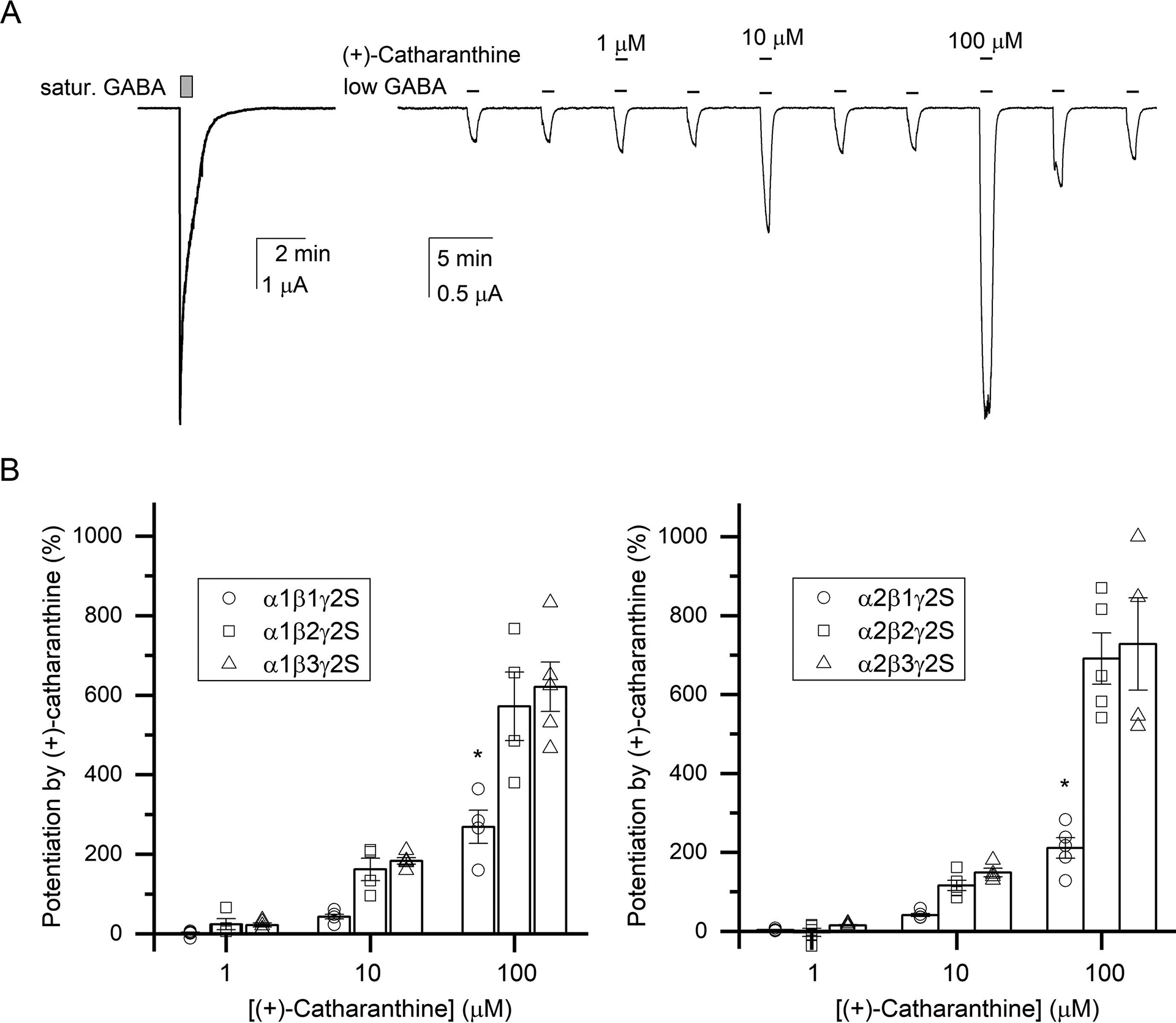
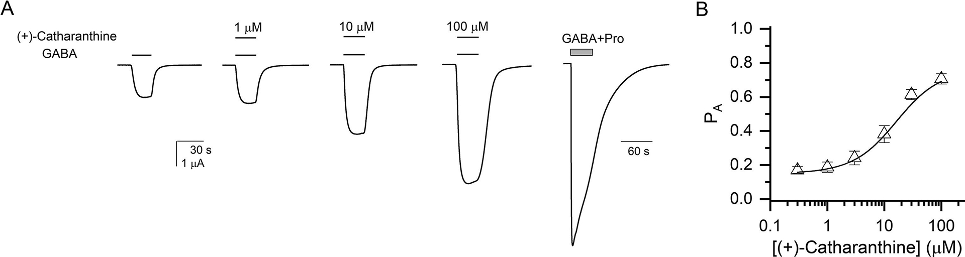
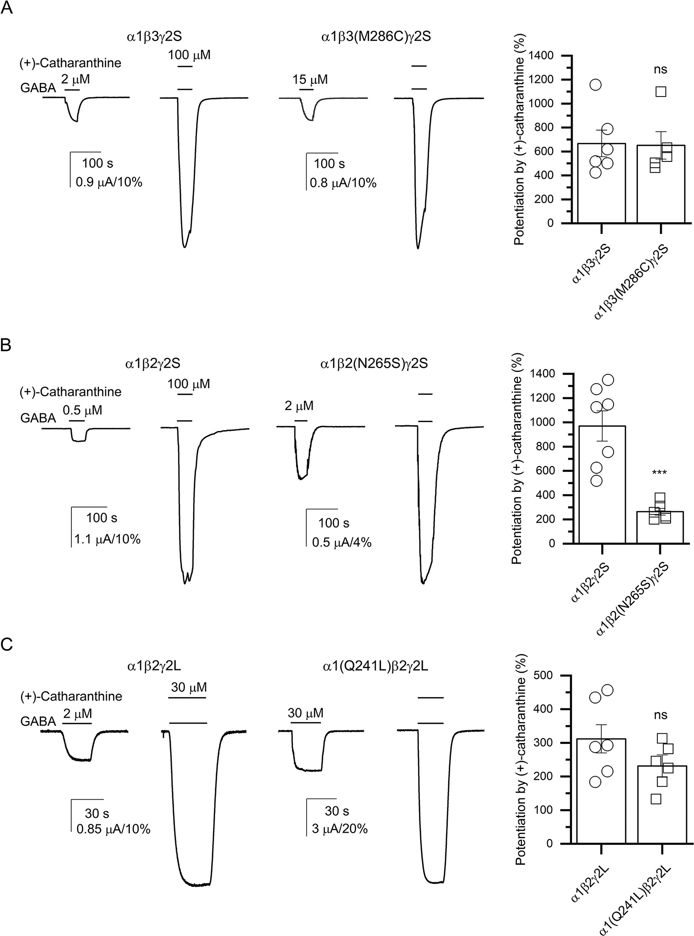
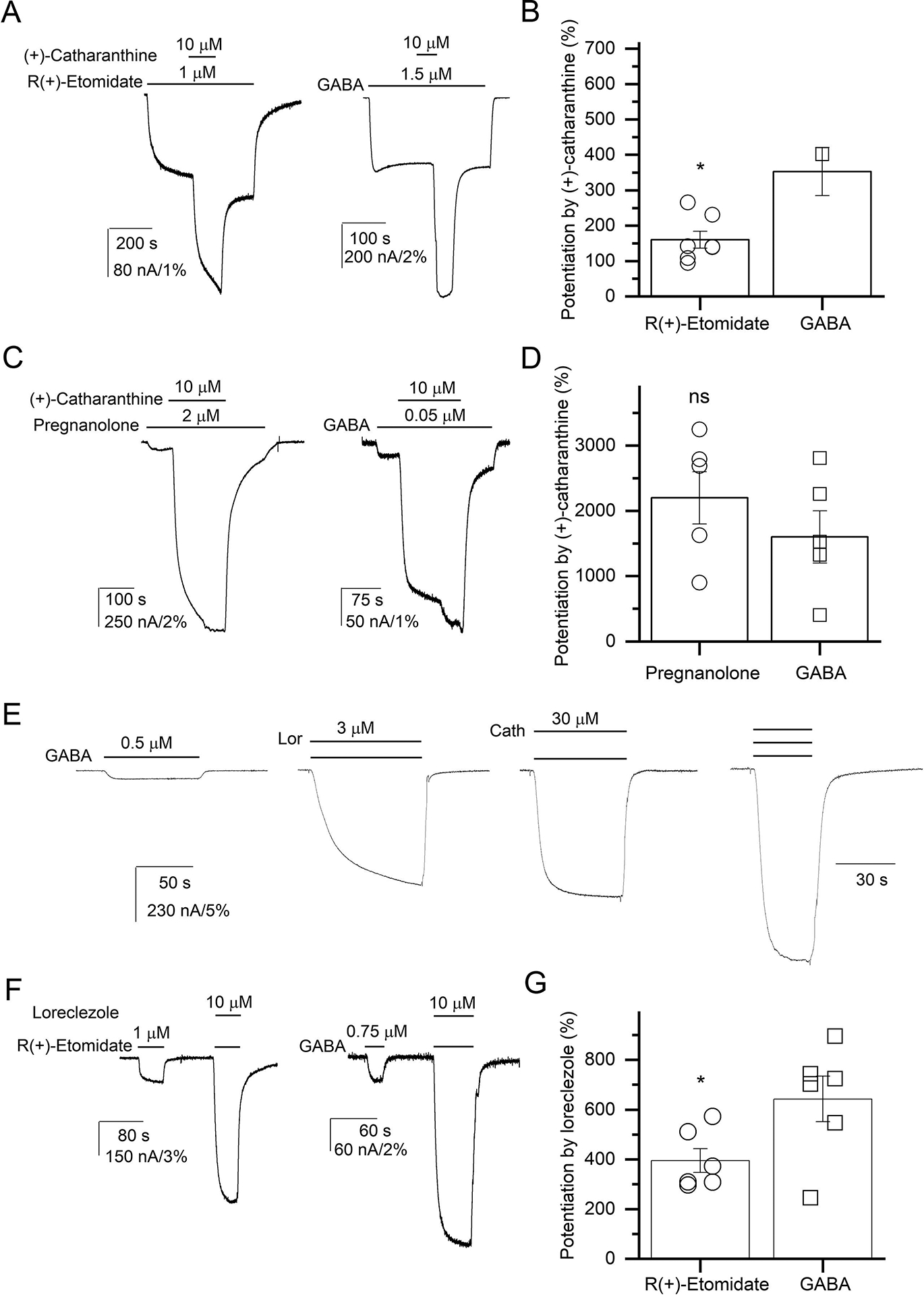
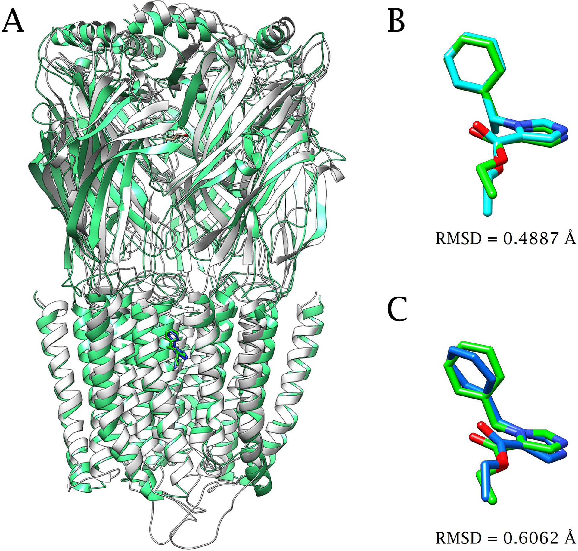
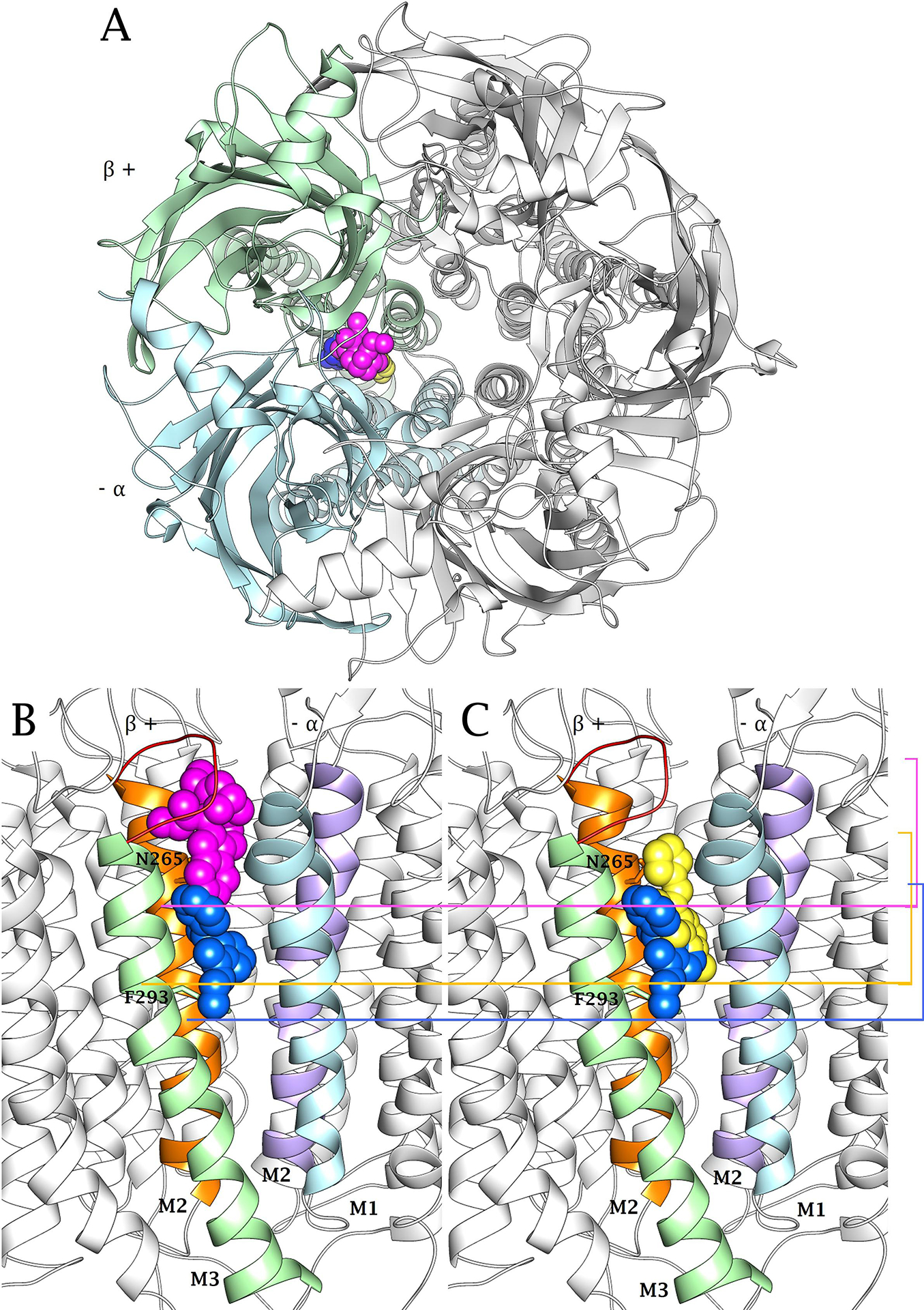
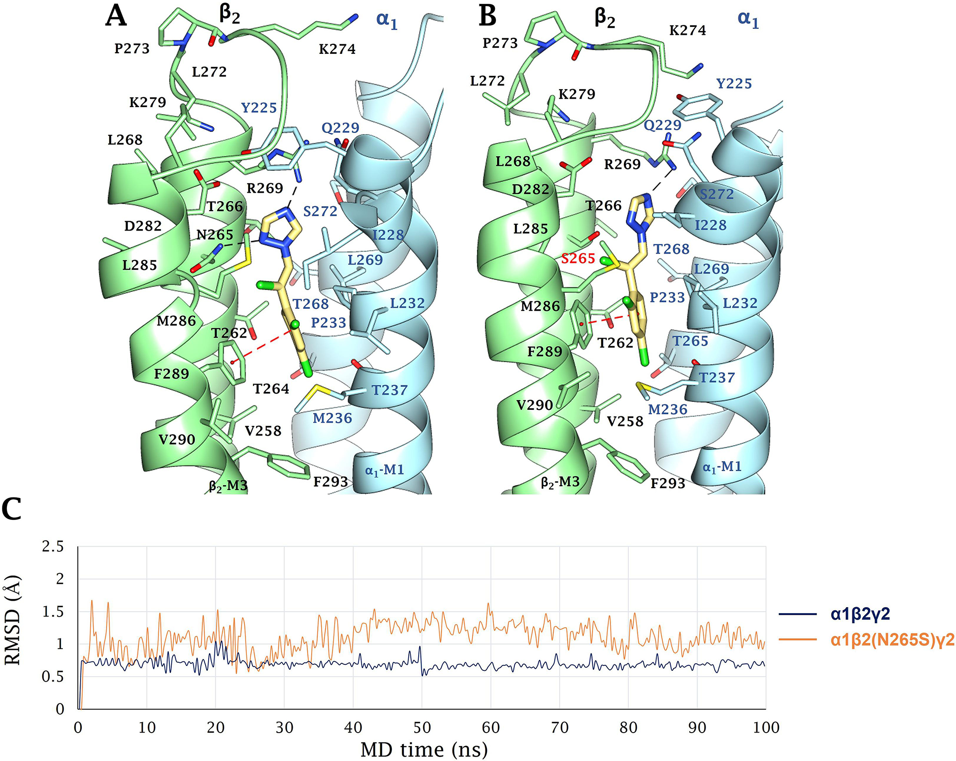
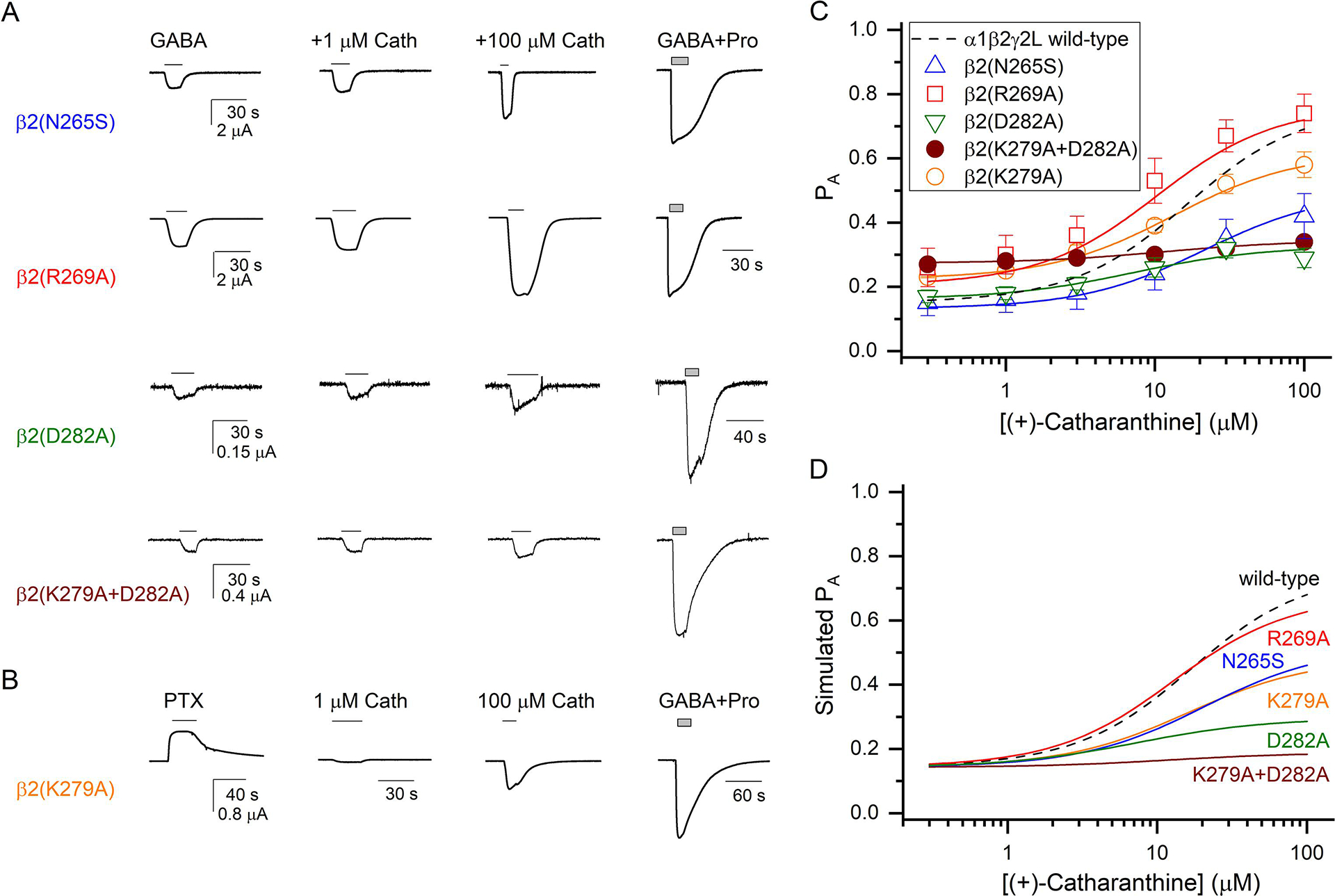
(+)-Catharanthine, a coronaridine congener, potentiates the γ-aminobutyric acid type A receptor (GABAAR) and induces sedation through a non-benzodiazepine mechanism, but the specific site of action and intrinsic mechanism have not beendefined. Here, we describe GABAAR subtype selectivity and location of the putative binding site for (+)-catharanthine using electrophysiological, site-directed mutagenesis, functional competition, and molecular docking experiments. Electrophysiological and in silico experiments showed that (+)-catharanthine potentiates the responses to low, subsaturating GABA at β2/3-containing GABAARs 2.4-3.5 times more efficaciously than at β1-containing GABAARs. The activity of (+)-catharanthine is reduced by the β2(N265S) mutation that decreases GABAAR potentiation by loreclezole, but not by the β3(M286C) or α1(Q241L) mutations that reduce receptor potentiation by R(+)-etomidate or neurosteroids, respectively. Competitive functional experiments indicated that the binding site for (+)-catharanthine overlaps that for loreclezole, but not those for R(+)-etomidate or potentiating neurosteroids. Molecular docking experiments suggested that (+)-catharanthine binds at the β(+)/α(-) intersubunit interface near the TM2-TM3 loop, where it forms H-bonds with β2-D282 (TM3), β2-K279 (TM2-TM3 loop), and β2-N265 and β2-R269 (TM2). Site-directed mutagenesis experiments supported the in silico results, demonstrating that the K279A and D282A substitutions, that lead to a loss of H-bonding ability of the mutated residue, and the N265S mutation, impair the gating efficacy of (+)-catharanthine. We infer that (+)-catharanthine potentiates the GABAAR through several H-bond interactions with a binding site located in the β(+)/α(-) interface in the transmembrane domain, near the TM2-TM3 loop, where it overlaps with loreclezole binding site.

Keywords: (+)-Catharanthine; Coronaridine congeners; Electrophysiology; Molecular docking; Molecular dynamics; Positive allosteric modulators.


Conflict of interest statement

Declarations of interest: none

Figures

Figure 1.
Figure 1.
(+)-Catharanthine structure. PubChem CID: 418553.
Figure 2.
Figure 2.
(+)-Catharanthine-induced potentiation of subsaturating GABA responses at GABAARs expressed in X. laevis oocytes. (A) Representative EC5 GABA (2 μM)-induced currents in α1β3γ2S GABAARs in the absence and presence of 1, 10, or 100 μM (+)-catharanthine, and a trace showing a response to 1 mM GABA in the same cell. The currents were recorded at −70 mV. (B) Summary of GABAAR potentiation by 1, 10, or 100 μM (+)-catharanthine. Data (n = 4–5 oocytes each) show the change of the GABA response as a percentage of the control GABA response [mean ± SEM of the EC5 GABA before and after the co-application with (+)-catharanthine]. Two-way ANOVA followed by Holm-Sidak’s multiple comparisons test analyses indicated that (+)-catharanthine-induced potentiation was higher at β2/β3- vs β1-containing GABAARs (*p < 0.0001), whereas no significant differences were observed between α1- and α2-containing GABAARs containing the same β subunit (the analysis was performed using all data but split in two graphs for clarity). The apparent potentiating efficacy of 100 μM (+)-catharanthine at each receptor subtype is summarized in Table 1.
Figure 3.
Figure 3.
(+)-Catharanthine potentiated α1β2γ2L GABAARs expressed in X. laevis oocytes in a concentration-dependent manner. (A) Representative α1β2γ2L GABAAR currents elicited by 2 μM GABA (PA = 0.18) in the absence or presence of 1, 10, and 100 μM (+)-catharanthine, and a trace showing a response to 1 mM GABA + 50 μM propofol (GABA+Pro; peak PA ~1). All responses are from the same oocyte. The currents were recorded at −60 mV. (B) Concentration-response relationship for potentiation of the α1β2γ2 GABAAR by (+)-catharanthine. The peak responses to GABA + (+)-catharanthine were normalized to the peak response to 1 mM GABA + 50 μM propofol (PA ~1) in the same cells. The data points give mean ± SEM from 5 oocytes. The curve represents a fit to (Eq. 1), yielding a KCath of 29.4 μM and a cCath of 0.231, with NCath constrained to 2 (Table 3). Fitting the concentration-response data to the Hill equation yielded an EC50 of 13.5 μM, a Hill coefficient of 1.66, and maximum potentiation of 341%.
Figure 4.
Figure 4.
The β2(N265S), but not the β3(M286C) or α1(Q241L), mutation, reduces (+)-catharanthine-induced potentiation. Representative current traces of wild-type or mutant receptors activated by (A) EC5 GABA [2 μM GABA in α1β3γ2S wild-type and 15 μM GABA in β3(M286C) mutant]; (B) EC5 GABA [0.5 μM GABA in α1β2γ2S wild-type and 2 μM GABA in β2(N265S) mutant], or (C) EC15 GABA [2 μM GABA in α1β2γ2L wild-type and 30 μM GABA in α1(Q241L) mutant], in the absence or presence of 100 μM (A and B) or 30 μM (+)-catharanthine (C). The calibration bars show current amplitude in μA, and in % of the peak response to saturating GABA for scaling purposes. The currents were recorded at −70 mV in A and B, and at −60 mV in C. A summary of wild-type and mutant receptor potentiation by (+)-catharanthine, expressed as mean ± SEM % change in GABA response, is shown next to the current traces. Student’s t-test analysis indicated that the β2(N265S) mutation reduces potentiation by (+)-catharanthine (p < 0.01; n = 6–7 oocytes), whereas neither the β3(M286C) nor α1(Q241L) mutation affected potentiation by (+)-catharanthine (p > 0.05; n = 5–6 oocytes for each receptor).
Figure 5.
Figure 5.
Steric vs allosteric interactions between (+)-catharanthine and R(+)-etomidate, (+)-catharanthine and loreclezole, and R(+)-etomidate and loreclezole. (A) Sample traces showing potentiation of 1 μM R(+)-etomidate and 1.5 μM GABA-activated α1β2γ2L GABAARs by 10 μM (+)-catharanthine. (B) Percent of change of R(+)-etomidate- or GABA-elicited responses by 10 μM (+)-catharanthine. Student’s t-test analysis of data indicated that the observed change is statistically significantly (p = 0.012; n = 5–6 oocytes). (C) Sample traces showing potentiation of 2 μM pregnanolone and 0.05 μM GABA-activated α1β2γ2L GABAARs by 10 μM (+)-catharanthine. (D) Percent of change of pregnanolone- or GABA-elicited responses by 10 μM (+)-catharanthine. Student’s t-test analysis of data indicated that the observed change is statistically significantly (p > 0.05; n = 5 oocytes per agonist). (E) Sample traces showing activation of α1β2γ2 GABAARs by 0.5 μM GABA, GABA + 3 μM loreclezole (Lor), GABA + 30 μM (+)-catharanthine (Cath), or GABA + loreclezole + (+)-catharanthine. (F) Sample traces showing potentiation of 1 μM R(+)-etomidate and 0.75 μM GABA-activated α1β2γ2 GABAARs by 10 μM loreclezole. (G) Percent of change of R(+)-etomidate- or GABA-elicited responses by 10 μM loreclezole. Student’s t-test analysis of data indicated that the observed change is statistically significant (p = 0.038; n = 6 oocytes for each receptor). All currents were recorded at −60 mV. The calibration bars show current amplitude in μA, and in % of the peak response to 1 mM GABA + 50 μM propofol for scaling purposes.
Figure 6.
Figure 6.
Etomidate docking results in cryo-EM structure and homology-built model (HM) of α1β2γ2 GABAAR. (A) Superposition of 6X3V (receptor and etomidate in green) and the adduct formed by etomidate (blue) docked within the HM built on 6HUJ (white). Ligand superposition and root mean squared deviation (RMSD) between the orientation of the cryo-EM R(+)-etomidate (green) and B. the docked conformer (cyano) within 6X3V, and C. the docking pose within the HM (blue). The docking poses obtained for R(+)-etomidate have an RMSD for the heavy atom of 0.489 Å (B) and 0.606 Å (C) from the original PDB coordinates and HM, respectively.
Figure 7.
Figure 7.
Predicted binding mode of loreclezole, R(+)-etomidate, and (+)-catharanthine to the α1β2γ2 GABAAR model as an example of β2/3-containing receptors. (A) Transversal view of the GABAAR model showing the β2(+)(light blue)/α1(−)(light green) interface comprising the binding sites for loreclezole (yellow), R(+)-etomidate (blue), and (+)-catharanthine (purple) (represented as spheres). (B,C) Longitudinal view of the GABAAR model showing the binding area for each ligand (represented as spheres). The bottom limits, showed as colored lines and brackets, support non-overlapping areas for (+)-catharanthine (purple) and R(+)-etomidate (blue) (B) as well as partially overlapping areas for loreclezole (yellow) and R(+)-etomidate (blue) (C), and for loreclezole (yellow) and (+)-catharanthine (purple) [compare (B) and (C)], respectively. Interestingly, only (+)-catharanthine interacted with the ECD-TMD junction (red coil). Similitudes and differences with the dockings to the α2β1γ2 GABAAR model as an example of β1-containing receptors are included in Table 2.
Figure 8.
Figure 8.
Predicted binding mode of (+)-catharanthine at the wild-type and mutant GABAAR models. (+)-Catharanthine interacts with the β2(+)/α1(−) interface, establishing vdW contacts with several α1 (light blue) and β2 (light green) residues, and additional H-bond interactions delineated as follow: (A) (+)-Catharanthine (purple) forms H-bonds with β2-N265 and β2-R269 (both at TM2), β2-D282 (TM3), and K279 (TM2-TM3 loop), respectively (black dashed lines). In the α1β2(N265S)γ2 (B), α1β2(D282A)γ2 (C), α1β2(R269A)γ2 (D), α1β2(K279A)γ2 (E), and α1β2(D282A+K279A)γ2 (F) mutants, the respective H-bonds with S265, A282, A269, and A279 (highlighted in red) are lost, although vdW interactions with S265 and A269 are maintained. In the case of the β2(N265S) mutation, even considering that Ser can form a H-bond, it is shorter than Asp, consequently the distance and the binding geometry were not optimal for the formation of a H-bond with (+)-catharanthine. (G) RMSD plots for the molecular dynamics (MD) simulations (100 ns) of (+)-catharanthine docked to the different receptors. Similitudes and differences with the dockings to the α2β1γ2 GABAAR model as an example of β1-containing receptors are included in Table 2.
Figure 9.
Figure 9.
Predicted binding mode of loreclezole at the wild-type and mutant GABAAR models. Loreclezole interacts with the β2(+)/α1(−) interface, establishing vdW contacts with several α1 (light blue) and β2 (light green) residues, and additional H-bond or π-π stacking interactions delineated as follow: (A) Loreclezole (yellow) forms H-bonds with β2-N265 and β2-R269 (black dashed lines), and a π-π stacking interaction with β2-F289 (TM3) (cyan dashed line). (B) In the α1β2(N265S)γ2 mutant, the respective H-bonds with β2-S265 (red) and β2-R269 lost stability over the MD course. (C) RMSD plots for the MD simulations (100 ns) of loreclezole docked to the different receptors. Similitudes and differences with the dockings to the α2β1γ2 GABAAR model as an example of β1-containing receptors are included in Table 2.
Figure 10.
Figure 10.
(+)-Catharanthine-induced potentiation and activation of mutated α1β2γ2L GABAARs. (A) Representative currents elicited by GABA (0.2–10 μM; PA = 0.12–0.25) alone or in the presence of 1 or 100 μM (+)-catharanthine at α1β2γ2L GABAARs containing the β2(N265S), β2(R269A), β2(D282A), or β2(K279A+D282A) mutation. For comparison, a trace showing the response to 1 mM GABA + 50 μM propofol (GABA+Pro) in the same cell is given for each mutant. (B) Representative currents elicited by 100 μM picrotoxin (PTX), 1 or 100 μM (+)-catharanthine, or 1 mM GABA + 50 μM propofol (GABA+Pro) at the α1β2(K279A)γ2L receptor. All currents were recorded at −60 mV. (C) Concentration-response relationships for (+)-catharanthine in the mutant receptors. The data points give mean ± SEM from 5 oocytes for each receptor. The curves show fits to (Eq. 1). The fitted KCath and cCath values are provided in Table 3. The curve for the wild-type α1β2γ2L GABAAR (dashed line) is reproduced from Fig. 2. (D)Simulated concentration-response relationships for (+)-catharanthine in the wild-type and mutant receptors. The simulations were done using the fitted KCath and cCath values (Table 3) at a fixed background PA of 0.15.

Similar articles

Cited by

References

    1. Maisonneuve IM, Glick SD, Anti-addictive actions of an iboga alkaloid congener: a novel mechanism for a novel treatment, Pharmacol Biochem Behav 75(3) (2003) 607–18. - PubMed
    1. Glick SD, Kuehne ME, Raucci J, Wilson TE, Larson D, Keller RW Jr., Carlson JN, Effects of iboga alkaloids on morphine and cocaine self-administration in rats: relationship to tremorigenic effects and to effects on dopamine release in nucleus accumbens and striatum, Brain Res 657(1–2) (1994) 14–22. - PubMed
    1. Arias HR, Jin X, Feuerbach D, Drenan RM, Selectivity of coronaridine congeners at nicotinic acetylcholine receptors and inhibitory activity on mouse medial habenula, Int J Biochem Cell Biol 92 (2017) 202–209. - PMC - PubMed
    1. Glue P, Cape G, Tunnicliff D, Lockhart M, Lam F, Hung N, Hung CT, Harland S, Devane J, Crockett RS, Howes J, Darpo B, Zhou M, Weis H, Friedhoff L, Ascending Single-Dose, Double-Blind, Placebo-Controlled Safety Study of Noribogaine in Opioid-Dependent Patients, Clin Pharmacol Drug Dev 5(6) (2016) 460–468. - PubMed
    1. Brown TK, Alper K, Treatment of opioid use disorder with ibogaine: detoxification and drug use outcomes, Am J Drug Alcohol Abuse 44(1) (2018) 24–36. - PubMed

Publication types

LinkOut - more resources