Review
. 2022 Jun:486:5-14.
doi: 10.1016/j.ydbio.2022.03.004. Epub 2022 Mar 17.

Spatial regulation of endosomes in growing dendrites

Affiliations
Review

Spatial regulation of endosomes in growing dendrites

Chan Choo Yap et al. Dev Biol. 2022 Jun.

Abstract

Many membrane proteins are highly enriched in either dendrites or axons. This non-uniform distribution is a critical feature of neuronal polarity and underlies neuronal function. The molecular mechanisms responsible for polarized distribution of membrane proteins has been studied for some time and many answers have emerged. A less well studied feature of neurons is that organelles are also frequently non-uniformly distributed. For instance, EEA1-positive early endosomes are somatodendritic whereas synaptic vesicles are axonal. In addition, some organelles are present in both axons and dendrites, but not distributed uniformly along the processes. One well known example are lysosomes which are abundant in the soma and proximal dendrite, but sparse in the distal dendrite and the distal axon. The mechanisms that determine the spatial distribution of organelles along dendrites are only starting to be studied. In this review, we will discuss the cell biological mechanisms of how the distribution of diverse sets of endosomes along the proximal-distal axis of dendrites might be regulated. In particular, we will focus on the regulation of bulk homeostatic mechanisms as opposed to local regulation. We posit that immature dendrites regulate organelle motility differently from mature dendrites in order to spatially organize dendrite growth, branching and sculpting.

Keywords: Dendrite; Dendritogenesis; Directional Transport; Endosome; Lysosome; Organelle Positioning.


Figures

Fig. 1.
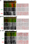
Fig. 1.. Different endosomal subtypes show distinct patterns of overall motility in dendrites of DIV8/9 cultured rat hippocampal neurons.
Dual live imaging of NSG1-cherry with the early endosome marker EEA1 (A), the recycling cargo Transferrin (Tfn) (B), and the late endosome marker Rab7 (C) in dendrites. Live imaging was carried out in DIV8/9 rat hippocampal neurons. Soma is oriented towards the right for all panels. Movies are displayed as kymographs in which a line scan along the dendrites for each movie frame is displayed to show changes in position (x-axis) over time (y-axis). Still frames of the first movie frame are shown above the kymographs. Capture rates were 2 fps (frames per second) for A and 1 fps for B and C. Early endosomes (EEs) are largely stationary (A) whereas recycling endosomes move rapidly and processively in both directions (B). Late endosomes (LEs) move intermittently with frequent reversals of direction (C). Some of the vesicle trajectories are traced in the right panels. Not all trajectories can be easily traced for Tfn because some of the motility is so fast that trajectories are often discontinuous.
Fig. 2.
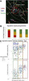
Fig. 2.. Degradative capacity is not uniform along the length of dendrites.
(A) DIV8/9 cultured hippocampal neurons were stained for the lysosomal enzyme Cathepsin B (blue), active Cathepsin D (bodipy-pepstatin; green), and LAMP1. Compartments with high degradative capacity (degradative lysosomes: containing Cathepsins) are spatially restricted to occupy mostly the soma and soma-near dendritic regions. LAMP1 is found more broadly distributed into more distal regions of dendrites (red). (B,C) Quantification of degradative capacity along dendrites was determined by feeding degradation-sensitive BSA (DQ-BSA) together with degradation-insensitive BSA (Alexa647-BSA) and determining the ratio of DQ-BSA/Alexa647-BSA. 20 μg/ml of Alexa647-BSA with 5 μg/ml of DQBSA were added to cultured rat hippocampal neurons on DIV8 and incubated overnight. Neurons were fixed and stained against either LAMP1 or CatD. Each Alexa647-BSA positive compartment was scored for intensity of Alexa647-BSA, DQ-BSA and their ratio is plotted along the X-axis. The intensity of the respective marker (LAMP1 or CatD) was determined in the same BSA-containing compartments and plotted on the Y-axis. The distance from the soma was determined for each compartment and is indicated by the symbols. Each compartment was thus scored for degradative capacity (DQ-BSA/Alexa 647-BSA ratio), intensity of LAMP1 or CatD, and for its location along the dendrite. Raw data points are shown in (C) whereas binned data (as demarked by green, yellow and red boxes in C) are shown in (B). More distal regions of dendrites have fewer highly degradative compartments (red) but still have compartments with moderate degradative capacity (B). Highly degradative compartments have high levels of LAMP1 and Cathepsin D. Moderately degradative compartments have high levels of LAMP1 but lower levels of Cathepsin D. Compartments with low/no degradative capacity have low levels of Cathepsin D, but can be LAMP1-positive or -negative (C). Highly degradative compartments are overwhelmingly found in the soma (orange circles) or in the proximal dendrite (within 25 μm of the soma along the dendrite; blue circles). Compartments with low degradative compartments can be found at all distances measured along the dendrite (squares, triangles) (C).
Fig. 3.
Fig. 3.. Late endosomes (GFP-Rab7) show strikingly different motility in axons and dendrites.
DIV8/9 hippocampal neurons were transfected with GFP-Rab7 to mark late endosomes and live imaged. Movies are displayed as kymographs in which a line scan along the dendrites for each movie frame is displayed to show changes in position (x-axis) over time (y-axis). Still frames of the first movie frame are shown above the kymographs. Capture rates were 1 fps (frames per second). Late endosomes in axons move processively retrogradely whereas late endosomes in dendrites move intermittently with frequent reversals of direction. Some of the vesicle trajectories are highlighted in the bottom panels: Red lines = stationary compartments. Blue lines = motile compartments.
Fig. 4.
Fig. 4.. Spatial regulation of endocytic flux in dendrites.
Endocytic cargos are endocytosed from the plasma membrane into early endosomes all along dendrites. Their transport along the endosomal pathway towards lysosomes (i.e. degradative flux) is regulated by the rate of maturation of early to late endosomes, rate of transport of late endosomes towards the soma, and fusion with degradative lysosomes. Many steps along this route are subject to regulation that is both temporally and spatially coordinated. Disruption of degradative flux can lead to many disturbances of signaling downstream of developmental cues and defects in dendrite growth. In older neurons, synapse formation and function as well as overall protein homeostasis are also affected by degradative flux. PM = plasma membrane. EE = early endosome. RE = recycling endosome. Tfn = transferrin. LE = late endosome. Lys = lysosome. Cat = cathepsin. TGN = trans-Golgi network. M6PR = mannose-6-phosphate receptor. LAMP = lysosome-associated membrane protein.

Similar articles

Cited by

References

    1. Adnan G, Rubikaite A, Khan M, et al., 2020. The gtpase arl8b plays a principle role in the positioning of interstitial axon branches by spatially controlling autophagosome and lysosome location. J. Neurosci. 40 (42), 8103–8118. - PMC - PubMed
    1. Aguirre-Chen C, Bülow HE, Kaprielian Z, 2011. C. elegans bicd-1, homolog of the Drosophila dynein accessory factor Bicaudal D, regulates the branching of PVD sensory neuron dendrites. Development 138 (3), 507–518. - PMC - PubMed
    1. Aiken J, Holzbaur ELF, 2021. Cytoskeletal regulation guides neuronal trafficking to effectively supply the synapse. Curr. Biol. 31 (10), R633–R650. - PMC - PubMed
    1. Al-Bassam S, Xu M, Wandless TJ, Arnold DB, 2012. Differential trafficking of transport vesicles contributes to the localization of dendritic proteins. Cell Rep. 2 (1), 89–100. - PMC - PubMed
    1. Alberi S, Boda B, Steiner P, Nikonenko I, Hirling H, Muller D, 2005. The endosomal protein NEEP21 regulates AMPA receptor-mediated synaptic transmission and plasticity in the hippocampus. Mol. Cell. Neurosci. 29 (2), 313–319. - PubMed

Substances

LinkOut - more resources