. 2022 Jun;381(3):204-216.
doi: 10.1124/jpet.121.001036. Epub 2022 Mar 19.

Neutrophil-Derived Myeloperoxidase and Hypochlorous Acid Critically Contribute to 20-Hydroxyeicosatetraenoic Acid Increases that Drive Postischemic Angiogenesis

Affiliations

Neutrophil-Derived Myeloperoxidase and Hypochlorous Acid Critically Contribute to 20-Hydroxyeicosatetraenoic Acid Increases that Drive Postischemic Angiogenesis

Juan A Azcona et al. J Pharmacol Exp Ther. 2022 Jun.

Abstract

Compensatory angiogenesis is an important adaptation for recovery from critical ischemia. We recently identified 20-hydroxyeicosatetraenoic acid (20-HETE) as a novel contributor of ischemia-induced angiogenesis. However, the precise mechanisms by which ischemia promotes 20-HETE increases that drive angiogenesis are unknown. This study aims to address the hypothesis that inflammatory neutrophil-derived myeloperoxidase (MPO) and hypochlorous acid (HOCl) critically contribute to 20-HETE increases leading to ischemic angiogenesis. Using Liquid Chromatography-Mass Spectrometry/Mass Spectrometry, Laser Doppler Perfusion Imaging, and Microvascular Density analysis, we found that neutrophil depletion and MPO knockout mitigate angiogenesis and 20-HETE production in the gracilis muscles of mice subjected to hindlimb ischemia. Furthermore, we found MPO and HOCl to be elevated in these tissues postischemia as assessed by immunofluorescence microscopy and in vivo live imaging of HOCl. Next, we demonstrated that the additions of either HOCl or an enzymatic system for generating HOCl to endothelial cells increase the expression of CYP4A11 and its product, 20-HETE. Finally, pharmacological interference of hypoxia inducible factor (HIF) signaling results in ablation of HOCl-induced CYP4A11 transcript and significant reductions in CYP4A11 protein. Collectively, we conclude that neutrophil-derived MPO and its product HOCl activate HIF-1α and CYP4A11 leading to increased 20-HETE production that drives postischemic compensatory angiogenesis. SIGNIFICANCE STATEMENT: Traditionally, neutrophil derived MPO and HOCl are exclusively associated in the innate immunity as potent bactericidal/virucidal factors. The present study establishes a novel paradigm by proposing a unique function for MPO/HOCl as signaling agents that drive critical physiological angiogenesis by activating the CYP4A11-20-HETE signaling axis via a HIF-1α-dependent mechanism. The findings from this study potentially identify novel therapeutic targets for the treatment of ischemia and other diseases associated with abnormal angiogenesis.


Figures

Fig. 1.
Fig. 1.
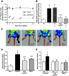
Neutrophil-derived MPO critically contributes to postischemic increases in 20-HETE production and angiogenesis. Femoral artery ligations were performed in Balb/C, Balb/C with neutrophils depleted by treating the animal with 0.5 mg of Ly6G/C antibody (i.p.), Balb/C depleted of macrophages/monocytes with 5 mg/ml Clodronate Liposomes (i.p.), and MPO−/− mice (global myeloperoxidase knock-out). (A) Flow cytometric quantitation of CD11b+Gr-1+ circulating neutrophils in blood isolated from untreated control mice and mice treated with Ly6G/C antibodies at days 0, 1-, 3-, 7-, and 14-days post ligation (mean ± S.E.M.; n = 4–6; *P < 0.05 versus corresponding controls, one-way ANOVA, repeated measures, Dunnett post hoc). (B) Hindlimb gracilis muscles were harvested 3 days post ligation and homogenized. Nonischemic muscles were used as controls. 20-HETE was measured by LC/MS/MS and samples were normalized to their corresponding nonischemic controls (mean ± S.E.M.; n = 6–8; *P < 0.05 versus corresponding NI control (9.2 ± 1.8 pg/mg protein) and #P < 0.05 versus ischemic controls (36.6 ± 4.7 pg/mg protein), one-way ANOVA, Tukey post hoc); (C) Laser Doppler Perfusion Imaging was performed to assess blood perfusion recovery in the ischemic hindlimbs at days 21 post ligation. Representative blood perfusion scans are shown for control, neutrophil-depleted, and MPO−/− mice (mean ± S.E.M.; n = 6–8; *P < 0.05 versus corresponding NI control and #P < 0.05 versus ischemic controls, two-way ANOVA, Tukey post hoc); (D) Quantitation of blood perfusion recovery to ischemic hindlimbs at days 21 (mean ± S.E.M.; n = 6–8; *P < 0.05 versus corresponding NI control and #P < 0.05 versus ischemic controls, one-way ANOVA, Tukey post hoc); (E) Ischemic gracilis muscles were also extracted at days 21 post ligation and frozen sectioned. Contralateral nonischemic gracilis muscles were used as controls. Immunofluorescent colocalization staining of tomato lectin (Green) and microvessel marker CD31 (Red) were carried out and the numbers of tomato lectin+CD31+ microvessels were counted for Microvascular Density analysis. I, Ischemic; NI, nonischemic. (mean ± S.E.M.; n = 6–8; *P < 0.05 versus corresponding NI control and #P < 0.05 versus ischemic controls, two-way ANOVA, Tukey post hoc).
Fig. 2.
Fig. 2.
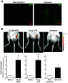
Ischemia leads to significant MPO deposition and HOCl formation in mouse hindlimb. Mice were again subjected to femoral artery ligation to induce hindlimb ischemia. (A) Gracilis muscles from nonischemic and ischemic hindlimbs were extracted and frozen sectioned 16 hours post ligation. Muscle samples were incubated with anti-MPO (1:250) antibody overnight, followed by incubation with anti-goat fluorescein isothiocyanate-conjugated secondary antibodies (1:1000) for 1 hour. Immunofluorescent microscopy was performed and representative images of MPO reactivity were shown (n = 3). Scale bar = 10 μm; (B) HOCl formation was determined and quantitated live at 30 minutes postischemia (n = 6) using the Xenogen In-vivo Imaging System (IVIS) Spectrum by injecting the FDOCl-1 HOCl-specific fluorescent probe (1 mM) directly into both hindlimb gracilis muscles for 5 minutes. In a different group of mice, HOCl (25 μM, n = 6) or lipopolysaccharide (10 μg, 1 hour, n = 4) was injected into hindlimb gracilis muscle as positive controls. Mock surgeries were performed on the contralateral hindlimbs with sterile saline injection serving as the nonischemic solvent control. Total epi fluorescence from each group was determined and quantitated compared with their respective nonischemic solvent controls. (mean ± S.E.M.; *P < 0.05, paired t test).
Fig. 3.
Fig. 3.
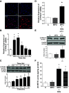
HOCl and catalytically active MPO significantly induce CYP4A11 mRNA and protein expression in HDMEC. (A) The kinetics of HOCl entry into HDMEC in vitro was determined live by immunofluorescence microscopy in cultures preloaded with FDOCl-1 (10 μM) dye. Hoescht 33342 were used to counter stain for the nuclei. The gradual increase of HOCl influx into the cultures was qualitatively assessed based on fluorescence intensity of FDOCl-1 in response to 10-minute HOCl incubation. Representative images of the time dependent HOCl entry into HDMEC are shown (n = 3). Another group of HDMEC were incubated with HOCl (15 μM) for 5 minutes. The effects of HOCl on (B) 20-HETE synthase CYP4A11 mRNA expression was assessed by quantitative real-time PCR and (C) CYP4A11 protein expression was determined using western blot analysis at 0.5, 1, 2, 4, and 16 hours post HOCl incubations (mean ± S.D.; n = 3–6; *P < 0.05 versus untreated control, one-way ANOVA, Dunnett post hoc). Additional HDMEC were also incubated in MPO alone or MPO + GOx + Glucose for 15 minutes to determine the effects of these treatments on the expression of CYP4A11 mRNA (D) and protein (E) at time points of maximal CYP4A11 expression for both parameters (1 hour for mRNA, 16 hours for protein). (mean ± S.D.; n = 3; *P < 0.05 versus untreated control, one-way ANOVA, Dunnett post hoc). Finally, (F) 20-HETE production in HDMEC exposed to MPO, MPO + GOx, or HOCl was measured as previously described using LC/MS/MS analysis. (mean ± S.D.; n = 3 in triplicates; *P < 0.05 versus vehicle controls, one-way ANOVA, Dunnett post hoc).
Fig. 4.
Fig. 4.
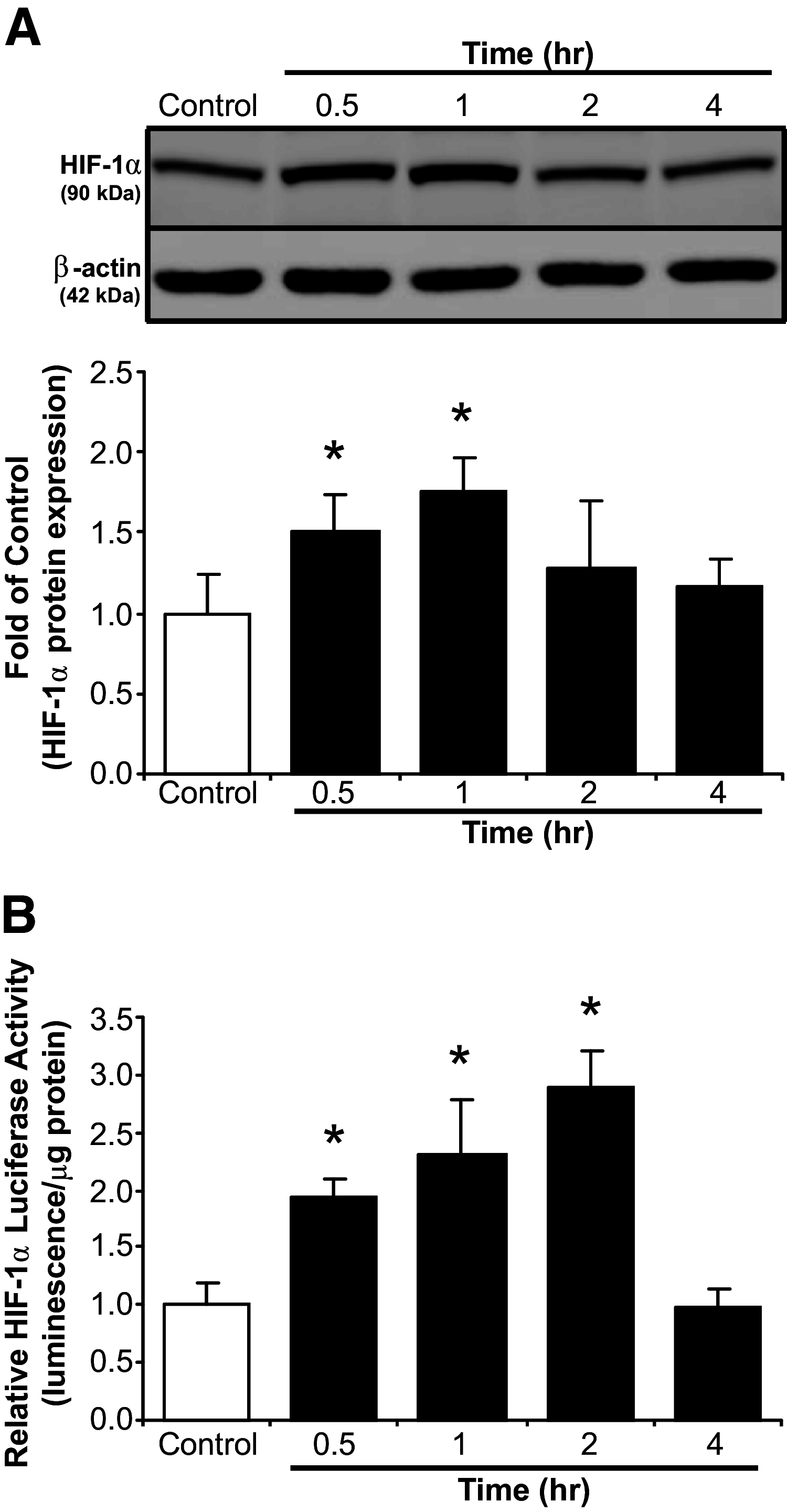
HOCl upregulates HIF1α protein expression and promotes HIF1α transcriptional activity in HDMEC. HDMEC were incubated with HOCl (15 μM) for 5 minutes. (A) HIF1α protein expression was determined at 0.5, 1, 2, and 4 hours after incubations by western blotting (mean ± S.D.; n = 3 in triplicates; *P < 0.05 versus untreated controls, one-way ANOVA, Dunnett post hoc); and (B) another group of HDMEC were transfected with a lentiviral vector carrying a luciferase reporter adjacent to multiple HIF responsive element (HRE) transcriptional sites as described in the methods. HIF transcriptional activity was measured by luciferase activity at 0.5, 1, 2, and 4 hours after 5-minute HOCl (15 μM) exposure (mean ± S.D.; n = 3; *P < 0.05 versus untreated control, one-way ANOVA, Dunnett post hoc). All data were normalized as fold of vehicle controls.
Fig. 5.
Fig. 5.
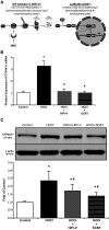
CYP4A11 induction by HOCl are HIF-dependent. HDMEC were pretreated with HIF inhibitors HIFI-V or SCBT for 16 hours (0.5 μM), and then exposed to HOCl (15 μM) for an additional 5 minutes. DMSO treated cultures were used as the vehicle control. (A) Schematic illustration of the differential mechanism of actions of HIF inhibitors HIFI-V and SCBT; (B) CYP4A11 mRNA expression was determined 1 hour after exposure to HOCl using quantitative real-time PCR and normalized to their vehicle control (mean ± S.D.; n = 3; *P < 0.05 versus vehicle controls and #P < 0.05 versus HOCl, one-way ANOVA, Tukey post hoc); and (C) CYP4A11 protein expression was examined and quantitated after 16 hours using western blot analysis. All data were normalized as fold of vehicle controls. (mean ± S.D.; n = 3–5; *P < 0.05 versus vehicle controls and #P < 0.05 versus HOCl, one-way ANOVA, Tukey post hoc).
Fig. 6.
Fig. 6.
Schematics by which neutrophil-derived MPO and HOCl contribute to 20-HETE increases that drive postischemic angiogenesis. Ischemic injury (1) such as femoral artery ligation results in recruitment of inflammatory neutrophils (2) to the target vasculature. Neutrophils then release MPO at the site of injured endothelium. MPO subsequently generates HOCl in the presence of H2O2 (3). Production of HOCl stimulates the activation of HIF-1α signaling (4), which in turn upregulates the 20-HETE synthase CYP4A11 (5). Consequently, increased CYP4A11 expression leads to increased endothelial 20-HETE production (6), which drives postischemic angiogenesis (7).

References

    1. Abbitt KB, Cotter MJ, Ridger VC, Crossman DC, Hellewell PG, Norman KE (2009) Antibody ligation of murine Ly-6G induces neutropenia, blood flow cessation, and death via complement-dependent and independent mechanisms. J Leukoc Biol 85:55–63 10.1189/jlb.0507305. - DOI - PubMed
    1. Ali MPulli BCourties GTricot BSebas MIwamoto YHilgendorf ISchob SDong AZheng W, et al. (2016) Myeloperoxidase inhibition improves ventricular function and remodeling after experimental myocardial infarction. JACC Basic Transl Sci 1:633–643 10.1016/J.JACBTS.2016.09.004. - DOI - PMC - PubMed
    1. Amaral SL, Maier KG, Schippers DN, Roman RJ, Greene AS (2003) CYP4A metabolites of arachidonic acid and VEGF are mediators of skeletal muscle angiogenesis. Am J Physiol Heart Circ Physiol 284:H1528–H1535 10.1152/ajpheart.00406.2002. - DOI - PubMed
    1. Antonelou M, Michaëlsson E, Evans RDR, Wang CJ, Henderson SR, Walker LSK, Unwin RJ, Salama AD; RAVE-ITN Investigators (2020) Therapeutic myeloperoxidase inhibition attenuates neutrophil activation, anca-mediated endothelial damage, and crescentic GN. J Am Soc Nephrol 31:350–364 10.1681/ASN.2019060618. - DOI - PMC - PubMed
    1. Banerjee D, Madhusoodanan UK, Sharanabasappa M, Ghosh S, Jacob J (2003) Measurement of plasma hydroperoxide concentration by FOX-1 assay in conjunction with triphenylphosphine. Clin Chim Acta 337:147–152 10.1016/j.cccn.2003.08.004. - DOI - PubMed

Publication types

Substances